After completing this protocol, identification of cells and tissues that are expressing the RNA probe of interest will be achieved. The representative results for this protocol are for AP-1, FosB, and TNFR41. These results, previously published by Traylor-Knowles et al.11, show spatial expression of RNA probes on adult corals that were exposed to heat stress. Two examples of different staining types are presented in Figure 1. Figure 1A is an example of diffuse tissue staining. The stain is found throughout the tissue, not only in specific cells. Here, the expression of anti-sense AP-1 is found throughout the epidermis and the oral gastrodermis11 (Figure 1A). The staining is diffuse and not segregated to one specific cell type. For this type of staining, it is particularly critical to run a sense control at the same time, as some of the results may be due to non-specific staining.
The second type of staining is cell-specific, in which the stain is only found in specific cell types. Both FosB (Figure 1B) and TNFR41 (Figure 1C) show this type of staining. Both of these probes were used on tissue samples of adult corals that had been exposed to a heat stress11. In the anti-sense panel, purple staining is observed. TNFR41 was expressed in different types of cnidocytes, a family of cell types that is only found within cnidarians (Figure 1). Specifically, it stained nematocytes that produce the microbasic mastigophore organelle (nematocysts) and spirocytes that produce spirocysts, all found within the epidermis of coral tissue. Nematocytes are also found in other tissue layers of the coral, such as the calicodermis; however, due to the sectioning performed on this particular sample, that information could not be gathered. FosB also stained cnidocytes and was specific to both spirocytes and nematocytes. For both of these probes, the sense control showed no staining, indicating that the staining for the anti-sense probe was indeed specific. These are just two types of representative results (diffuse tissue staining and cell-specific staining) of the staining techniques that may be present with different types of probes. Due to this variability, it is critical to use a sense control to determine non-specific staining.
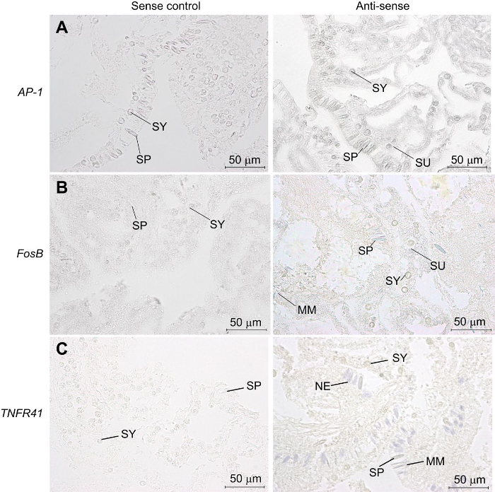
Figure 1: Representative outcome of ISH that stained specific cells. (A) In the first panel, sense control for the AP-1 probe shows that little staining is present within the slide. In the second panel, the anti-sense probe for AP-1 shows that staining with this probe is diffuse throughout the tissue. The stain is specific to the oral gastrodermis and epidermis. With this type of staining, it is critical to run a sense control to ensure that the observed staining is specific. (B) A sense control for the FosB probe in the first panel shows little staining present. In the second panel, the anti-sense probe for FosB shows staining specific to the spirocytes and nematocytes, with diffuse staining throughout the epidermis and oral gastrodermis. (C) In the first panel, the sense control for the TNFR 41 probe shows that little staining is present on the slide. In the second panel, the anti-sense probe for TNFR 41 shows specific staining within the spirocytes, nematocytes, and microblastic mastigophores. Abbreviations: spirocytes (SP), symbiodium (SY), nematocyte (NE), microbasic mastigophores (MM), symbiont-containing gastrodermal cell (SU). This figure was reproduced with permission11. Please click here to view a larger version of this figure.
| Note: All dilutions should be done with sterile, RNase Free water in autoclaved glassware. | |||
| Prehybe Buffer (50 mL) | ADD | ||
| Formamide | 25 mL | ||
| 20X SSC (pH 4.5) | 12.5 mL | ||
| 20 mg/mL heparin | 0.1 mL | ||
| 50X Denhardts | 5 mL | ||
| 20% Tween-20 | 0.5 mL | ||
| 20% SDS | 0.5 mL | ||
| 10 mg/mL Salmon Sperm DNA (denature before adding) | 2 mL | ||
| RNase Free water | 4.4 mL | ||
| Note: Solution can be made in a disposible 50 mL tube. Salmon Sperm should be denatured by boiling on a heat block for 5 minutes before adding to solution. | |||
| Hybridization Buffer (47 mL) | ADD | ||
| Formamide | 25 mL | ||
| 20X SSC (pH 4.5) | 12.5 mL | ||
| 20 mg/mL heparin | 0.1 mL | ||
| 50X Denhardts | 5 mL | ||
| 20% Tween-20 | 0.5 mL | ||
| 20% SDS | 0.5 mL | ||
| 10 mg/mL Salmon Sperm DNA (denature before adding) | 2 mL | ||
| RNase Free water | 1 mL | ||
| Note: Solution can be made in a disposible 50 mL tube. Salmon Sperm should be denatured by boiling on a heat block for 5 minutes before adding to solution. | |||
| 10X PBS (1.0 L) | ADD | ||
| 18.6 mM NaH2PO4 | 2.56 g | ||
| 84.1 mM Na2HPO4 | 11.94 g | ||
| 1,750 mM NaCl | 102.2 g | ||
| Notes: Mix phosphates in about 800 mL of water for a 1.0 L volume. Check pH. It should be 7.4 ± 0.4. Otherwise adjust pH to 7.4 with NaOH of HCl. Add NaCl and rest of water. After pH is adjusted autoclave. | |||
| 20X SSC (1.0 L) | ADD | ||
| 3 M NaCl | 175.3 g | ||
| 0.3 M Na Citrate | 88.2 g | ||
| Notes: Mix in about 800 mL of RNase free water to bring to a 1.0 L volume. Adjust pH to 4.5 and autoclave. | |||
| Alkaline Phosphatase (AP) buffer w/o MgCl2 (50 mL) | ADD | ||
| 1 M NaCl | 5.0 mL | ||
| 1 M Tris, pH 9.5 | 5.0 mL | ||
| 20% Tween-20 | 1.25 mL | ||
| RNase Free water | 38.75 mL | ||
| Notes: Prepare just prior to use in a 50 mL tube. | |||
| Alkaline Phosphatase (AP) buffer (100 mL) | ADD | ||
| 1 M NaCl | 10.0 mL | ||
| 1 M MgCl2 | 5 mL | ||
| 1 M Tris, pH 9.5 | 10 mL | ||
| 20% Tween-20 | 2.5 mL | ||
| RNase Free water | 72.5 mL | ||
| Notes: Prepare just prior to use in a 50 mL tube. The solution will become cloudy after a few hours and will no longer work for the enzymatic reaction. | |||
| AP Substrate Solution | ADD | ||
| AP Buffer | 25 mL | ||
| NBT | 82.5 uL | ||
| BCIP | 82.5 uL | ||
| Notes: This can be used instead of BM Purple. Prepare just prior to use in a 50 mL tube. Keep this solution in the dark by covering tube in foil, and preparing in low light. | |||
| Proteinase K Stock Solution (10 mg/mL) | ADD | ||
| Proteinase K | 10 mg | ||
| RNase Free water | 10 mL | ||
| Notes: Aliquot and store at -20 °C. | |||
| Proteinase K Working Solution | ADD | ||
| Proteinase K stock solution | 90 ul | ||
| 1X PBS | 18 mL | ||
| Notes: Make just prior to use for best results. | |||
| 0.2% glycine-PBS solution | ADD | ||
| 10X PBS | 45 mL | ||
| RNase Free Water | 405 mL | ||
| Glycine | 1 g | ||
| Notes: Prepare in autoclaved glass container. Mix at room temperature on a stirer plate until glycine is fully dissolved. | |||
| Boehringer-Mannheim Blocking Buffer (30 mL) (Part of the DIG Wash and Block Buffer Set) | ADD | ||
| 10x Blocking solution | 3 mL | ||
| 1x maleic acid buffer | 27 mL | ||
| Notes: Make just prior to use for best results. Stock Blocking solution and maleic acid buffer were used from the “DIG Wash and Block -Buffer Set". | |||
| 4X SSC—50% formamide (30 mL) | ADD | ||
| 20X SSC | 6 mL | ||
| 50% formamide | 24 mL | ||
| Notes: Prepare just prior to use in 50 mL tube. Prepare under laboratory hood. | |||
| Glycerol Mounting Media | ADD | ||
| 50 mM Tris, pH 8.4 | 80 µL | ||
| Glycerol | 20 µL | ||
Table 1: Stock solutions for performing in situ hybridization. The table is a list of the different stock solutions needed for this protocol. Total amounts are estimated for performance of approximately 2 slide mailers. It is advised to adjust total amounts based on the amount of slides that are being processed.