1. Culturing ES-D3 cells
2. Generation of GM-CSF expression plasmid
NOTE: Generate the transfection plasmid pEF1α-mGM-CSF-IRES-hrGFP to overexpress GM-CSF in ES-D3 cells. In this plasmid, expression of murine GM-CSF cDNA along with the marker protein humanized Renilla reniformis GFP (hrGFP) is driven by the human polypeptide chain elongation factor 1α (EF1α) promoter17,18.
3. Generation of ES-D3 cells overexpressing GM-CSF
NOTE: Transfect the ES-D3 cells with the plasmid pEF1α-mGM-CSF-IRES-hrGFP to overexpress GM-CS. Cotransfect the plasmid pBabe-Neo into ES-D3 cells to facilitate selection of stably transfected cells18,20.
4. Isolation of exosome-enriched extracellular vesicles
5. Characterization of exosome-enriched extracellular vesicles by transmission electron microscopy
NOTE: Investigate the composition and the structure of the exosome-enriched EVs isolated from ESCs using transmission electron microscopy (TEM)5.
6. Evaluation of exosome-enriched extracellular vesicles by western blot analysis
7. Determining GM-CSF concentrations in exosome-enriched extracellular vesicles by ELISA
NOTE: Evaluate the amounts of GM-CSF in exosome-enriched EVs by ELISA using a kit for murine GM-CSF, following manufacturer's protocol with some modifications.
GM-CSF is overexpressed in murine ESCs.
To stably overexpress GM-CSF in ES-D3 cells, murine GM-CSF cDNA was cloned into a transfection vector to generate the expression vector pEF1α-mGM-CSF-IRES-hrGFP (Figure 1A). GM-CSF was overexpressed in ES-D3 cells by transfection, and about 20% of transiently transfected ES-D3 cells were GFP-positive. Cell clones stably overexpressing GM-CSF or the empty vector control were acquired by FACS. As shown in Figure 1B, the GFP fluorescence intensity of a GM-CSF-expressing ES-D3 cell line or an ES-D3 cell line expressing the empty vector was much higher than that of their parental counterparts. An ELISA assay was carried out to evaluate the GM-CSF concentrations in the cell culture supernatant of different cell lines (Figure 2). ES-D3 cells expressing GM-CSF produced markedly higher levels of GM-CSF in the cell culture supernatant than their empty vector control. Furthermore, the amount of GM-CSF generated by GM-CSF-expressing ES-D3 cells was similar to that of STO fibroblasts expressing GM-CSF, as reported previously19.
Exosomes are enriched in extracellular vesicles derived from murine ESCs.
Vector control and GM-CSF-expressing ES-D3 cell cultures were expanded, and cell culture supernatant was collected. EVs were isolated after several steps of centrifugation. Single EVs were first evaluated by TEM (Figure 3). As shown in the TEM images, isolated EVs contained vesicles of different sizes, which is commonly observed in exosomal preparations5. Importantly, the diameters of the individual vesicles were 30-100 nm, consistent with earlier reports describing exosomes21. Furthermore, the presence of exosomes in EVs was examined by Western blotting (Figure 4). The expression of exosomal markers, including CD81, annexin V, and Flotillin-1, was markedly enhanced in EVs isolated from ES-D3 cells compared with corresponding whole cell extracts (WCE). Importantly, the presence of other subcellular compartment markers in ES-D3-derived EVs was not detected, including (1) the endoplasmic reticulum (ER) marker protein disulfide isomerase (PDI), (2) the mitochondrial markers cytochrome c and COX IV-subunit IV, and (3) the cytosolic marker GAPDH. Overall, these data demonstrate that exosomes were highly enriched in EVs derived from ES-D3 cells.
GM-CSF is localized inside exosome-enriched extracellular vesicles isolated from ESCs.
To determine whether exosome-enriched EVs contain GM-CSF molecules, ELISA assay was conducted to evaluate the levels of GM-CSF in exosome-enriched EVs acquired from ES-D3 cells with or without GM-CSF expression (Figure 5). To further investigate GM-CSF protein localization within exosome-enriched EVs, the GM-CSF levels were quantitated in exosome-enriched EVs under different washing conditions by ELISA. For this purpose, the detergent Tween-20 (0.05%) was first employed to permeabilize the exosomal membranes, and ELISA assays were carried out in the buffers with or without 0.05% Tween-20. Because Tween-20 is known to reduce protein-protein interactions, the background GM-CSF levels detected in the control EVs were significantly reduced by Tween-20 in the washing buffer. In contrast, GM-CSF levels in the EVs of GM-CSF-expressing cells were significantly increased by Tween-20. These results demonstrate that Tween-20-induced exosomal membrane permeabilization makes GM-CSF molecules inside the vesicles accessible for antibody recognition, providing evidence that the majority of exosomal GM-CSF molecules are localized inside the lumen of isolated vesicles.
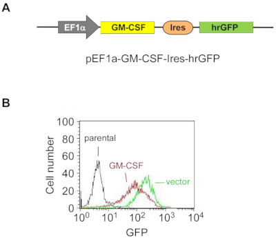
Figure 1: Exogenous GM-CSF is stably overexpressed in ES-D3 cells.
(A) The schematic diagram of the plasmid for overexpressing murine GM-CSF in ES-D3 cells, in which an EF1α promoter drives GM-CSF expression and hrGFP serves as an expression marker.
(B) Fluorescence intensity of GFP in GM-CSF-expressing ES-D3 cells or their empty vector control counterparts was determined by FACS. Please click here to view a larger version of this figure.
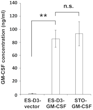
Figure 2: ES-D3 cells overexpressing GM-CSF produce high levels of GM-CSF.
GM-CSF concentrations in the medium of the indicated cells were measured by ELISA. The data are presented as mean ± standard deviations (mean ± SD) of three independent ELISA measurements: **p < 0.001, NS = not significant, ANOVA with Tukey's multiple comparison test. Please click here to view a larger version of this figure.
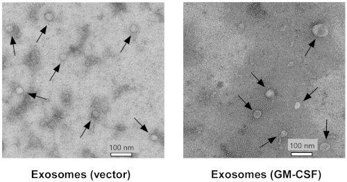
Figure 3: ES-D3-derived extracellular vesicles are examined by transmission electron microscopy.
Extracellular vesicles were prepared from ES-D3 cells transfected with the plasmid expressing GM-CSF or its empty vector counterpart. Arrows indicate individual vesicles. Scale bar = 100 nm. Please click here to view a larger version of this figure.
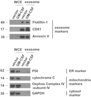
Figure 4: Exosomal markers are highly concentrated in extracellular vesicles isolated from ES-D3 cells.
The amounts of markers for exosomes, endoplasmic reticulum (ER), mitochondria, and cytosol in the indicated whole cell extracts (WCE) and EVs were evaluated by Western blotting. PDI = protein disulfide isomerase. Molecular weights markers (kD) are on the left. Please click here to view a larger version of this figure.
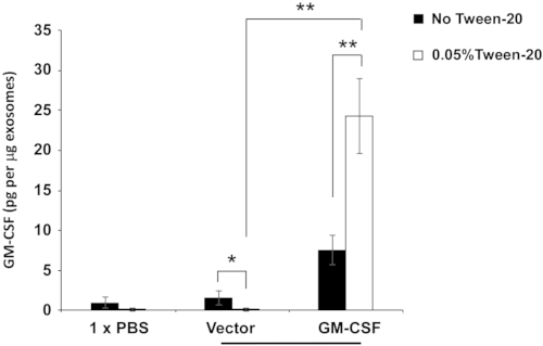
Figure 5: Evaluation of GM-CSF levels in exosome-enriched extracellular vesicles.
The levels of GM-CSF in the indicated exosome-enriched EVs were determined under different ELISA conditions. Exosome-enriched EVs were pretreated with or without 0.05% Tween-20. ELISA was carried out using washing buffer containing either PBS only or PBS + 0.05% Tween-20. The data are presented as the mean ± SD of three independent ELISA assays. *p < 0.05, **p < 0.005, ANOVA with Tukey's multiple comparison test. Please click here to view a larger version of this figure.
Alkaline phosphate, Calf Intestinal | New England Biolabs | M0290S | Dephosphorylating DNA plasmid |
anti-Annexin V mAb | Santa Cruz Biotechnology | clone H-3, sc-74438 | Western blot, RRID:AB_1118989 |
anti-CD81 mAb | Santa Cruz Biotechnology | clone B-11, sc-166029 | Western blot, RRID:AB_2275892 |
anti-cytochrome c mAb | Santa Cruz Biotechnology | clone A-8, sc-13156 | Western blot, RRID:AB_627385 |
anti-Flotillin-1 mAb | Santa Cruz Biotechnology | clone C-2; sc-74566 | Western blot, RRID:AB_2106563 |
anti-GAPDH pAb | Rockland | 600-401-A33S | Western blot, RRID:AB_11182910 |
anti-mouse IgG, goat, peroxidase-conjugated | Thermo Fisher | 31430 | Western blot, RRID:AB_228307 |
anti-Oxphos COX IV-subunit IV mAb | Thermo Fisher | clone 20E8C12 A21348 | Western blot, RRID:AB_221509 |
anti-protein disulfide isomerase (PDI) pAb | Enzo | ADI-SPA-890 | Western blot, RRID:AB_10616242 |
anti-rabbit IgG, goat, peroxidase-conjugated | Thermo Fisher | 31460 | Western blot, RRID:AB_228341 |
BCA (bicinchoninic acid) assay | Thermo Fisher | 23223 | Determining protein concentrations |
Bis-Tris PAGE Gel, ExpressPlus, 4-20% | Genscript | M42015 | Western blot |
Carbenicillin, Disodium Salt | Thermo Fisher | 10177012 | Selecting E. coli colonies |
Centrifuge, Avanti J-26 XPI | Beckman Coulter | Low speed centrifugation | |
Centrifuge rotor, JA-10 | Beckman Coulter | 09U1597 | Low speed centrifugation |
Centrifuge bottle, Nalgene PPCO | Thermo Fisher | 3120-0500PK | Low speed centrifugation |
Cu grids with carbon support film | Electron Microscopy Sciences | FF200-Cu | Acquiring electron microscopy images |
EcoRI | New England Biolabs | R0101 | Digesting DNA plasmid |
Enhanced chemiluminescence detection system | Thermo Fisher | 32106 | Western blot |
FACScalibur flow cytometer | Becton Dickinson | Examining GFP levels of ES-D3 cells | |
Fetal bovine serum | ATCC | SCRR-30-2020 | Medium for ES-D3 cells |
Fisherbrand Sterile Cell Strainers; Mesh Size: 40μm | Thermo Fisher | 22-363-547 | Filtering ES-D3 cells for FACS sorting |
Gelatin (0.1%) | Thermo Fisher | ES006B | Culturing ES-D3 cells |
GM-CSF ELISA kit | Thermo Fisher | 88733422 | Determining GM-CSF concentrations |
KnockOut Dulbecco’s Modified Eagle’s Medium | Thermo Fisher | 10-829-018 | Medium for ES-D3 cells |
Leukemia Inhibitory Factor | Thermo Fisher | ESG1106 | Medium for ES-D3 cells |
L-glutamine | VWR | VWRL0131-0100 | Medium for ES-D3 cells |
Lipofectamine 2000 transfection reagent | Thermo Fisher | 11668019 | Transfecting ES-D3 cells |
Microplate reader, PowerWave XS | BioTek | Determining GM-CSF concentrations | |
MoFlo XDP high-speed cell sorter | Beckman Coulter | Isolating single ES-D3 cell clones | |
NEB 5-alpha Competent E. coli | New England Biolabs | C2988J | Generating GM-CSF expression plasmid |
Neomycin | Thermo Fisher | 10-131-035 | Selecting ES-D3 clones |
Non-essential amino acids | Thermo Fisher | SH3023801 | Medium for ES-D3 cells |
Non-fat dry milk | Thermo Fisher | NC9022655 | Western blot |
Opti-MEM I Reduced Serum Medium | Thermo Fisher | 31985062 | Transfecting ES-D3 cells |
Paraformaldehyde | Electron Microscopy Sciences | 15710 | Acquiring electron microscopy images |
Penicillin/streptomycin | VWR | sc45000-652 | Medium for ES-D3 cells |
Plasmid pEF1a-FD3ER-IRES-hrGFP | Addgene | 37270 | Generating GM-CSF expression plasmid |
PVDF membranes | Millipore EMD | IPVH00010 | Western blot |
QIAprep Spin Miniprep Kit (250) | QIAGEN | 27106 | Generating GM-CSF expression plasmid |
QIAquick Gel Extraction Kit (50) | QIAGEN | 28704 | Generating GM-CSF expression plasmid |
Quick Ligation Kit | New England Biolabs | M2200S | Generating GM-CSF expression plasmid |
Transmission electron microscope | Hitachi | HT7700 | Acquiring electron microscopy images |
Trypsin | VWR | 45000-660 | Culturing ES-D3 cells |
Ultracentrifuge, OptimaTM L-100 XP | Beckman Coulter | High speed centrifugation | |
Ultracentrifuge rotor, 45Ti | Beckman Coulter | 09U4454 | High speed centrifugation |
Ultracentrifuge polycarbonate bottle | Beckman Coulter | 355622 | High speed centrifugation |
UranyLess staining solution | Electron Microscopy Sciences | 22409 | Acquiring electron microscopy images |
Embryonic stem cells (ESCs) are pluripotent stem cells capable of self-renewal and differentiation into all types of embryonic cells. Like many other cell types, ESCs release small membrane vesicles, such as exosomes, to the extracellular environment. Exosomes serve as essential mediators of intercellular communication and play a basic role in many (patho)physiological processes. Granulocyte-macrophage colony-stimulating factor (GM-CSF) functions as a cytokine to modulate the immune response. The presence of GM-CSF in exosomes has the potential to boost their immune-regulatory function. Here, GM-CSF was stably overexpressed in the murine ESC cell line ES-D3. A protocol was developed to isolate high-quality exosome-enriched extracellular vesicles (EVs) from ES-D3 cells overexpressing GM-CSF. Isolated exosome-enriched EVs were characterized by a variety of experimental approaches. Importantly, significant amounts of GM-CSF were found to be present in exosome-enriched EVs. Overall, GM-CSF-bearing exosome-enriched EVs from ESCs might function as cell-free vesicles to exert their immune-regulatory activities.