At the onset of experiments, mice were at least 6 weeks of age and were maintained under specific pathogen-free conditions. The present protocol complies with the Institutional ethical standards and prevailing local regulations. Animal studies were approved by the local regulatory agency (Regierung von Oberbayern, Munich, Germany). Possible sex-related biases were not investigated in these studies.
1. Generation and isolation of EVs derived from tumor cells after chemotherapy exposure
2. Immunization of mice with EVs
3. Flow cytometry analysis of splenic T cells
This protocol is intended to facilitate the straightforward and easily reproducible assessment of the immunogenicity of tumor-derived EVs. Hereby, mice are inoculated with EVs derived from in vitro cultures of tumor cells expressing the model antigen chicken ovalbumin (OVA). The subsequent immune response is analyzed in splenic T cells via flow cytometry.
Figure 1 gives an overview of the practical steps of the entire protocol. Since the work focuses on immunogenic cell death, cross-presentation, and EV-induced anti-tumor immunity, this protocol is restricted to the function of CD8+ cytotoxic T cells. As displayed in Figure 2, cells were gated as single cells, lymphocyte subset (by size and granularity), viable cells (excluding a life/dead marker), and CD3+ CD4– CD8+ cytotoxic T cells. Intracellular accumulation of IFN-γ was assessed as a surrogate marker for activation. Possible additional markers regarding T cell differentiation and exhaustion are discussed below.
Using the method described here, mice were immunized with EVs derived from OVA-expressing tumor cells cultured either under steady-state (untreated) or genotoxic stress conditions (oxaliplatin-treated). Only mice injected with EVs derived from tumor cells under genotoxic stress conditions induced potent activation of splenic cytotoxic T cells in recipient animals (Figure 3A). Injection of EVs derived from tumors under steady-state conditions resulted in some T cell activation, but that was not significantly different from T cell activation in mice injected with the PBS vehicle. These data show that under genotoxic stress, tumor cells can release potently immunogenic EVs. The production of IFN-γ was particularly increased when splenocytes of tumor EV-treated animals were ex vivo restimulated with the model tumor antigen OVA before analysis (Figure 3B). These data suggest that tumor-derived EVs can induce tumor antigen-specific immune responses. Interestingly, IFN-γ-production – even though to a much lesser extent – is also detected in the absence of antigen-specific restimulation. Possibly, other melanoma-associated antigens, such as the differentiation antigen TRP225, may be targeted by some part of the EV-induced T cell response.
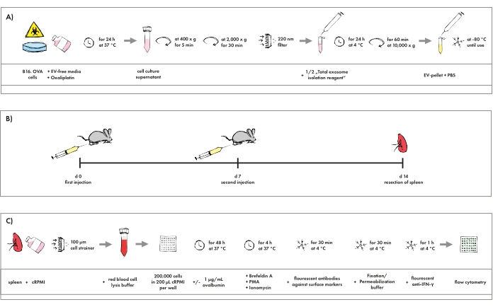
Figure 1: Pictographic overview of the protocol. (A) Isolation procedure of EVs generated in tumor cell cultures resembling chemotherapy. (B) Schedule for the immunization of mice with EVs. (C) Staining protocol for flow cytometry analysis of cytotoxic T cells. Please click here to view a larger version of this figure.
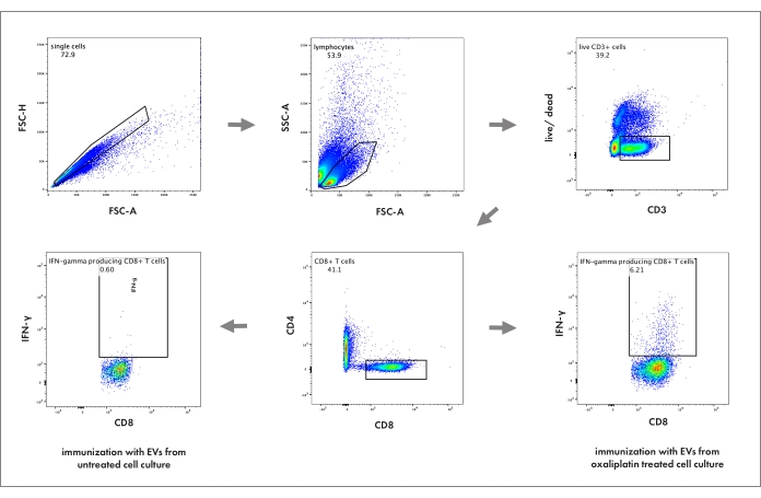
Figure 2: Flow cytometry gating strategy to analyze cytotoxic T cell activation in the spleen. The numbers represent the percentage of its respective parent population. FSC-A: forward scatter area; FSC-H: forward scatter height; SSC: sideward scatter; live/dead: cell death marker. Please click here to view a larger version of this figure.
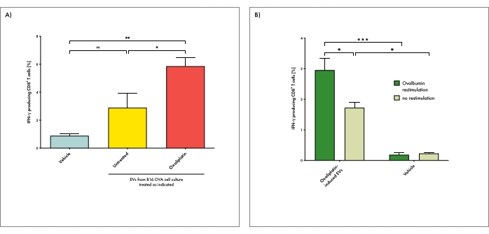
Figure 3: EVs derived from tumor cells under genotoxic stress can induce antigen-specific T cell responses in recipient animals. (A) Mice were immunized with EVs derived from tumor cells cultured either under steady-state (untreated) or genotoxic stress conditions (oxaliplatin-treated). Vehicle (PBS) injections were used as a negative control. IFN-γ production by cytotoxic T cells in the spleen upon EV immunization was determined. With this, splenic cell suspensions were restimulated with ovalbumin ex vivo before analysis. (B) Mice were treated with EVs derived from tumor cells under genotoxic stress conditions as described above. Splenic T cell activation was determined after ex vivo restimulation either in the presence or absence of ovalbumin. Bars depict the mean per group and whiskers its standard error. The one-way analysis of variance (ANOVA) test with Bonferroni posttest was used for multiple statistical comparisons of a dataset. The significance level was set at P < 0.05, P < 0.01, and P < 0.001 and is indicated here with asterisks (*, **, and ***). Please click here to view a larger version of this figure.
Anti-CD3 FITC | Biolegend | 100204 | Clone 17A2 |
Anti-CD4 PacBlue | Biolegend | 100428 | Clone GK1.5 |
Anti-CD8 APC | Biolegend | 100712 | Clone 53-6.7 |
Anti-IFNγ PE | eBioscience | RM90022 | Clone XMG1.2 |
Brefeldin A | Biolegend | 420601 | Brefeldin A Solution (1,000x) |
Cell Strainer, 100 µm | Greiner | 542000 | EASYstrainer 100 µm |
DMEM | Sigma-Aldrich | D6429 | Dulbecco's Modified Eagle's Medium with D-glucose (4.5 g/L) and L-glutamine (4 mM) |
FBS Good Forte | PAN BIOTECH | P40-47500 | Fetal Calf Serum (FCS) |
Fixable Viability Dye eFluor 506 | eBioscience, division of Thermo Fischer Scientific | 65-0866-14 | |
Fixation/Permeabilization Concentrate | eBioscience | 00-5123-43 | Fixation/Permeabilization Concentrate (10x) |
Fixation/Permeabilization Diluent | eBioscience | 00-5223-56 | |
Ionomycin | Sigma-Aldrich | 407952 | From Streptomyces conglobatus – CAS 56092-82-1, ≥ 97% (HPLC) |
L-Glutamine | Gibco | 25030-032 | L-Glutamine (200 mM) |
Ovalbumin | InvivoGen | vac-pova | Ovalbumine with < 1 EU/mg endotoxin – CAS 9006-59-1 |
Oxaliplatin | Pharmacy of MRI hospital | ||
PBS | Sigma-Aldrich | D8537 | Phosphate Buffered Saline without calcium chloride and magnesium chloride |
Penicillin-Streptomycin | Gibco | 1514-122 | Mixture of penicillin (10,000 U/mL) and streptomycin (10,000 ug/mL) |
PMA | Sigma-Aldrich | P1585 | Phorbol 12-myristate 13-acetate, ≥ 99% (HPLC) |
PVDF filter, 0,22 µm, for syringes | Merck Millipore | SLGV033RS | Millex-GV Filter Unit 0.22 µm Durapore PVDF Membrane |
Red Blood Cell Lysis Buffer | Invitrogen | 00-4333-57 | |
RPMI 1640 | Thermo Fischer Scientific | 11875 | Roswell Park Memorial Institute 1640 Medium with D-glucose (2.00 g/L) and L-glutamine (300 mg/L), without HEPES |
Syringe, 26 G | BD Biosciences | 305501 | 1 mL Sub-Q Syringes with needle (0.45 mm x 12.7 mm) |
Total Exosome Isolation Reagent | Invitrogen | 4478359 | For isolation from cell culture media |
β-Mercaptoethanol | Thermo Fischer Scientific | 31350 | β-Mercaptoethanol (50 mM) |
Immunogenic cell death of tumors, caused by chemotherapy or irradiation, can trigger tumor-specific T cell responses by releasing danger-associated molecular patterns and inducing the production of type I interferon. Immunotherapies, including checkpoint inhibition, primarily rely on preexisting tumor-specific T cells to unfold a therapeutic effect. Thus, synergistic therapeutic approaches that exploit immunogenic cell death as an intrinsic anti-cancer vaccine may improve their responsiveness. However, the spectrum of immunogenic factors released by cells under therapy-induced stress remains incompletely characterized, especially regarding extracellular vesicles (EVs). EVs, nano-scale membranous particles emitted from virtually all cells, are considered to facilitate intercellular communication and, in cancer, have been shown to mediate cross-priming against tumor antigens. To assess the immunogenic effect of EVs derived from tumors under various conditions, adaptable, scalable, and valid methods are sought-for. Therefore, herein a relatively easy and robust approach is presented to assess EVs’ in vivo immunogenicity. The protocol is based on flow cytometry analysis of splenic T cells after in vivo immunization of mice with EVs, isolated by precipitation-based assays from tumor cell cultures under therapy or steady-state conditions. For example, this work shows that oxaliplatin exposure of B16-OVA murine melanoma cells resulted in the release of immunogenic EVs that can mediate the activation of tumor-reactive cytotoxic T cells. Hence, screening of EVs via in vivo immunization and flow cytometry identifies conditions under which immunogenic EVs can emerge. Identifying conditions of immunogenic EV release provides an essential prerequisite to testing EVs’ therapeutic efficacy against cancer and exploring the underlying molecular mechanisms to ultimately unveil new insights into EVs’ role in cancer immunology.