This study was carried out in accordance with the clinical ethics committee guidelines of the First Affiliated Hospital of Zhejiang University. Written informed consent was obtained from all participating patients. The OLIF L5-S1 technique is indicated for patients with various spinal pathologies of L5-S1 involving degenerative disc disease, lumbar spondylosis, low-grade spondylolisthesis, and scoliosis. Patients with a history of trauma, neoplasia, or infection were excluded.
1. Patient position
2. Approaching the L5-S1 intervertebral disc between the Psoas muscle and the great vessels
3. Exposure of surgical field at the L5-S1 disc
4. Discectomy and cage insertion
5. Pedicle screws fixation
6. Postoperative period
7. Radiographic and clinical evaluation
Clinical outcomes
A total of 20 patients underwent OLIF L5-S1 via a retroperitoneal oblique corridor between the Psoas muscle and the great vessels. The study population exhibited female predominance (n=12, 60%), with a mean age of 55.4 ± 6.8 years. OLIF L5-S1 procedures were performed on patients with isthmic spondylolisthesis (n=10), degenerative disc disease (n=6), and degenerative spondylolisthesis (n=4). The procedures included single-level (n=16) and two-level cases (n=4), involving L4-L5 (n=4) and L5-S1 (n=20). The mean blood loss volume was 133.4 ± 48.5 mL, and the average operative time was 153.6 ± 38.3 min. Based on preoperative axial MR images, the left common iliac vein (LCIV) was categorized into three types according to the difficulty of mobilization: type I – no requirement for mobilization; LCIV runs laterally for more than two-thirds of the length of the left side of the L5-S1 disc, type II – easy mobilization; LCIV obstructs the L5-S1 disc space, but the perivascular adipose tissue is present under the LCIV, and type III – potentially difficult mobilization; no perivascular adipose tissue under the LCIV. In our series, patients were classified as type I (n=3), type II (n=14), and type III (n=3) LCIV. For two patients with type III LCIV, no other perioperative complications were observed apart from iliolumbar vein lacerations during exposure.
The preoperative VAS score for lower back pain was 6.3 ± 1.5, significantly decreasing to 1.2 ± 0.8 at 12 months. The VAS score for lower limb pain was 5.6 ± 1.4 preoperatively and significantly decreased to 0.8 ± 0.3 at 12 months (Figure 3). The preoperative Oswestry disability index improved from 82.4% ± 16.2% preoperatively to 8.1% ± 2.0% at 12 months (Figure 4).
Radiographic outcomes
Postoperative radiographic examinations confirmed improved reconstruction at the lumbosacral junction for all patients. At the final follow-up, bony fusion was observed in all patients (Figure 5). No cage retropulsion or pedicle screw loosening cases were observed.
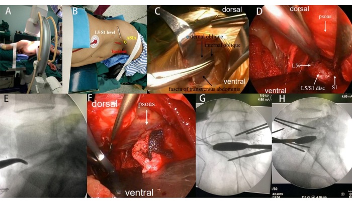
Figure 1: Intraoperative photographs. (A-B) The patient's lateral decubitus position and skin marking for the incision. (C) Blunt dissection involving the external oblique, internal oblique, and transversus abdominis muscles. (D) Exposure of the L5-S1 disc space using handheld retractors and Kirschner wire pins. (E) Intraoperative confirmation of the L5-S1 disc space by fluoroscopy. (F-H) Sequential use of different trial sizes to distract the disc space and release the contralateral annulus. Please click here to view a larger version of this figure.
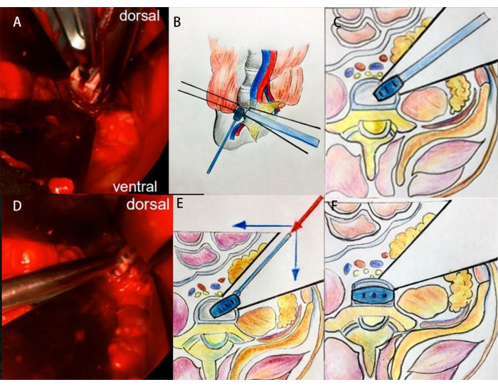
Figure 2: Intraoperative clinical images and schematic diagrams illustrating the technique for cage insertion at the L5-S1 segment. (A-C) First step of cage insertion. (D-F) The second step of cage insertion. Please click here to view a larger version of this figure.
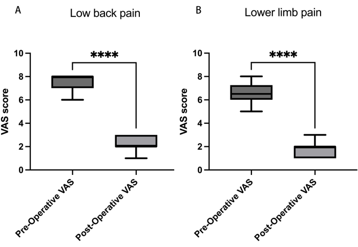
Figure 3: Visual analog scores (VAS) for low back and leg pain. (A) Over 12 months, the VAS score for lower back pain reduced from 6.3 ± 1.5 to 1.2 ± 0.8, and (B) for lower limb pain, the VAS score decreased from 5.6 ± 1.4 preoperatively to 0.8 ± 0.3. Data presented as mean ± SD. ****: Signifies a significant difference by Student's t-tests (p < 0.001). Please click here to view a larger version of this figure.
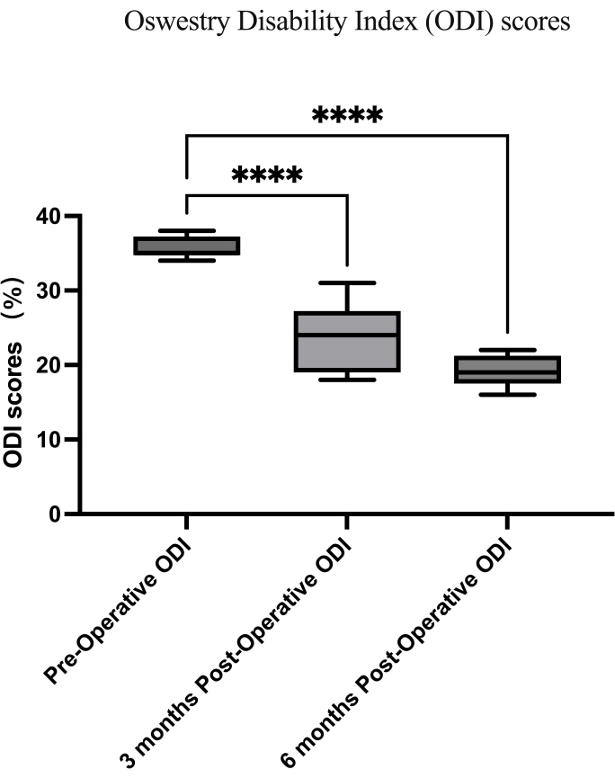
Figure 4: Oswestry disability index scores. The preoperative Oswestry disability index (ODI) improved from 82.4% ± 16.2% to 8.1% ± 2.0% at 12 months. Data presented as mean ± SD. ****: Signifies a significant difference by Student's t-tests (p < 0.001). Please click here to view a larger version of this figure.
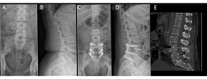
Figure 5: A 51-year-old male undergoing OLIF from L4-5 to L5-S1. (A, B) Preoperative anteroposterior and lateral radiographs displaying spondylolytic spondylolisthesis at L4-L5 and L5-S1. (C, D) Postoperative anteroposterior and lateral radiographs reveal satisfactory L4-S1 reconstruction. (E) Bony union was observed at the final follow-up. Please click here to view a larger version of this figure.
Fluoroscopy System | Allengers | ||
Handheld retractor | gSource | gS 36.9362 | |
Kirschner wire | Sklar surgical instruments | SKU 40-1535 | |
OLIF cages | Medtronic Sofamor Danek, Memphis, Tennessee, USA | ||
Pedicle screws | Beijing Fule Technology Development Co. , Ltd China | ||
Tonsil sponge | teleflex | MC-008133 | |
Vascular clamp |
Over the years, the oblique lateral interbody fusion (OLIF) technique has gained significant recognition for treating various spinal conditions in lumbar segments L2-L5. However, the adoption of OLIF for the L5-S1 segment has not been widely embraced by the spinal surgery community, given that significant concerns remain regarding the applicability of OLIF for lumbosacral fusion. In this study, a cohort of 20 patients underwent interbody fusion at the L5-S1 level using the OLIF technique through a single retroperitoneal oblique approach positioned between the Psoas muscle and the great vessels. The procedure involved discectomy and endplate preparation accomplished through a surgical window created on the anterolateral side of the L5-S1 disc. For secure interbody fusion cage placement, a supplementary cage insertion approach was employed. All patients were followed up for a minimum of 12 months. The mean preoperative visual analog scale (VAS) score for lower back pain was 6.3 ± 1.5 and experienced a significant reduction to 1.2 ± 0.8 at 12 months. The VAS score for lower limb pain significantly decreased from 5.6 ± 1.4 preoperatively to 0.8 ± 0.3 at 12 months after the surgery. Furthermore, the preoperative Oswestry disability index (ODI) improved from 82.4% ± 16.2% to 8.1% ± 2.0% at 12 months. Radiographic evaluations after surgery confirmed improved lumbosacral junction reconstruction for all patients. At the final follow-up, successful bony fusion was observed in all cases. Based on these findings, the OLIF technique for L5-S1 fusion represents an attainable approach for lumbosacral reconstruction. The procedure's success hinges on a comprehensive preoperative plan and precise intraoperative techniques.