1. Prepare the Dissecting Solutions and Enzyme Solutions
- Make the Artificial Cerebral Spinal Fluid (ACSF) and the Serum-free Media ahead of time and refrigerate.
- Weigh out the Kynurenic Acid (0.2 mg/mL)and dissolve in 10 mL of hilo ACSF in a 37° C waterbath ahead of time since it does not readily dissolve.
- Weigh out the Trypsin (1.33 mg/mL) and Hyaluronidase (0.67 mg/mL) and place in one 15 mL tube and keep it at -20°C until needed. Add the Kynurenic Acid solution to this tube and filter using a 22 μm syringe filter just prior to use.
- Weigh out Trypsin Inhibitor (Ovamucoid: 1 mg/mL) and dissolve in warm Serum-free Media and filter using 22μm syringe filter.
- Make the plating media: Serum-free Media containing FGF2 (10 ng/mL) and Heparin (2 μg/mL).
2. Isolating Retinal Stem Cells from the Mouse Eye
- Isolation of the retinal stem cells is performed in a specific sterile room dedicated to primary culture experiments. Prior to starting this procedure, set up the dissecting microscope and cold-light source, and then lay out the sterile dissecting instruments. Each hood has a hot bead sterilizer for sterilizing the instruments between steps.
- We will isolate retinal stem cells from eyes that have been placed immediately in a sterile Petri dish containing Artificial Cerebral Spinal Fluid (ACSF) after their removal from mice sacrificed according to approved animal ethics protocols.
- Under the dissecting microscope, clean the eye with forceps: get rid of the hair and the connective tissue that is attached to the cornea/scleral border. Then transfer the eye to a new dish with ACSF.
- While gently holding the eye stationary with serrated forceps, use angled micro-dissecting scissors to remove the ocular muscles. Remove the optic nerve as well if it is still attached to the eye. Try not to squish the eye during this process. Transfer eyes to a new dish with ACSF.
- Next use curved micro-dissecting scissors to cut the eye in half: begin at the optic nerve and cut through the middle of the cornea, meeting back at the optic nerve where the cut was initiated. It is ideal if the two pieces of eye are roughly equivalent in size because this will make the next step easier.
- Holding the corneas using two forceps, gently peel the two eye halves apart. Remove and discard the lens, viscera, and the neural retinal from the eye shells. Transfer the shells to a new dish with ACSF.
- Now we are ready to isolate the ciliary epithelium. To begin, orient the eye shell so that the cornea is on your right and the Retinal Pigmented Epithelium (RPE) is on the left. Gently pin the eye shell down with straight forceps on the RPE side.
- Next, use a scalpel to cut the cornea and iris away from the ciliary epithelium of the eye by gently pushing down on the scalpel rather than using sawing motions.
- After that, cut the ciliary epithelium away from the RPE. Use non-serrated forceps to transfer the strip of ciliary epithelium to a new 35-mm dish containing ACSF.
- In the same way, isolate the ciliary epithelium from the other eye shell.
- Once both ciliary epithelial strips have been collected, transfer the strips into a 35-mm dish containing 2 mL of Dispase. Place the dish with the strips in a 37° C incubator for 10 minutes.
- After 10 minutes, transfer the strips from the Dispase into a dish containing 2 mL of filtered Trypsin, Hyaluronidase, and Kynurenic Acid. Place the dish at 37 °C for 10 minutes.
- Now return to the dissecting microscope. While holding the sclera down with straight forceps, use the bottom of the curved non-serrated forceps to gently scrape the ciliary epithelium away from the sclera.
- Remove the sclera from the dish. All that should be left in the dish now are the cells of interest and the enzyme solution.
- Using a fire-polished cotton-plugged pipette, transfer this solution into a 14-ml tube. Triturate this solution 30 times to break apart the epithelial cells by gently forcing the solution in and out of the pipette.
- Centrifuge the tube for 5 minutes at 1500 RPM. When the centrifugation is done, remove the tube from the centrifuge carefully since the cells are prone to coming off the bottom of the tube at this stage.
- Gently aspirate the majority of the supernatant using a fire-polished pipette, and then add 1 mL of Trypsin inhibitor in Serum-free Media to the cells. Use a small borehole cotton-plugged pipette to triturate the sample approximately 50 times until it is a single-cell suspension.
- Centrifuge the tube again for 5 minutes at 1500 RPM.
- Remove the supernatant and replace it with 1 mL of your plating medium. Triturate gently using a fire-polished glass pipette to resuspend the cells.
- After counting the cells, plate them at the desired density in a 24-well plate. We usually plate 10 cells/μL by filling each well first with medium and then adding the cell suspension to get a final volume of 500 μL of media.
- Place the plate in a 37° C CO2 incubator where it will not be moved until you count the spheres that arise after 7 days.
3. Results of Isolating Retinal Stem Cells
- When this procedure is performed successfully, the dissected ciliary epithelial cells should look like this after being dissociated and plated at low density (Figure 1).
- After 7 days in culture, the retinal stem cell spheres that arise are counted. A sphere should be over 75 μm in diameter and free-floating to be counted as a stem cell derived sphere. (Figure 2). However, some cells will have limited proliferation and form spheroids that do not meet size criterion and will not be counted as stem cell derived spheres; an example of such a spheroid is shown here (Figure 2).
4. Representative Results
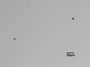
Figure 1.
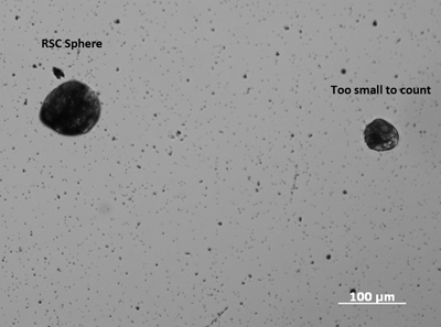
Figure 2.
