Epigenetic mechanisms are frequently altered in glioma. Chromatin immunoprecipitation could be used to study the consequences of genetic alterations in glioma that result from changes in histone modifications which regulate chromatin structure and gene transcription. This protocol describes native chromatin immunoprecipitation on murine brain tumor neurospheres.
Epigenetic modifications may be involved in the development and progression of glioma. Changes in methylation and acetylation of promoters and regulatory regions of oncogenes and tumor suppressors can lead to changes in gene expression and play an important role in the pathogenesis of brain tumors. Native chromatin immunoprecipitation (ChIP) is a popular technique that allows the detection of modifications or other proteins tightly bound to DNA. In contrast to cross-linked ChIP, in native ChIP, cells are not treated with formaldehyde to covalently link protein to DNA. This is advantageous because sometimes crosslinking may fix proteins that only transiently interact with DNA and do not have functional significance in gene regulation. In addition, antibodies are generally raised against unfixed peptides. Therefore, antibody specificity is increased in native ChIP. However, it is important to keep in mind that native ChIP is only applicable to study histones or other proteins that bind tightly to DNA. This protocol describes the native chromatin immunoprecipitation on murine brain tumor neurospheres.
Epigenetic events are frequent in gliomas and likely play an important role in tumor pathogenesis. Indeed, in pediatric high-grade glioma, mutations in genes encoding histone variants H3.3 and H3.1 occur frequently1. The mutations affect histone modifications and have major epigenetic consequences2,3. In the adolescent to adult spectrum, recurrent mutations in isocitrate dehydrogenase gene 1/2 (IDH1/2), a mutation that inhibits α-KG dependent histone and DNA de-methylasaes, and genetic alterations in other chromatin regulators such as ATRX and DAXX occur4. Therefore, it is of critical importance to study how mutations that affect epigenetic regulators alter chromatin structure and regulatory histone modifications, which, in turn, have a dramatic impact of the tumor cells' transcriptome.
Chromatin immunoprecipitation (ChIP) is a powerful tool used to evaluate the impact of epigenetic modifications in the genome5,6,7. In native ChIP, chromatin is digested with micrococcal nuclease (MNase), immunoprecipitated using an antibody raised against the protein of interest, and then DNA is purified from the immunoprecipitated chromatin complex6. Cells are not fixed during the procedure so this technique is only applicable for the study of proteins that interact tightly with DNA6. The absence of cross-linking aids antibody specificity since antibodies are usually raised against unfixed peptides or proteins7. In addition, since there is no cross-linking step, this reduces the chances of fixing transient protein-DNA interactions that are non-specific and not regulatory7,8. ChIP can be used to identify the enrichment of histone modifications in a specific genomic region. Here, we detail a protocol for performing native ChIP in neurospheres (NS) generated from genetically engineered mouse models of glioma.
All methods described here have been approved by the Institutional Animal Care and Use Committee (IACUC) of the University of Michigan.
1. Generation of brain tumor NS and culturing conditions.
2. Native ChIP
A schematic representation of tumor NS generated from a brain tumor where brain tumor cells are Katushka positive is presented in Figure 1. Figure 2 is a schematic representation of the entire ChIP technique. Figure 3 shows the representative results of chromatin from brain tumor NS digested with MNase for 12 min, yielding a majority of mono, di-, and tri- nucleosomes. Following ChIP, a qPCR may be performed on the ChIP and input DNA samples. Figure 4 shows representative ChIP qPCR data from a qPCR for glyceraldehyde-3-phosphate dehydrogenase (Gapdh). Gapdh is a housekeeping gene that is enriched with H3K4me3, a modification that is associated with active transcription. The results show that Gapdh is enriched with H3k4me3 and are not enriched with H3K27me3 which is a modification associated with regions of repressed chromatin.
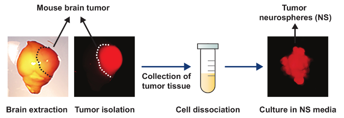
Figure 1: Schematic of tumor NS generated from a Katushka positive brain tumor. A bright field and fluorescent image of a brain harvested from a mouse that reached the end point stage. The tumor area is delineated by the dotted line and is positive for the fluorescent reporter, Katushka. The tumor is then isolated and the tumor tissue is collected in a 1.5 mL tube. The cells are then dissociated and cultured in NSC medium and tumor NS form. Please click here to view a larger version of this figure.
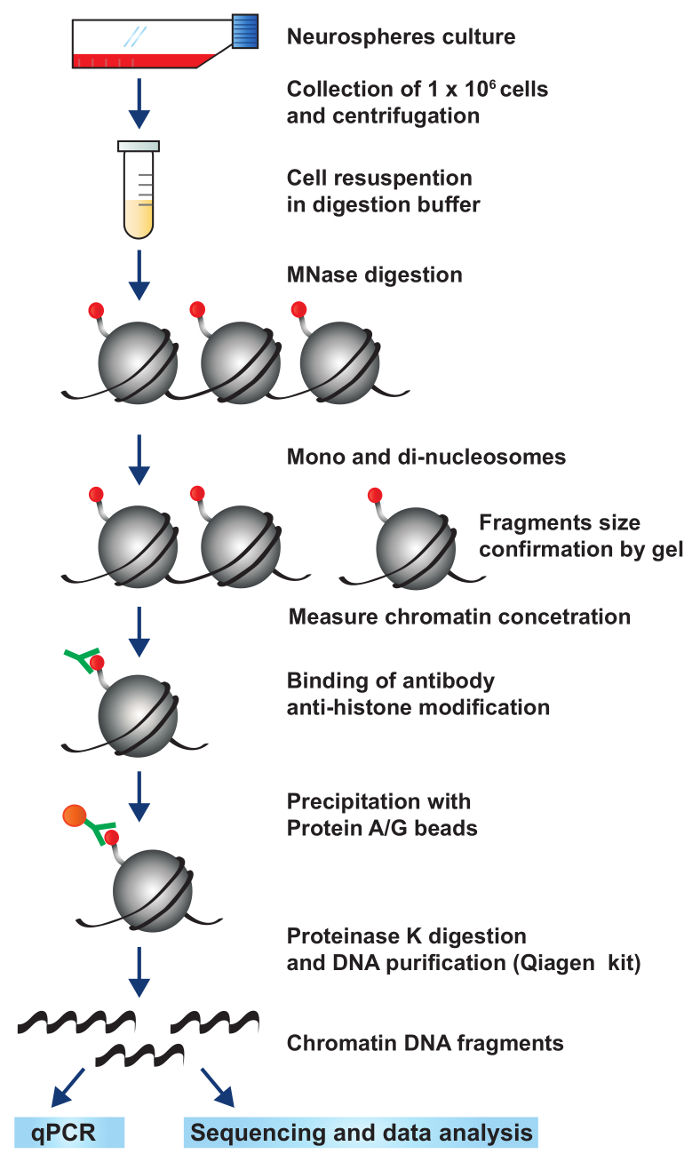
Figure 2: A schematic representation of the workflow for native ChIP. Tumor NS are cultured and expanded. Each IP is performed with 1 X 10⁶ cells. Chromatin is fragmented using by MNase digestion to obtain mono, di-, and tri- nucleosomes. The chromatin is incubated with an antibody specific for a histone modification or DNA-associated protein of interest. The antibody-DNA complex is immunoprecipitated with magnetic protein A/G beads. Finally, protein is digested and DNA is purified to obtain only DNA enriched with histone modification or DNA-associated protein of interest. Please click here to view a larger version of this figure.
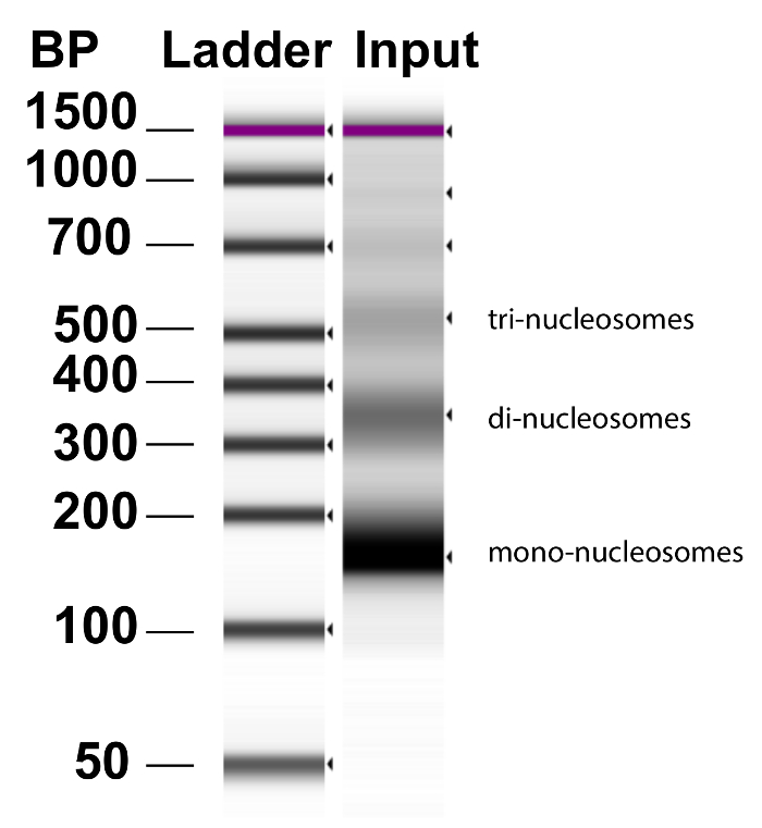
Figure 3: Chromatin fragmentation by MNase. Chromatin was prepared from brain tumor NS by the addition MNase and incubation at 37 °C for exactly 12 min. Representative DNA results from Bioanalyzer analysis of an input sample demonstrate that the majority of the DNA has been fragmented into mono-, di-, and tri- nucleosomes. Lane 1 is the ladder in base pairs (BP). Please click here to view a larger version of this figure.
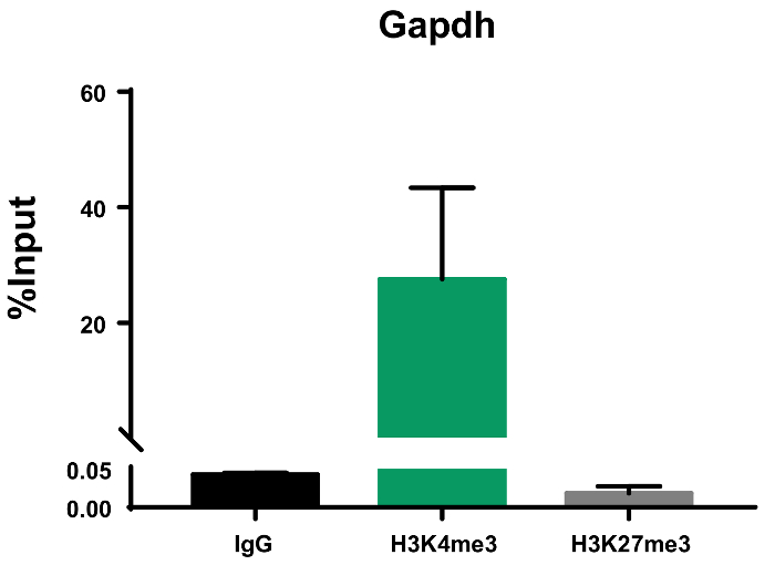
Figure 4: Representative ChIP qPCR data presented as percent input. qPCR was performed with IgG, H3K4me3, and H3K27me3 ChIP DNA using primers for glyceraldehyde-3-phosphate dehydrogenase (Gapdh). Representative results demonstrate that Gapdh, a housekeeping gene, is only enriched with the H3K4me3 modification, associated with active transcription, and not the H3K27me3 modification, associated with repressed chromatin. This graph represents the results of two biological experiments run with three replicate wells each. Error bars represent standard error of the mean (SEM). Please click here to view a larger version of this figure.
Gene | Forward primer | Reverse primer |
Gapdh | TCCCCTCCCCCTATCAGTTC | GACCCGCCTCATTTTTGAAA |
Table 1: Primers used for ChIP qPCR experiments. The sequences for primers used for Gapdh are provided in this table.
mean Ct (input) | Standard Deviation | Adjusted Input | |
24.265 | 0.071 | 20.943 | |
Percent input | |||
Sample | Raw Mean Ct | Standard Deviation | Percent input = 100*2^ (adjusted input-Ct(IP)) |
IgG | 32.23 | 0.112 | 0.04 |
H3K4me3 | 22.148 | 0.128 | 43.37 |
H3k27me3 | 32.79 | 0.519 | 0.027 |
NTC | undetermined | undetermined | undetermined |
Table 2: Sample calculation of the percent input method for ChIP qPCR analysis. Sample calculation of percent input for ChIP performed with 10% starting input; DF = 10. The numbers in this table illustrate the raw values of one biological experiment with 3 replicate wells run for each sample.
The protocol presented here will enable the user to perform native ChIP on NS derived from genetically engineered brain tumors. In contrast to cross-linking ChIP, this protocol is limited for the study of proteins that associate tightly with DNA6. The number of cells used can be modified as necessary and the protocol can be scaled up. We used 1 X 10⁶ cells per IP, however, native ChIP may also be performed with as little as 4 x 10⁴ cells5. In this protocol, we analyzed ChIP DNA via qPCR; however, this protocol may also be combined with next generation sequencing to study protein-DNA interactions on a genome wide level. We have used this ChIP protocol successfully to analyze the effect of glioma oncogenes on the deposition of histone modifications within specific regions of the tumor cells genome.
There are a few critical steps when performing native ChIP. First, the digestion conditions for each cell type must be empirically determined. The majority of the chromatin should be mono-nucleosomes (150 bp) with some di- and tri- nucleosome peaks (Figure 3). Our protocol results in more than 70% mono-, di-, or tri- nucleosome peaks, however, some undigested DNA remains. If the protocol will be used for ChIP-seq, additional steps will be required to remove undigested DNA. In addition, it is important to keep in mind that each batch of MNase purchased may differ in activity and thus require optimization of the digestion conditions. Second, the quality of a ChIP depends highly on the specificity of the antibody used11. A high-quality ChIP antibody should have high reactivity against the intended target and low cross reactivity with other DNA-associated proteins or off target histone modifications11. For this reason, we recommend testing antibody specificity using a peptide binding test. ENCODE guidelines suggest that a ten-fold enrichment should be observed for the modification of interest relative to other modifications11. It is also critical to perform necessary control immunoprecipations, i.e., pre-immune or IgG control. When these considerations are met, native ChIP data is a strong reliable method to study epigenetic mechanisms regulated by protein-DNA interactions.
The authors have nothing to disclose.
This work was supported by National Institutes of Health/National Institute of Neurological Disorders & Stroke (NIH/NINDS) Grants R37-NS094804, R01-NS074387, R21-NS091555 to M.G.C.; NIH/NINDS Grants R01-NS076991, R01-NS082311, and R01-NS096756 to P.R.L.; NIH/NINDS R01-EB022563; the Department of Neurosurgery; Leah's Happy Hearts, and Chad Though Foundation to M.G.C. and P.R.L. RNA Biomedicine Grant F046166 to M.G.C. F.M.M. is supported by an F31 NIH/NINDS-F31NS103500. R.I.Z.-V. is supported by the NIH/ NIGMS grant 5T34GM007821-37.
Agilent 2100 Bioanalyzer | Agilent | G2946-90004 | bioanalyzer |
AccuSpin Micro 17R, refrigerated | Fisher Scientific | 13-100-676 | |
C57BL/6 | Taconic | B6-f | C57BL/6 mouse |
Calcium Chloride | Aldrich | 22350-6 | buffer reagent |
D-Luciferin, Potassium Salt | Goldbio | LuckK-1g | |
DiaMag1.5 magnetic rack | Diagenode | B04000003 | for magnetic bead washes |
DiaMag Rotator EU | Diagenode | B05000001 | rotator |
DMEM/F-12 | Gibco | 11330-057 | NSC component |
Dynabeads Protein A | Thermo Fisher Scientific | 10001D | protein A magnetic beads |
Dynabeads Protein G | Thermo Fisher Scientific | 10003D | protein G magnetic beads |
EGF | PeproTech | AF-100-15 | prepare 20 μg/mL stock in 0.1% BSA and aliquot. |
Ethylenediaminetetraacetic acid (EDTA) | Sigma | E-4884 | buffer reagent |
ethylene glycol-bis(β-aminoethyl ether)-N,N,N',N'-tetraacetic acid) (EGTA) | Sigma | E-4378 | buffer reagent |
Fast SYBR Green Master Mix | Applied Biosystems | 4385612 | qPCR reagent |
Fetal Bovine Serum | Gibco | 10437028 | for freezing cells |
FGF | PeproTech | 100-18b | Prepare 20 μg/mL stock in 0.1% BSA and aliquot. |
Forceps | Fine Science Tools | 11008-13 | for dissection of tumor |
Glycerol, MB Grade | EMD- Millipore | 356352 | |
H3K4me3 | Abcam | Ab8580 | |
H3K27me3 | Millipore | 07-449 | |
HBSS | Gibco | 14175-103 | balanced salt solution |
Fluriso | VETone | 501017 | inhalation anesthetic |
Ivis Spectrum | Perkin-Elmer | 124262 | in vivo optical imaging system |
Hyqtase | HyClon | SV3003001 | cell detachment media |
Lithium Chloride | Sigma | L8895 | buffer reagent |
Low binding microtubes | Corning Costar | CLS3207 | low protein binding microcentrifuge tube |
Microcentrifuge tube | Fisher | 21-402-903 | regular microcentrifuge tube |
Micrococcal Nuclease | Thermo Fisher Scientific, Affymetrix | 70196Y | each batch may differ; purchase sufficient amount for experiments and aliquot. |
N2 | Gibco | 17502-048 | NSC component |
Normal Rabbit IgG | Millipore | 12-372 | |
Normocin | Invivogen | NOL-36-063 | anti-microbial agent, use at 0.1 mg/mL. |
NP-40 (Igepal CA-630) | Sigma | 18896-50ML | buffer reagent |
Kimble Kontes Pellet Pestle | Fisher Scientific | K749515-0000 | |
Protease Inhibitor Cocktail | Sigma-Aldrich | P8340 | aliquot and store at -20 °C. |
Protinase K | Sigma-Aldrich | P2308 | make 10 mg/mL stock in water; aliquot and store at -20 °C. |
QIAquick PCR Purification Kit | Qiagen | 28104 | DNA purification kit |
Scalpel | Fine Science Tools | 10007-16 | for dissection of tumor |
Sodium Chloride | VWR | 0241-5KG | buffer reagent |
Sodium Deoxycholate | Sigma-Aldrich | D670-25G | buffer reagent |
Sodium Dodecyl sulfate (SDS) | Sigma | L-4390 | buffer reagent |
Tris Base | Thermo Fisher Scientific | Bp152-1 | buffer reagent |
Triton X-100 | Thermo Fisher Scientific | BP 151-500 | polyethylene glycol octylphenyl ether |
Standard Mini Centrifuge | Fisherbrand | 12-006-901 | standard mini centrifuge |
SZX16 microscope | Olympus | SZX16 | flourescent dissecting microscope |
ViiA 7 Real-Time PCR System with Fast 96-Well Block | Applied Biosystems | 4453535 | |
Nanodrop One | Thermo-Fisher Scientific | ND-ONEC-W |