Intraspinal injection of recombinase dependent recombinant adeno-associated virus (rAAV) can be used to manipulate any genetically labelled cell type in the spinal cord. Here we describe how to transduce neurons in the dorsal horn of the lumbar spinal cord. This technique enables functional interrogation of the manipulated neuron subtype.
Selective manipulation of spinal neuronal subpopulations has mainly been achieved by two different methods: 1) Intersectional genetics, whereby double or triple transgenic mice are generated in order to achieve selective expression of a reporter or effector gene (e.g., from the Rosa26 locus) in the desired spinal population. 2) Intraspinal injection of Cre-dependent recombinant adeno-associated virus (rAAV); here Cre-dependent AAV vectors coding for the reporter or effector gene of choice are injected into the spinal cord of mice expressing Cre recombinase in the desired neuronal subpopulation. This protocol describes how to generate Cre-dependent rAAV vectors and how to transduce neurons in the dorsal horn of the lumbar spinal cord segments L3-L5 with rAAVs. As the lumbar spinal segments L3-L5 are innervated by those peripheral sensory neurons that transmit sensory information from the hindlimbs, spontaneous behavior and responses to sensory tests applied to the hindlimb ipsilateral to the injection side can be analyzed in order to interrogate the function of the manipulated neurons in sensory processing. We provide examples of how this technique can be used to analyze genetically defined subsets of spinal neurons. The main advantages of virus-mediated transgene expression in Cre transgenic mice compared to classical reporter mouse-induced transgene expression are the following: 1) Different Cre-dependent rAAVs encoding various reporter or effector proteins can be injected into a single Cre transgenic line, thus overcoming the need to create several multiple transgenic mouse lines. 2) Intraspinal injection limits manipulation of Cre-expressing cells to the injection site and to the time after injection. The main disadvantages are: 1) Reporter gene expression from rAAVs is more variable. 2) Surgery is required to transduce the spinal neurons of interest. Which of the two methods is more appropriate depends on the neuron population and research question to be addressed.
The dorsal spinal cord is essential for information exchange between the periphery of the body and the brain. Sensory stimuli such as heat, cold, touch, or noxious stimuli are detected by specialized peripheral neurons, which convey this information to neurons of the spinal cord dorsal horn. Here, a complex network of inhibitory and excitatory interneurons modulates and eventually relays sensory information via spinal projection neurons to supraspinal sites1,2. The computations carried out by spinal inter- and projection neurons gate sensory information, thus determining which information is suppressed or relayed at which intensity. Changes in the integration of sensory stimuli, such as an altered balance between inhibition and excitation, can cause sensory dysfunctions such as hypersensitivity or allodynia (painful sensations after normally non-painful stimulation). These changes are thought to be the underlying cause of various chronic pain states3,4. Thus, spinal circuits are of high importance in sensory processing and consequently in the perception of an organism's environment and self. With the recent advent and combination of molecular, genetic, and surgical techniques that allow the precise manipulation of genetically identified spinal neuron subpopulations, scientists are now beginning to understand the underlying spinal circuits responsible for the processing of distinct sensory modalities.
Intraspinal injection of rAAV into wild-type or transgenic mice has greatly contributed to the manipulation, analysis, and understanding of the function of specific subsets of spinal neurons5,6,7,8,9,10,11. This technique allows the delivery of marker proteins (such as GFP/ GFP fusion proteins), reporter proteins (such as GCaMP), or effector proteins (such as bacterial toxins, channelrhodopsin, or pharmacogenetic receptors) in a spatially restricted manner to spinal neurons. Local injection of Cre-dependent rAAVs into transgenic mice expressing Cre recombinase in a specific subset of spinal neurons allows the specific analysis of the respective neuronal population. We have employed this technique to label, ablate, inhibit or activate spinal glycinergic neurons demonstrating that they are an essential part of the spinal gate controlling pain and itch transmission7. In these experiments, intraspinal injection of Cre-dependent rAAV into GlyT2::Cre mice enabled the selective manipulation of glycinergic neurons in the lumbar spinal cord. Thereby, simultaneous manipulation of supraspinal circuits that contain glycinergic neurons critical for the survival of the animal can be avoided.
While an intraspinal injection of rAAVs limits infection to the site of injection, viral transduction can occur not only in local neurons but also in neurons that connect to the injection site via axonal projections. The latter is often used to trace CNS areas providing neuronal input to a particular nucleus in the brain. The infection of axonal projections can, however, also be a confounding factor when a defined population of neurons shall be studied at a particular site. To address these issues, we have recently conducted a comprehensive analysis of AAV serotypes and expression cassettes to identify serotypes and promoters that can be used to either minimize or maximize retrograde transduction. In the context of this specific research in spinal circuits, we analyzed the ability of different serotypes and promoters to retrogradely transduce neurons in the dorsal root ganglia (DRG), the rostral ventromedial medulla (RVM), and the somatosensory cortex12. The technique outlined in this protocol can therefore be used either to analyze spinal neurons at the injection site or to analyze projection neurons that provide input to the injected site of the spinal cord. In the protocol described here, three injections of rAAV into the left side of the lumbar spinal cord are performed to enable transduction of neurons in the three lumbar segments (L3-L5). The L3-L5 segments receive the majority of the sensory input from the hindlimb ipsilateral to the injection site. We demonstrate that functional manipulation of genetically labeled neurons in L3-L5 is sufficient to evoke robust behavioral changes, thus providing functional evidence for the circuit function of such a genetically labeled neuron subtype.
All animal experiments were approved by the Swiss cantonal veterinary office (Zurich) and are in accordance and compliance with all relevant regulatory and institutional guidelines.
NOTE: All materials along with respective manufacturers and/or vendors are listed in the Table of Materials.
1. Generation of Cre-dependent AAV Vectors
NOTE: A variety of Cre-dependent vectors with different promoters can be purchased (see Table of Materials) or, if the desired expression construct is not available, it can be generated by modifying existing AAV constructs. Note, the promoter and serotype can have an impact on the spread of viral transduction (see 12). The first part of this protocol briefly describes the generation of two different Cre-dependent AAV vectors suitable for gain and loss of function experiments, respectively.
2. Transduction of Spinal Cells
3. Behavioral and Morphological Analyses
In order to illustrate the expression levels that can be obtained by the intraspinal injection of rAAV encoding a marker protein, we first injected AAV1.CAG.eGFP into the lumbar spinal cord of wild-type mice. Three injections spaced approximately 1 mm apart produced a nearly continuous infection of lumbar spinal segments L3 to L5 (Figure 1A-C). Virus injection at a depth of 300 µm from the spinal surface leads to predominant infection of cells in the spinal cord dorsal horn. However, infected cells could also be found in the ventral horn (Figure 1D). Next, the goal was to illustrate the difference in rAAV-mediated expression when injecting a Cre-dependent rAAV into a Cre transgenic mouse. We therefore injected the Cre-dependent AAV1.CAG.flex.eGFP vector into the spinal cord of GlyT2::Cre transgenic mice. As before, eGFP expression was observed in the dorsal and ventral horn. However, as expected, the expression of eGFP became more restricted, reflecting the distribution of GlyT2+ neurons, i.e. the relatively sparse expression in the superficial dorsal horn and dense expression in the deep dorsal horn (Figure 1E).
Next, to demonstrate the feasibility of addressing circuit function of spinal neuronal subpopulations, two different Cre-dependent AAVs coding for different effector proteins (DTA and hM3Dq) were injected into GlyT2::Cre transgenic mice. Analogous to the viral expression observed after three injections of AAV1.CAG.eGFP into wild-type mice, there was robust ablation of inhibitory neurons (Pax2+) in lumbar segments L3-L5 after the injection of AAV1.EF1a.flex.DTA into Glyt2::Cre mice (Figure 2A,C). Loss of glycinergic inhibitory neurons in these segments evoked a marked mechanical hypersensitivity (Figure 2D) and spontaneous aversive behavior directed towards the ipsilateral hindlimb (Figure 2E). The aversive behavior led to self-inflicted lesions, which could be observed on the paws, calf, and thigh (for data, see Foster et al.7) Opposite effects were observed when activating glycinergic neurons through injection of AAV1.hSyn.flex.hM3Dq and subsequent intraperitoneal injection of CNO (Figure 2B). Mice became desensitized to noxious mechanical stimulation (Figure 2F) and other noxious stimuli (see Foster et al.7) In addition, when treated with the pruritogens histamine or chloroquine, hM3Dq-mediated activation of glycinergic neurons efficiently suppressed prurifensive responses (Figure 2G). These results demonstrate that three injections of rAAV into the lumbar spinal cord are able to transduce an area of the lumbar spinal cord sufficient to observe robust behavioral changes evoked by stimulation of the corresponding hindlimb.
In a final set of experiments, we wanted to demonstrate the potential differences in using Cre reporter mice compared to Cre reporter viruses. Therefore, a Cre driver gene was chosen that has previously been described as displaying a restricted expression pattern in the mouse spinal cord. The gene RORβ has been suggested to be expressed predominantly in inhibitory interneurons of the deep dorsal horn13,14. This study used RORβCre knock-in mice and crossed them to Rosa26lox-STOP-lox-tdTomato (R26Tom) Cre reporter mice, which lead to the expression of tdTomato in all cells displaying Cre expression at any time point before analysis (Figure 3A). Characterization of tdTomato+ cells in RORβCre; R26Tom mice revealed expression in neurons and astrocytes of the dorsal horn (Figure 3B,C). In fact, quantification of the tdTomato+ cells suggested that the majority of cells undergoing Cre-mediated recombination (58%) were non-neuronal. We then injected a Cre-dependent tdTomato reporter rAAV (AAV1.CAG.flex.tdTomato) into the spinal cord of P40 RORβCre mice (Figure 3D). In contrast to R26Tom-mediated reporter expression, we found all tdTomato+ cells labeled by the reporter virus to be neurons (Figure 3E,F). Finally, the identity of the tdTomato+ neurons was analyzed in both sets of mice (RORβCre; R26Tom and RORβCre injected with AAV1.CAG.flex.tdTomato). In both cases, the majority of neurons were inhibitory (>85%) and the minority of neurons excitatory (<20%) (Figure 3 G-I), which is in agreement with previous assessments of the identity of RORβ+ neurons13,14.
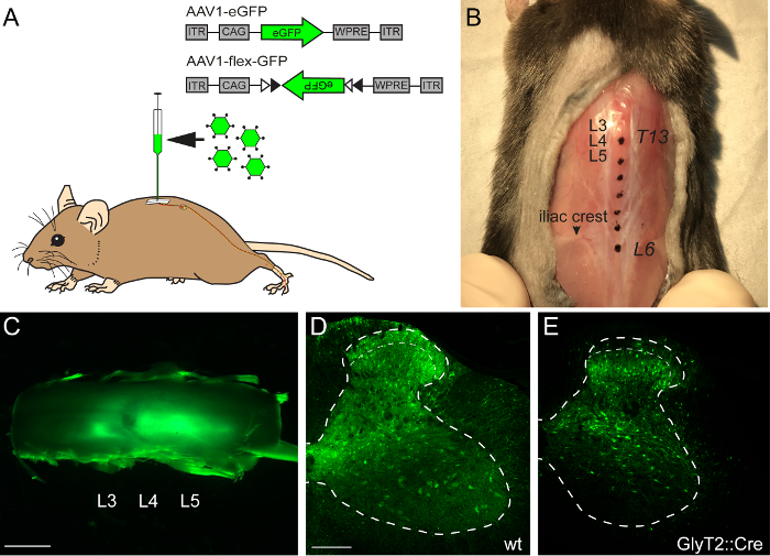
Figure 1: Intraspinal Injection of AAV1-eGFP/AAV1-flex-eGFP
(A) Schematic illustration of an intraspinal injection of an AAV1.CAG.eGFP (AAV1-eGFP) into the lumbar spinal cord, which is innervated from the hindlimb. (B) Anatomical location of the lumbar spinal cord segments L3-L4 can be seen in a top down view of the back of a mouse. The skin was opened to expose the vertebral column. Spinal processes of vertebrae T13-L6 are colored as anatomical references and an arrow indicates the iliac crest. (C) Representative image of a whole mount lumbar spinal cord. Green fluorescence indicates virus-transduced areas of the spinal cord. (D) Representative image of a cross section through an AAV1-eGFP-transduced lumbar spinal cord from a wild-type mouse. (E) Representative image of a cross section of an AAV1.CAG.flex.eGFP-transduced spinal cord of a GlyT2::Cre mouse. Dashed lines represent outline of the gray matter and superficial dorsal horn of the spinal cord. Scale bars C = 1 mm, D = 100 µm. Please click here to view a larger version of this figure.
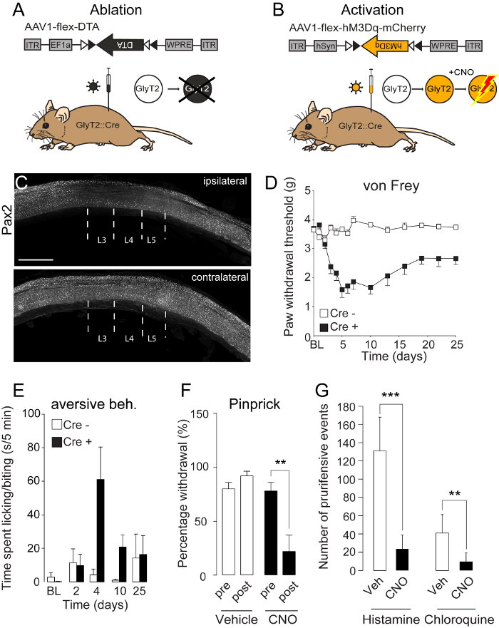
Figure 2: Functional Manipulation of Spinal Cre-expressing Glycinergic Neurons
(A+B) Schematic illustration of an intraspinal injection of an AAV1.EF1a.flex.DTA (A) or an AAV1.EF1a.flex.hM3Dq (B) into the lumbar spinal cord. Cre-dependent expression of diphtheria toxin fragment A (DTA) will lead to ablation of the glycinergic neurons (GlyT2+) (A), while Cre-dependent expression of the pharmacogenetic designer receptor hM3Dq will render GlyT2 neurons activatable by clozapine-N-oxide (CNO) (B). (C) Three injections of AAV1.EF1a.flex.DTA into the L3-L5 segments of GlyT2::Cre mice led to a marked loss of inhibitory neurons in the respective segments of the ipsilateral but not of the contralateral side. (D) Loss of GlyT2 neurons evoked a long-lasting hypersensitivity to mechanical von Frey stimulation in the ipsilateral hind paw of GlyT2::Cre mice injected with AAV1.EF1a.flex.DTA, but no change was observed if Cre-negative mice were injected. (E) Loss of GlyT2 neurons evoked spontaneous aversive behavior reminiscent of chronic itch. (F) hM3Dq-mediated activation of GlyT2 neurons alleviated noxious mechanical pain evoked by pinprick stimulation. (G) hM3Dq-mediated activation of GlyT2 neurons reduced pruritogen (chloroquine or histamine)-evoked aversive behavior. Data are represented as mean ± SEM. *** p < 0.001; ** p < 0.01. Scale bar C = 1 mm. g = grams. Images are re-used and modified from Foster et al.7 Please click here to view a larger version of this figure.
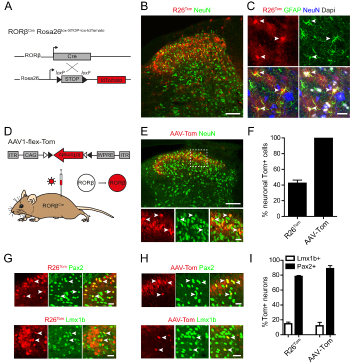
Figure 3: Genetic and Virus-mediated Labeling of RORβ-expressing Cells. (A) Diagram showing the strategy for Cre-dependent expression of fluorescent tdTomato reporter in RORβCre; Rosa26lox-STOP-lox-tdTomato (R26Tom) mice. (B) Immunostaining on spinal cord sections of RORβCre; R26Tom mice revealed that NeuN+ RORβ-Tom neurons can be found in laminae I-IV. (C) Cre-dependent expression of tdTomato was also observed in GFAP+ cells of RORβCre; R26Tom mice, suggesting that RORβ is expressed in astrocytes during development. Arrowheads indicate double-labeled astrocytes in lamina III. (D) Diagram showing intraspinal injection of AAV-flex-Tom (rAAV1.CAG.flex.tdTomato) virus into RORβCre mice to drive local Cre-dependent expression of tdTomato. (E) Immunostaining on spinal cord sections of RORβCre mice injected with AAV-flex-Tom virus. RORβ-Tom neurons were localized to the superficial laminae of the spinal dorsal horn and expression of tdTomato was absent from astrocytes. (F) Percentage of RORβ-Tom cells expressing the neuronal marker NeuN in spinal sections of RORβCre; R26Tom mice and RORβCre mice injected with AAV-flex-Tom virus. (G-H) Immunostaining on spinal cord sections of (G) RORβCre; R26Tom mice and (H) RORβCre mice injected with AAV-flex-Tom virus showing colocalization between RORβ-Tom neurons and the inhibitory marker Pax2 or the excitatory marker Lmx1b. (I) Percentage of RORβ-Tom neurons expressing Lmx1b and Pax2 in RORβCre; R26Tom mice and RORβCre mice injected with AAV-Tom virus. Data are represented as mean ± SEM. Data are from 2-3 mice and 1-3 sections per mouse. Scale bars represent 100 µm (B,E) and 20 µm (C,E in high magnification images, G and H). Please click here to view a larger version of this figure.
Variable | Set value | Unit |
heat (H) | 450 | – (value proportional to power of the radiated heat) |
force preliminary pull (F(TH)) | 20 | – (value proportional to voltage applied to force coil) |
distance threshold (s(TH)) | 25 | 0.12 mm |
delay heatstop (t(H)) | 30 | 0.5 ms |
distance heatstop (s(H)) | 0 | 0.12 mm |
delay pull 1 (t(F1)) | 200 | 0.5 ms |
force pull 1 (F1) | 300 | – (value proportional to voltage applied to force coil) |
distance pull 2 (s(F2)) | 30 | 0.12 mm |
force pull 2 (F2) | 600 | – (value proportional to voltage applied to force coil) |
adjust (AD) | 0 | – |
Table 1: Puller Settings
Intraspinal injection of AAVs may become a powerful technique in a research laboratory, enabling the analysis of spinal cells with a high temporal and spatial solution. This protocol enables the transduction of the three main spinal segments innervated by sensory neurons extending their peripheral axons to the hindlimb. Transducing three segments produces robust and reproducible behavioral data. It also enables testing of a larger sensory area than possible after a single intraspinal injection. For example, the same injection regime allows for testing the paw (von Frey, Hargreaves, etc.) and the thigh (intradermal injections, mechano-receptor stimulation), thus expanding the sensory modalities that can be addressed.
Critical Steps:
There are several steps critical for obtaining robust morphological and behavioral data and for avoiding artefacts. The design of the viral vector and the choice of the promoter and serotype can have a strong impact on transduction efficiency and extent of the transduced area, and therefore on behavioral results (for details on viral spread and transduction efficiencies, see 12). High quality viral preparations are required to minimize side effects of the virus injection, which can occur if contaminants or cell debris are present in the virus solution. The surgery that is required to inject the spinal cord has to be undertaken with great care in order to avoid morphological and behavioral artefacts introduced by the surgery. Particular attention is required in handling the spinal cord, especially during the laminectomy and during the perforation of the dura with the needle. Damage to the spinal cord could impair somatic sensations and motor coordination of the mouse, thus compromising any planned behavioral experiments. In addition, it is important to assemble the syringe carefully. Improper assembly or clogging of the micropipette can hamper the experiment. Spinal tissue at the injection site has to be examined at the end of the experiment in order to verify successful injection and transduction of the injected area and to exclude excessive tissue damage as a result of the surgery. This protocol has used viral titers up to a concentration of 1×10¹³ GC/mL without apparent toxicity, yet higher titers of virus might become toxic at some time point. In addition, toxicity will most likely also depend on the protein encoded by the injected AAV.
Modifications:
Several parameters in the protocol can be adapted to the neuronal population and research question to be studied. Injection of 300 nL virus solution at a depth of 300 µm leads to transduction of neurons throughout, and predominantly in the dorsal horn. If the aim is to target neurons further ventral, the depth can be adjusted. Injection of larger volumes of virus (we have used up to 500 nL per injection) will lead to a larger spread in dorsoventral and rostrocaudal directions. This may be beneficial to achieve targeting of additional cells in the rostrocaudal extent, but can increase the spillover to the contralateral side, which may hamper the use of the contralateral side as an internal control. In addition, the dorsoventral extent of transduced cells will increase, which may be the desired effect or should be avoided. Injection of 300 nL at a depth of 300 µm avoided side effects on motor control in GlyT2::Cre mice, while injection of 500 nL or injection at a greater depth occasionally produced motor phenotypes, such as spastic extension of the hindlimb (data not shown). Injection of smaller volumes can be used to restrict targeting to even more confined areas5. The viral titer is another parameter that can be modified and should in fact be determined for each virus. For bacterial toxins, such as the highly efficient DTA, low expression levels may be sufficient to induce the desired effect, whereas for fluorescent reporters and DREADDs, higher titers may be necessary for detectable or effective expression.
Significance of the Method with Respect to Existing/Alternative Methods:
To date, mainly two techniques have been used to functionally interrogate the neuronal circuits required for transmission of sensory signals. Many researchers have used intraspinal injections of rAAV coding for reporter and effector proteins into recombinase-expressing mice in order to label and manipulate spinal neuronal subpopulations. Intraspinal injection as a technique to manipulate spinal cells has previously been described15,16. The protocol outlined here was specifically designed to transduce genetically labeled neurons in three consecutive segments of the lumbar spinal cord while minimizing surgical manipulation. This is achieved by placing two of the three injections in the intervertebral space rostral and caudal of the T13 vertebrae and second, by drilling a hole into T13 for the third injection instead of removing the vertebrae. The targeted L3-L5 segments are the main termination area of sensory neurons innervating the hindlimb. We demonstrate that this type of transduction is sufficient to produce robust behavioral changes after the manipulation of glycinergic neurons and suggest that it is suitable for the analysis of a variety of different genetically labeled spinal neurons.
The second technique that has been used in order to manipulate the function of spinal neuron subsets is based on crossing transgenic animals. Here, mice expressing a recombinase in a specific subset of spinal neurons have to be crossed to reporter mice in order to achieve expression of the desired marker or effector protein in the respective neuronal subset17,18,19. To illustrate some of the differences that can occur when comparing results obtained by using reporter mice or intraspinal virus injections we either crossed RORβCre knock-in animals to a Cre-dependent reporter mouse or injected RORβCre mice with a Cre-dependent reporter rAAV. We compared reporter gene expression obtained from the rAAV to reporter gene expression obtained in RORβCre; R26Tom mice. Reporter gene expression obtained from the intraspinal injection of a Cre-dependent rAAV was restricted to neurons of the spinal cord, while R26Tom reporter mouse-evoked tdTomato expression is also found in astrocytes. This suggests that RORβ is transiently expressed in astrocytes. Similarly, Gutierrez-Mecinas et al. found a more restricted reporter expression (spatially and cell-type specific) after viral injection compared to mouse-evoked reporter expression in Tac1Cre mice20. By using intraspinal injections of a rAAV reporter into the spinal cord of adult mice, reporter and/or effector gene expression can efficiently be restricted to the population of cells that express the gene in the adult and thus avoid recombination/expression in those populations with earlier transient gene activity.
A second main advantage of intraspinal rAAV injections is the ability to restrict the expression of the rAAV-encoded genes to the injection site. In transgenic mice, Cre driver-mediated expression is often not only found in the neuronal subpopulation of interest, but also in other populations within the nervous system. Dymecki and colleagues, as well as the groups of Ma and Goulding, have developed elegant ways of using intersectional reporter mouse-based approaches that avoid recombination in parts of the nervous system that shall remain unaltered17,18,19,21,22,23. Using knock-in mice to obtain expression of marker or effector proteins can also be assumed to be less variable, as there is only one genomic copy of the respective expression cassette per cell and the expression cassette is present in all cells in the region of interest. In contrast, the presence of a respective expression cassette and also the copy number of the expression cassette after viral transduction is dependent on the transduction efficiency, which may vary from cell to cell, from injection to injection, and depends on the virus lot. Finally, the main disadvantage of virus-mediated gene expression over traditional genetic methods is the necessity of surgery. Surgery-mediated injury/inflammation may affect neurons and circuits directly. Inclusion of control mice that have undergone the same surgery is therefore mandatory.
Future Applications:
Intraspinal injection can be used to analyze and manipulate any genetically identified spinal subpopulation through overexpression of marker and effector proteins. It is therefore likely in the future that many will adopt this technique to study neuronal populations in their specific focus. Moreover, besides using intraspinal injection of rAAVs to achieve overexpression of exogenous effector proteins, this technique can also be used to silence or overexpress endogenous proteins and therefore to study the function of any given gene with high spatial and temporal resolution.
The authors have nothing to disclose.
We thank Hanns Ulrich Zeilhofer for generously supporting this work. Hendrik Wildner was supported by the Olga Mayenfisch foundation. We thank Carmen Birchmeier for the Lmx1b antibody.
Equipment | |||
micropipette puller: DMZ-Universal-Electrode-Puller | Zeitz | NA | |
anesthesia unit: Oxymat3 oxygen concentrator | Weinmann | NA | |
anesthesia unit: VIP 3000 Veterinary Vaporizer | Midmark | NA | |
Heat mat: Mio Star Thermocare 100 | Migros | 717614700000 | |
Electric shaver | Philips | BT9290 | |
surgical microscope (OPMI pico) | Zeiss | NA | |
Small animal stereotaxic apparatus | Kopf | NA | |
Neurostar StereoDrive (optional) | Neurostar | NA | |
Model 51690 Cunningham mouse spinal adaptor | Harvard Apparatus | 72-4811 | |
PHD Ultra syringe pump with nanomite | Harvard Apparatus | 70-3601 | |
Hamilton 701 RN 10 μl glass microliter syringe | Hamilton | 7635-01 | |
Hamilton Removable needle (RN) compression fitting 1 mm | Hamilton | 55750-01 | |
fine dentistry drilling apparatus: Osada success 40 | Osada | OS-40 | |
spherical cutter, 0.5mm | Busch | 12001005B | |
electronic von Frey anesthesiometer | IITC | 23905 | |
flexible von Frey hairs | IITC | #7 | |
LSM710 Pascal confocal microscope | Zeiss | NA | |
0.8 NA × 20 Plan-apochromat objective | Zeiss | NA | |
1.3 NA × 40 EC Plan-Neofluar oil-immersion objective | Zeiss | NA | |
Name | Company | Catalog Number | Comments |
Surgical Tools | |||
Scalpel Handle #4, 13cm | Fine Science Tools | 10004-13 | |
Extra Fine Bonn Scissors | Fine Science Tools | 14084-08 | |
Adson forceps, 1 x 2 teeth, 12 cm | Fine Science Tools | 11027-12 | |
Friedman-Pearson rongeurs, curved, 0.7 mm cup | Fine Science Tools | 16121-14 | |
Dumont #2 laminectomy forceps | Fine Science Tools | 11223-20 | |
Olsen-Hegar needle holders, serrated, 8.5 mm clamp length | Fine Science Tools | 12002-12 | |
Fine forceps #5 | Fine Science Tools | 11254-20 | |
Name | Company | Catalog Number | Comments |
Consumables and Chemicals | |||
Thin-wall glass capillary, 1mm outside diameter | World Precision Instruments | TW 100-3 | |
Syringes (1, 5 and 20 ml) | B. Braun | (9166917V, 4606051V, 4606205V) | |
26G beveled needle | B. Braun | 4665457 | |
Sterile scalpel blades | B. Braun | BB523 | |
Surgical sutures Safil Quick+ 4/0, absorbable | B. Braun | C1046220 | |
Surgical sutures Premilene 5/0, non-absorbable | B. Braun | C0932191 | |
Sterile PBS or saline (0.9%) | NA | ||
Ethanol, 70% (disinfectant) | NA | ||
Iodine solution (e.g. Braunol) | B. Braun | 18380 | |
Anaesthetics (e.g. Attane isoflurane) | Provet | 2222 | |
Aldasorber | Provet | 333526 | |
analgesics (e.g. buprenorphine: temgesic) | Indivior | GTIN: 7680419310018 | |
Ophthalmic ointment (e.g. vita-pos) | Pharma medica | GTIN: 4031626710635 | |
Cotton swabs (e.g. from) | IVF Hartmann | 1628100 | |
Facial tissues (e.g. from) | Uehlinger AG | 2015.10018 | |
Superfrost plus microscope slides | ThermoScientific | J1800AMNZ | |
Name | Company | Catalog Number | Comments |
Mice | |||
C57BL/6J mice (wildtype) | The Jackson Laboratory | RRID:IMSR_JAX:000664 | |
Rorbtm1.1(cre)Hze/J mice (RORβCre) | The Jackson Laboratory | RRID:IMSR_JAX:023526 | |
Gt(ROSA)26Sortm14(CAG-tdTomato)Hze/J mice (R26Tom) | The Jackson Laboratory | RRID: IMSR_JAX:007914 | |
Name | Company | Catalog Number | Comments |
Viral vectors | |||
AAV1.CB7.CI.eGFP.WPRE.rBG (AAV1.CAG.eGFP) | Penn Vector Core | AV-1-PV1963 | |
AAV1.CAG.flex.eGFP.WPRE.bGH (AAV1.CAG.flex.eGFP) | Penn Vector Core | AV-1-ALL854 | |
AAV1.CAG.flex.tdTomato.WPRE.bGH (AAV1.CAG.flex.tdTomato) | Penn Vector Core | AV-1-ALL864 | |
AAV1.EF1a.flex.DTA.hGH (AAV1.EF1a.flex.DTA) | Penn Vector Core | Custom production | |
AAV1.hSyn.DIO.hM3D(Gq)-mCherry.hGH (AAV.flex.hM3D(Gi)) | Penn Vector Core | Custom production | |
Name | Company | Catalog Number | Comments |
Plasmids | |||
pAAV.hSyn.flex.hM3D(Gq)-mCherry | Addgene | 44361 | |
pAAV.EF1α.flex.hChR2(H134R)-eYFP | Addgene | 20298 | |
Name | Company | Catalog Number | Comments |
Bacteria | |||
MDS42 | ScarabGenomics | ||
Stbl3 | ThermoScientific | C737303 | |
Name | Company | Catalog Number | Comments |
Reagents | |||
EndoFree Plasmid Maxi Kit | Quiagen | 12362 | |
NucleoBond PC 500 | Machery & Nagel | 740574 | |
clozapine-N-oxide (CNO) | Enzo Life Sciences | BBL-NS105-0025 | |
chloroquine diphosphate salt | Sigma | C6628 | |
histamine | Sigma | H7125 | |
Dapi | Invitrogen | D3571 | |
Name | Company | Catalog Number | Comments |
Antibodies (dilution) | |||
Rabbit anti-GFP (1:1000) | Molecular Probes | RRID:AB_221570 | |
Rabbit anti-NeuN (1:3000) | Abcam | RRID:AB_10711153 | |
Goat anti-Pax2 (1 : 200) | R & D Systems | RRID:AB_10889828 | |
Guinea pig anti-Lmx1b (1 : 10 000) | Dr Carmen Birchmeier | Muller et al. 2002 | |
Rabbit anti-GFAP (1 : 1000) | DakoCytomation | RRID:AB_10013382 | |
Secondary antibodies raised in donkey (1:800) | Jackson ImmunoResearch Laboratories | NA |