In vitro use of degenerative tenocytes is essential when investigating the efficacy of novel treatment on tendinopathy. However, most research studies use only the animal model or a healthy tenocyte. We propose the following protocol to isolate human degenerative tenocytes during surgery.
Tendinopathy, a painful condition that develops in response to tendon degeneration, is on the rise in the developed world due to increasing physical activity and longer life expectancy. Despite its increasing prevalence, the underlying pathogenesis still remains unclear, and treatment is generally symptomatic. Recently, numerous therapeutic options, including growth factors, stem cells, and gene therapy, were investigated in hopes of enhancing the healing potency of the degenerative tendon. However, the majority of these research studies were conducted only on animal models or healthy human tenocytes. Despite some studies using pathological tenocytes, to the best of our knowledge there is currently no protocol describing how to obtain human degenerative tenocytes. The aim of this study is to describe a standard protocol for acquiring human degenerative tenocytes. Initially, the tendon tissue was harvested from a patient with lateral epicondylitis during surgery. Then biopsy samples were taken from the extensor carpi radialis brevis tendon corresponding to structural changes observed at the time of surgery. All of the harvested tendons appeared to be dull, gray, friable, and edematous, which made them visually distinct from the healthy ones. Tenocytes were cultured and used for experiments. Meanwhile, half of the harvested tissues were analyzed histologically, and it was shown that they shared the same key features of tendinopathy (angiofibroblastic dysplasia or hyperplasia). A secondary analysis by immunocytochemistry confirmed that the cultured cells were tenocytes with the majority of the cells having positive stains for mohawk and tenomodulin proteins. The qualities of the degenerative nature of tenocytes were then determined by comparing the cells with the healthy control using a proliferation assay or qRT-PCR. The degenerative tenocyte displayed a higher proliferation rate and similar gene expression patterns of tendinopathy that matched previous reports. Overall, this new protocol might provide a useful tool for future studies of tendinopathy.
Tendinopathy is a chronic degenerative musculoskeletal condition that develops in various parts of the body. Recently, the number of cases of tendinopathy has increased greatly in the developed world due to growing participation in recreational sports and increased life expectancy1,2. The cause of tendinopathy is considered to be multifactorial and these causes include ischemia, oxygen free radical injuries, an imbalance between vasoconstrictor and vasodilator innervations, internal micro-tears, and changes to neuro-regulation3,4,5,6,7,8. Most treatments for tendinopathy only relieve its symptoms. Moreover, treatments without tissue regeneration require a long time for rehabilitation and achieve a limited response from injured tendons, which imposes a clinical challenge for physicians9.
The incompetence of current treatment options along with the lack of the degenerative tendon's ability to self-heal has lead researchers to take interest in exploring alternative treatment strategies. Recently, new studies reported many promising results for enhancing the healing efficacy of the tendinopathy tendons using growth factors, stem cell based therapy, and gene therapy10,11,12.
Through a literature review, we found that the involved studies may be divided into two categories based on their analysis materials: animal models such as a rat, a mouse, or a rabbit; and human models. With regard to the animal model, currently there are two popular techniques to generate tendinopathy: chemical induction of injury or mechanical overloading the model. However, each animal model was limited in reproducing the complex human tendinopathy pathology13,14.
Most papers using human samples were analyzed histologically or performed the in vitro experiment based on a healthy human tenocyte instead of a degenerative tenocyte15,16,17,18,19,20,21. Only a few papers reported that they used a human degenerative tenocyte, but they did not describe in detail the protocol used to get the degenerative tenocyte from the human22,23. In this context, it should be noted that successful results from either the animal model or the healthy tissue/tenocyte may not necessarily predict human efficacy or efficacious dosing because tendon degeneration is a complicated process and the pathogenesis is still not fully understood.
Collectively, it is necessary to describe the standard protocol for obtaining the degenerative tenocyte from human tissue without causing adverse effects to the donor. This article describes a protocol on how to acquire the human degenerative tenocyte. To validate the protocol, the harvested tissues were analyzed histologically. Then, the cultured cell was confirmed as the degenerative tenocyte by using immunocytochemistry (ICC), a quantitative real time-polymerase chain reaction (qRT-PCR), and a viability assay.
The protocol was conducted in accordance with the Declaration of Helsinki and the protocol was approved by the Institutional Review Board at CHA Bundang Medical Center.
1. Degenerative Tendon Tissue Harvest from the Patient
2. Histology
3. Tenocyte Culture
4. Immunocytochemistry (ICC)
5. Proliferation Assay
6. Statistical Analysis
Histological analyses revealed that the harvested tissue from lateral epicondylitis had the characteristics of a tendinopathic tendon.H&E section of tendinopathy degenerative tendon revealed a disorganized collagen bundle with a loss of polarity and fine straight, strongly packed parallel fiber structures. Histological signs suggestive of degeneration such as higher cellularity and enlarged nuclei without the typical spindle shape were common in samples. Additionally, collagen bundles of the degenerated tendon were relatively weak eosin stained by H&E but were positively stained by Alcian blue indicating increased proteoglycans and glycosaminoglycans substance.
In all cases, blood vessels were increased in number and aggregates occupying spaces embedded between fibers were compared to normal slit-like blood vessels. In the IHC staining, cells of degenerated tendons showed cytoplasmic granular positive reaction to VEGF in comparison to the negative reaction of control ones. Collectively, these findings confirmed that the harvested tissues from lateral epicondylitis were degenerative tissues and suitable for the degenerative tenocyte culture (see Figure 2).
The cultured cells were confirmed as tenocytes by ICC and the harvested tissues were confirmed by IHC as tendon tissue. Most of the cultured cells and cells in tendon tissue showed a positive stain for the representative tenocyte markers known as mohawk (green) proteins and tenomodulin (red) proteins with an elongated appearance under microscopy (see Figure 3). Also, the harvested tissues had the positive staining for mohawk proteins and tenomodulin proteins immunohistochemically (see Supplementary Figure 1). These suggested that the harvested tissues were composed by the tendon tissue.
Cultured tenocytes from the harvested tissue of lateral epicondylitis had the characteristic features that correspond to tendinopathy. Both degenerative and healthy tenocytes were cultured for 4 days under the same condition. At each time point, the proliferation rate was measured by BrdU assay and WST-1 assay. The results showed that the proliferation rate of the degenerative cells was significantly higher compared to the controls as previously reported (p <0.05) (see Figure 4A).
Five genes which are known to be up-regulated in the degenerative tendon were analyzed by qRT-PCR. Expressions of the genes, COL3A1, ACTA2, TAC1 (SP), TAC receptor 1, and PTGS2 (COX2) were increased more significantly in the cultured cells from the degenerative tendon than in the cells from the healthy ones. The relative expressions for all genes are shown in Figure 4B and Supplemental Figure 2.
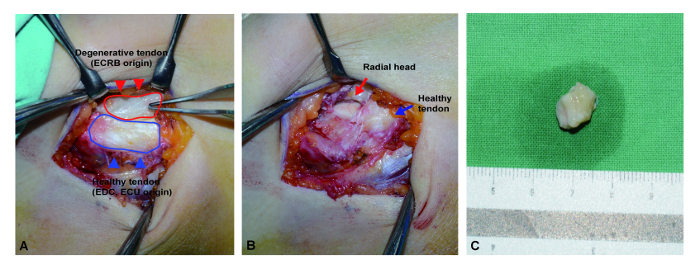
Figure 1: Surgical photo of degenerative tendon harvest. (A) After skin incision and dissection, the pathologic degenerative tissue on the common extensor origin was exposed, showing a characteristic dull-grayish color with edematous change (red line). Conversely, the general appearance of normal tendon tissue was shiny and firm with a slightly grayish tone (black line). (B) All identified degenerative tissues were resected sharply and then the radial head bone was exposed. (C) All resected tissues were embedded into the phosphate buffered saline immediately and the surrounding non-tendinous tissue was carefully dissected as soon as possible. ECRB: extensor carpi radialis brevis. EDC: extensor digitorum communis. ECU: extensor carpi ulnaris. Please click here to view a larger version of this figure.
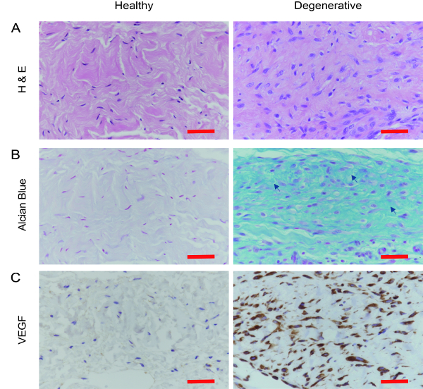
Figure 2: Confirmation of degenerative tendon histologically. (A) In the H & E staining, degenerative samples from the lateral epicondylitis had disorganized collagen bundles with loss of polarity and increased cell number compared to the healthy control. (B). Alcian blue staining showed that the degenerative tendon had increased ground substance consisting of proteoglycans and glycosaminoglycans with several mucoid patches and vacuoles between fibers (Arrow). (C). Additionally, VEGF immunohistochemistry demonstrated increased staining in vessel formation in degenerative tendon samples compared to the control samples. VEGF: Vascular endothelial growth factor. Scale bar = 50 µm. Please click here to view a larger version of this figure.
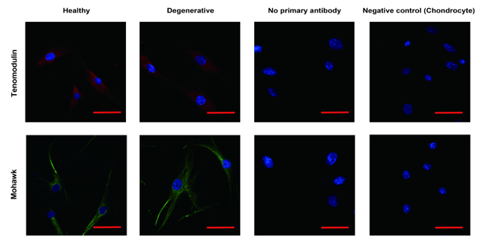
Figure 3: Identification of tenocyte by ICC. The cultured cells and cells in tendon tissue were confirmed as tenocyte by ICC. Most of the cells showed positive stain for the representative tenocyte marker, including mohawk (green) and tenomodulin (red) with elongated appearance under microscopy. Chondrocyte was used as the negative control. ICC: Immunocytochemistry. Scale bar = 50 µm. Please click here to view a larger version of this figure.
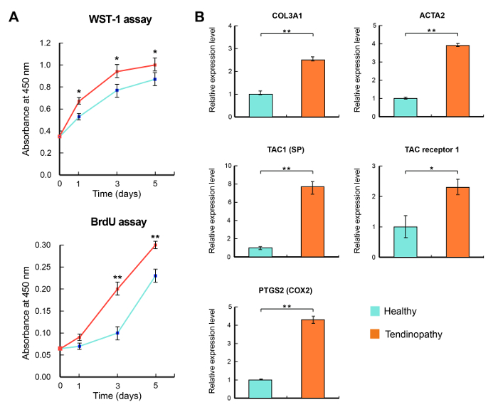
Figure 4: Characteristics of degenerative tenocyte. (A, B) Degenerative tenocytes (red) had a higher proliferation rate with a notable increase in cellularity. (C) Five genes that are known to be related in the development of tendinopathy were significantly elevated in the degenerative tenocytes. Gene expression levels were normalized against GAPDH. *: mean <0.05, **: mean <0.01, respectively. SP: substance P. Please click here to view a larger version of this figure.
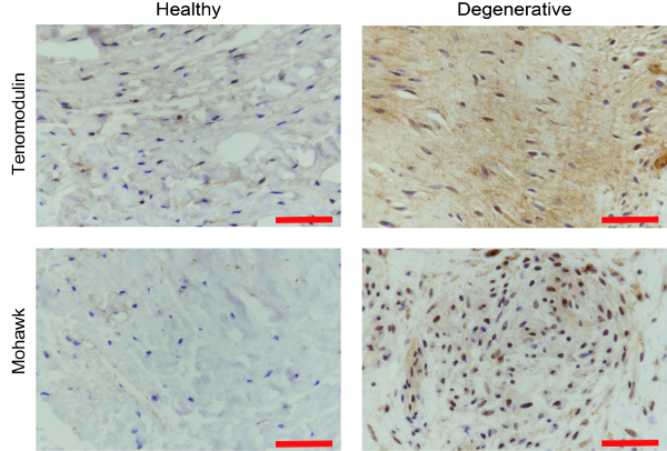
Supplementary Figure 1: Histological confirmation of the harvested tissue as the tendon tissue. The harvested tissue was also analyzed by IHC for mohawk and tenomodulin to determine whether the harvested tissue had the adequate characteristics of tendon tissue. IHC: Immunohistochemistry. Scale bar = 50 µm. Please click here to view a larger version of this figure.
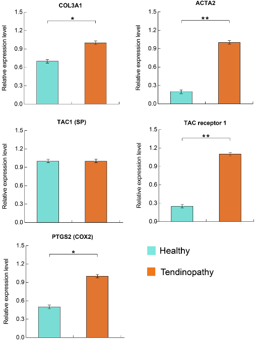
Supplementary Figure 2: Expression of tendinopathy related genes in tenocyte. The transcripts of five genes normalized by actin expression were significantly elevated in the degenerative tenocytes and have similar trends to Figure 4. Gene expression levels were normalized against actin with a mean < 0.05 and < 0.01, respectively. SP: substance P. Please click here to view a larger version of this figure.
A number of previous studies have reported how to create chronic tendinopathic animal models using different procedures such as collagenase or kartogenin injection, treadmill running, and more26,27. Although numerous studies showed promising therapeutic effects based on these animal models, experiments using the human degenerative tenocyte would be crucial in the field of tendinopathy in order to reproduce the efficacy of treatment. In this article, we established a protocol to successfully harvest and culture human degenerative tenocytes consistently. Both harvested tendon tissue and the cultured tenocyte showed the common characteristics of the degenerative tendon and tenocyte7,28.
In our experience, for the successful tissue harvest, it is essential to comprehend the morphological changes frequently associated with degenerative tendon and to accurately delineate that area during surgery. Degenerative tissue is typically grey or brown in color and the tissue is thin, fragile, and disorganized with loose texture. The inclusion and exclusion criteria of the protocol should be strictly kept with careful evaluation of previous treatments before harvest.
Tissue size is also important for a successful cell culture. We recommend harvesting more than 1 cm3. Usually, as long as all of the degenerative area is removed, the amount of collected tissue would be enough for the cell culture. Lastly, we did not use tenocytes over passage six in all experiments. Until passage six, we believe that the cultured cells still had the characteristics of degeneration.
Our protocol has several advantages. These advantages include no harm to the patient since the degenerative tissue was supposed to be removed during surgery, efficiency since the amount of tissue required is enough for the experiment, highly ethical due to no violation of healthy tissues, reproducibility in that pathological tissues from humans can be readily obtained during surgery, and, lastly, ease of application once surgical techniques become familiar.
However, we acknowledge that our experiment has several limitations. First, we cannot completely exclude the remnant effect of previous corticosteroid or platelet rich plasma injection even though we included only patients who had passed at least three months from receiving any previous treatment. In addition, in this protocol, we only harvested the ECRB tendon and used it to culture degenerative tenocytes since the ECRB tendon is a common tendinopathy site for surgery along with Achilles and rotator cuff tendons. Further studies should be carried out with a larger sample size focusing on any differences between various tendinopathy sites.
In conclusion, this validated protocol for acquiring human degenerative tenocytes will be a useful tool for future investigations in tendon biology and for testing novel therapeutic interventions.
The authors have nothing to disclose.
This research was supported by a grant from the Korea Health Technology R&D Project through the Korea Health Industry Development Institute (KHIDI), which was funded by the Ministry of Health & Welfare, Republic of Korea (grant number: HI16C1559).
Scalpel | Kisanbio | KS-Q0306-15 | No. 15 |
Mini-blade | Beaver | 374769 | |
Dulbecco's modified Eagle's medium (DMEM) | Gibco | 11995065 | |
Collagenase Ⅱ | Sigma-Aldrich | C6885 | |
PBS | Gibco | 14190250 | |
fetal bovine serum (FBS) | Gibco | 16000044 | |
50 mM ascorbic acid-2-phosphate | Sigma-Aldrich | A5960 | |
Antibiotic-Antimycotic solution | Gibco | 15240062 | |
4% formaldehyde | Bio-solution | BP031 | |
Triton X-100 | Sigma-Aldrich | X100-100ml | |
BSA | Rdtech | C0082 | |
TWEEN 20 | Sigma-Aldrich | P9416-100ml | |
MKX (C-5) | Santa cruz biotechnology | sc-515878 | |
Tenomodulin (N-14) | Santa cruz biotechnology | sc-49325 | |
Fluorescence Mounting Medium | DAKO | S3023 | |
DAPI (4',6-Diamidino-2-Phenylindole, Dihydrochloride) | Thermo Fisher Scientific | D1306 | |
WST-1 | Dojindo Molecular Technologies | CK04 | |
BrdU Cell Proliferation Assay Kit | Cell Signaling Technology | #6813 | |
TRIzol Reagent | Invitrogen | 15596018 | |
iScript cDNA Synthesis Kit | Bio-Rad | 170-8891 | |
TaqMan Gene Expression Master Mix | Applied Biosystems | 4369016 | |
GAPDH | Thermo Fisher Scientific | Hs02786624_g1 | |
COL3A1 | Thermo Fisher Scientific | Hs00943809_m1 | |
ACTA2 | Thermo Fisher Scientific | Hs00426835_g1 | |
TAC1 | Thermo Fisher Scientific | Hs00243225_m1 | |
TACR1 | Thermo Fisher Scientific | Hs00185530_m1 | |
PTGS2 | Thermo Fisher Scientific | Hs00153133_m1 | |
ACTB | Thermo Fisher Scientific | Hs99999903_m1 | |
Cell Strainers (100 µm) | Corning | 352360 | |
100mm culture dish | Thermo Fisher Scientific | 8188207 | |
8-well Chamber Slide | Thermo Fisher Scientific | 154534 | |
96 Well Clear Flat Bottom Polystyrene TC-Treated Microplates | Corning | 3596 | |
Nikon Eclipse 50i Microscope | Nikon | ||
VERSA max microplate reader | Molecular Devices | ||
CFX96 Real-Time PCR Detection System | Bio-Rad | ||
Formalin solution, neutral buffered, 10% | Sigma-Aldrich | HT501128 | |
Paraffins | Leica Biosystems | 3801340 | |
Ethanol | JUNSEI CHEMICAL | 90303-2185 | |
Hematoxylin | DAKO | CS70030-2 | |
Eosin | DAKO | CS70130-2 | |
Alcian blue | DAKO | AR16011-2 | |
Citric acid | Sigma-Aldrich | 251275 | |
Xylene | JUNSEI CHEMICAL | 25165-0430 | |
Endogenous peroxidases | DAKO | S200380-2 | |
Canada balsam | JUNSEI CHEMICAL | 23255-1210 | |
Microtome Blade | FEATHER | A35 | |
Slide glass | SUPERIOR | 1000612 | |
Cover glass | Marienfeld-Superior | 101050 | |
VEGF | Santa cruz biotechnology | sc-7269 | |
SPSS Software | IBM | Ver. 18.0 | |
Multi-purpose Centrifuge | LABOGENE | 1248R |