Phenol red-free/fetal bovine serum-free medium is a better option than advanced RPMI to eliminate exogenous hormones without altering the normal function of conjunctival goblet cells in the study of sex-based differences.
Dry eye is a multi-factorial disease affecting ocular surface health, with a profoundly higher prevalence in women. Disruption of the gel-forming mucin that is secreted by conjunctival goblet cells (CGCs) onto the ocular surface contributes to multiple ocular surface diseases. The elimination of exogenous sex hormones is essential to obtain consistent results during in vitro study of sex-based differences in CGCs. This paper describes a method to minimize the presence of exogenous hormones in the study of sex-based differences in CGCs while maintaining their physiological function. CGCs from postmortem human donors of both sexes were cultured from pieces of the conjunctiva in RPMI medium with 10% fetal bovine serum (FBS) (referred to as the complete medium) until confluency. Nearly 48 h before the start of the experiments, CGCs were transferred to RPMI medium without phenol red or FBS but with 1% BSA (referred to as phenol-red-free medium). The normal cellular function was studied by measuring the increase in intracellular [Ca2+] ([Ca2+]i) after carbachol (Cch, 1 x 10⁻⁴ M) stimulation using fura 2/acetoxymethyl (AM) microscopy. The result shows that CGCs maintained normal function in the phenol-red-free media after 48 h. No significant difference in [Ca2+]i response was observed between phenol red-free RPMI medium and complete medium upon Cch stimulation. Therefore, we recommend using the phenol-red free RPMI medium with 1% BSA to eliminate exogenous hormones without altering the normal function of CGCs in the study of sex-based differences.
Sex-based differences affect multiple processes of the ocular surface1,2,3. The clinical manifestation of these sex-based differences is the difference in the prevalence of many ocular surface diseases between men and women, such as dry eye and conjunctivitis4,5,6. Evidence suggests that sex-based differences arise from multiple biological levels, including the different profiles of genes on X and Y chromosomes7 and the effects of hormones8. Studying the molecular basis of sex-based differences can provide a better understanding of disease and, eventually, improve personalized medicine.
The ocular surface comprises the overlying tear film, cornea, and conjunctiva. Sex-based differences are observed in multiple components of the ocular surface, including the tear film9,10, cornea11, the lacrimal gland12,13, and meibomian glands that also secrete tears12. Numerous mechanistic studies have investigated the effect of sex hormones on the cornea and its associated components14,15; however, little is known about the sex-based differences in the conjunctiva and its goblet cells. The conjunctiva is a mucous membrane that covers the sclera and the inner surface of the eyelid. The epithelium of the conjunctiva is comprised of nonkeratinizing, multi-layered, stratified squamous cells16.
Among the stratified squamous cells of the conjunctiva, there are goblet cells (CGCs) interspersed at the apical surface of the epithelium. These goblet cells are characterized by the large number of secretory granules located at the apical pole17. CGCs synthesize and secrete the gel-forming mucin MUC5AC to moisturize the ocular surface and lubricate it during blinking17. Mucin secretion is tightly regulated by the intracellular [Ca2+] ([Ca2+]i) and the activation of the Ras-dependent extracellular signal-regulated kinase (ERK1/2)18. Inability to secrete mucin results in dryness of the ocular surface and sequelae of pathological abnormalities. On an inflamed ocular surface, however, extensive mucin secretion stimulated by inflammatory mediators leads to a perception of stickiness and itchiness of the eye19. These conditions with disturbed mucin secretion will eventually lead to deterioration of the ocular surface.
The role of goblet cells as the major source of ocular mucin has long been recognized20, however, the sex-based differences in mucin regulation in both physiological and pathological states remain undiscovered. An in vitro system would be useful to monitor the function of goblet cells without the hormonal effect or with a precisely controlled level of sex hormones. Even though a conjunctival epithelial cell line has developed21, there is no goblet cell line with functional mucin secretion available. Therefore, we modified our developed primary human CGC culture to establish a method to analyze the sex-based difference in vitro16, and present it as below.
All human tissue was donated to the eye bank with prior informed consent and authorization of the donor for use in scientific research. Use of the human conjunctival tissue was reviewed by the Massachusetts Eye and Ear Human Studies Committee and determined to be exempt and not meeting the definition of research with human subjects.
1. Primary human goblet cell culture
2. Human conjunctival goblet cell passage and experimental preparation
3. Fura-2/acetoxymethyl (AM) assay for [Ca2+]i measurement
Human CGCs in primary culture grow to 80% confluency in approximately 14 days. The cell type was confirmed by immunofluorescence staining with antibodies to the goblet cell markers CK7 and HPA-125 (Figure 1). Even though the removal of FBS from the medium can eliminate the sex hormones, the lack of FBS could potentially affect the cellular response. To verify the hormone elimination method, a cholinergic agonist (carbachol, Cch 1 × 10⁻⁴ M) was used as the stimulus to mimic the physiological goblet cell secretion mediated by different nerve endings surrounding the goblet cells26. No difference in the magnitude of the change in calcium was observed from the control complete RPMI medium, indicating that this method can be applied to study the sex-based difference in cell response.
The Fura-2/AM assay described in the protocol section was performed on dishes treated with complete RPMI + 10% FBS medium, advanced RPMI medium (RPMI supplied a proprietary concentration of insulin) supplemented with 1% BSA, and phenol red-free RPMI with 1% BSA. The results are obtained from four individuals. No significant difference was found when comparing the cells incubated in phenol red-free RPMI with 1% BSA and complete RPMI medium. However, a significantly increased [Ca2+]i response to Cch 1 × 10⁻⁴ M was observed when comparing the advanced RPMI medium group to the complete RPMI medium group (Figure 2). In summary, phenol red-free RPMI with 1% BSA does not affect the cellular response of goblet cells, even outperforming the advanced RPMI medium in the current research setting.
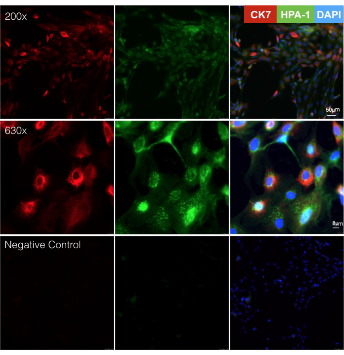
Figure 1: Human conjunctival goblet cell primary culture. The conjunctival goblet cells were passaged onto coverslips after 14 days in culture, and immunofluorescence staining using primary antibody against CK7 (red), and fluorescein-conjugated lectin HPA-1 (green) was performed. (A) Images were taken under a 200x magnification, and (B) images were taken under a 630x magnification. CK7 presents as a perinuclear fluorescent signal (left panel); HPA-1 presents a punctuate pattern perinuclearly (middle panel). Positive immunofluorescence signal of both CK7 and HPA-1 indicates a goblet cell. Negative control is incubation in the absence of the primary antibody (C). Scale bars = 50 µm (A) and 8 µm (B). Please click here to view a larger version of this figure.
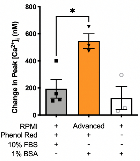
Figure 2: Effects of different media on Cch-induced [Ca2+]i response. Cells were incubated with phenol red-free RPMI with 1% BSA, RPMI media with 10% FBS, or advanced RPMI with 1% BSA for 48 h incubation before the addition of 1 × 10⁻⁴ M Cch. [Ca2+]i was measured by Fura2 assay and the change in peak [Ca2+]i was calculated and shown in the graph. The black bar indicates complete medium with phenol red and 10% FBS. The orange bar indicates advanced medium with 1% BSA, and the grey bar indicates RPMI without phenol red and with 1% BSA. Data are mean ± SEM, n = 4. One-way ANOVA with Dunnett correction was used in multiple group comparisons, p < 0.05 was set as statistically significant. * indicates significant difference between the groups. Abbreviations: Cch = carbachol; [Ca2+]i = intracellular Ca2+ concentration. Please click here to view a larger version of this figure.
Stock Component | Amount for 250 mL of KRB | Final Concentration |
Glucose | 0.25 g | 0.1% w/v |
1 M NaCl | 30 mL | 119 mM |
0.5 M NaHCO3 | 12.5 mL | 25 mM |
1 M 4-(2-hydroxyethyl)-1- piperazineethanesulfonic acid (HEPES) | 2.5 mL | 10 mM |
1 M KCl | 1.2 mL | 4.8 mM |
1 M KH2PO4 | 300 µL | 1.2 mM |
1 M MgSO4 | 300 µL | 1.2 mM |
1 M CaCl2 | 250 µL | 1 mM |
Table 1: Material and formula for KRB. Abbreviation: w/v, weight/volume.
Investigating the sex-based differences in ocular tissues helps understand the processes of diseases, especially dry eye and allergic conjunctivitis, which disproportionately affect one sex4,5,6. Even though animal models can be used for these studies, data obtained directly from human tissue are essential due to the highest similarity to the human cells in vivo. The conjunctival tissues used in the current experimental setting are from deceased donors with an age range of 70-85 years (postmenopausal), making it easier to control the hormonal effect ante mortem. Here, we presented a cost-efficient method to study sex-based differences in human CGCs.
The critical step for acquiring consistent data is the removal of FBS and phenol red for 48 h before experiments to eliminate the effects of sex hormones. The actions of sex hormones in cells can be categorized into non-transcriptional and transcriptional effects. The non-transcriptional effects are rapidly mediated through G-proteins in a few seconds to minutes, and the transcriptional effect in which certain genes get activated takes 30 min to several hours27; the half-life of estrogen-induced estrogen receptor alpha (ERα) is 2-6 h28,29. Even though the turnover rates of other types of sex hormone-induced proteins are rarely reported, results from the existing studies indicate that sex hormone-induced proteins are synthesized and degraded within hours. Therefore, 48 h is enough time to wash out the potential effect of hormones contained in the FBS while maintaining normal cellular function. Using the current method, we successfully reported sex-based differences in the expression of sex hormone receptors and cellular functions in CGCs30.
FBS is an essential factor for goblet cell cultures to remain healthy and viable. Replacing the normal heat-inactivated FBS with charcoal-stripped FBS is another possible way to eliminate sex hormones in the culture system. The limitation of using charcoal stripped FBS is the cost and the complex composition of the FBS. Completely removing FBS also eliminates the potential inconsistency from the undefined medium components of FBS.
The critical step for [Ca2+]i measurement is to stabilize the pH and temperature. Despite the HEPES supplement, we have noticed a shift of pH in the KRB buffer over time when stored at room temperature. Thus, a readjustment of the pH of the buffer is recommended for a long experiment, approximately every 3 h. The same [Ca2+]i method also applies to other types of cells such as stratified squamous cells of the conjunctiva, vascular endothelium cells, microglial cells, and neurons to study the action of different stimuli.
Binding of Fura-2 to free calcium in the cell causes a change in its peak absorption wavelength from 380 nm (unbound) to 340 nm (bound). However, the emission wavelength remains at 510 nm and these emissions are captured by the camera as black and white images. The ratio of emission intensity excited by 340 nm and 380 nm light is recorded. Besides comparing the 340/380 ratio to a stand curve to calculate a [Ca2+]i concentration, one may also report the fold change of the ratio without having to create a standard curve, therefore, without an actual concentration. Investigators can choose either way based on their equipment setup and the experimental design.
In conclusion, cultured primary conjunctival goblet cells can be used as a model to study the sex-based differences of ocular surface diseases. Incubation for 48 h in phenol red-free RPMI medium with 1% BSA before performing any experiments is a consistent and cost-efficient method to eliminate the potential effects of hormones in culture media and can be used as a basal medium to control hormone levels.
The authors have nothing to disclose.
The work is funded by the National Eye Institute Grant EY019470 (D.A.D).
0.05% trypsin with 1x EDTA | Gibco (Grand Island, NY) | 25300-054 | |
4-(2-hydroxyethyl)-1- piperazineethanesulfonic acid | Fisher Bioreagent (Pittsburgh, PA) | BP310-500 | |
Advanced RPMI media | Gibco (Grand Island, NY) | 12633020 | |
carbachol | Cayman Chemical (Ann Arbor, MI) | 144.86 | |
Fetal Bovin Serum | R&D (Minneapolis, MN) | S11150H | |
Fura-2- acetoxymethyl ester | Thermo Fisher Scientific (Waltham, MA, USA) | F1221 | |
Human conjunctival tissue | Eversight Eye Bank (Ann Arbor, MI) | N/A | |
inorganic salt for KRB buffer | Sigma-Aldrich (St. Louis, MO) | Any brand will work | |
L-glutamine | Lonza Group (Basel, Switzerland) | 17-605F | |
non-essential amino acids | Gibco (Grand Island, NY) | 11140-050 | |
penicillin/streptomycin | Gibco (Grand Island, NY) | 15140-122 | |
phenol red-free RPMI media | Gibco (Grand Island, NY) | 11835055 | |
Pluronic acid F127 | MilliporeSigma (Burlington, MA, USA) | P2443-250G | |
RPMI-1640 culture medium | Gibco (Grand Island, NY) | 21875034 | |
scalpel | Thermo Fisher Scientific (Waltham, MA, USA) | 12460451 | Any sterile surgical scalpel can work |
sodium pyruvate | Gibco (Grand Island, NY) | 11360-070 | |
sulfinpyrazone | MilliporeSigma (Burlington, MA, USA) | S9509-5G |