Here, we present the application of atomic force microscopy (AFM) as a simple and fast method for bacterial characterization and analyze details such as the bacterial size and shape, bacterial culture biofilms, and the activity of nanoparticles as bactericides.
Electron microscopy is one of the tools required to characterize cellular structures. However, the procedure is complicated and expensive due to the sample preparation for observation. Atomic force microscopy (AFM) is a very useful characterization technique due to its high resolution in three dimensions and because of the absence of any requirement for vacuum and sample conductivity. AFM can image a wide variety of samples with different topographies and different types of materials.
AFM provides high-resolution 3D topography information from the angstrom level to the micron scale. Unlike traditional microscopy, AFM uses a probe to generate an image of the surface topography of a sample. In this protocol, the use of this type of microscopy is suggested for the morphological and cell damage characterization of bacteria fixed on a support. Strains of Staphylococcus aureus (ATCC 25923), Escherichia coli (ATCC 25922), and Pseudomonas hunanensis (isolated from garlic bulb samples) were used. In this work, bacterial cells were grown in specific culture media. To observe cell damage, Staphylococcus aureus and Escherichia coli were incubated with different concentrations of nanoparticles (NPs).
A drop of bacterial suspension was fixed on a glass support, and images were taken with AFM at different scales. The images obtained showed the morphological characteristics of the bacteria. Further, employing AFM, it was possible to observe the damage to the cellular structure caused by the effect of NPs. Based on the images obtained, contact AFM can be used to characterize the morphology of bacterial cells fixed on a support. AFM is also a suitable tool for the investigation of the effects of NPs on bacteria. Compared to electron microscopy, AFM is an inexpensive and easy-to-use technique.
Different bacterial shapes were first noted by Antony van Leeuwenhoek in the 17th century1. Bacteria have existed in a great diversity of shapes since ancient times, ranging from spheres to branching cells2. Cell shape is a fundamental condition for bacterial taxonomists to describe and classify each bacterial species, mainly for the morphological separation of gram-positive and gram-negative phyla3. Several elements are known to determine bacterial cell forms, all of which are involved in the cell covers and support as components of the cell wall and membrane, as well as in the cytoskeleton. In this way, scientists are still elucidating the chemical, biochemical, and physical mechanisms and processes implicated in determining bacterial cell forms, all of which are defined by clusters of genes that define bacterial shapes2,4.
Additionally, scientists have shown that the rod shape is likely the ancestral form of bacterial cells, since this cell shape appears optimal in cell-significant parameters. Thus, cocci, spiral, vibrio, filamentous, and other forms are regarded as adaptations to various environments; indeed, particular morphologies have evolved independently multiple times, suggesting that the shapes of bacteria could be adaptations to particular environments3,5. However, throughout the bacterial cell life cycle, the cell shape changes, and this also occurs as a genetic response to damaging environmental conditions3. The bacterial cell shape and size strongly determine the stiffness, robustness, and surface-to-volume ratio of the bacteria, and this characteristic can be exploited for biotechnological processes6.
Electronic microscopy is used to study biological samples due to the high magnification that can be reached beyond light-based microscopes. Transmission electron microscopy (TEM) and scanning electron microscopy (SEM) are the most commonly used techniques for this purpose; however, samples require some treatments before they are placed into the chamber of the microscope in order to obtain appropriate images. A gold cover on the samples is required, and the time used for total image acquisition should not be too long. In contrast, atomic force microscopy (AFM) is a technique widely used in the analysis of surfaces but is also employed in the study of biological samples.
There are several types of AFM modes used in surface analysis, such as contact mode, non-contact mode or tapping, magnetic force microscopy (MFM), conductive AFM, piezoelectric force microscopy (PFM), peak force tapping (PFT), contact resonance, and force volume. Each mode is used in the analysis of materials and provides different information about the surface of the materials and their mechanical and physical properties. However, some AFM modes are used for the analysis of biological samples in vitro, such as PFT, because PFT allows for obtaining topographical and mechanical data on cells in a liquid medium7.
In this work, we used the most basic mode included in every old and simple AFM model-the contact mode. AFM uses a sharp probe (around <50 nm in diameter) to scan areas less than 100 µm. The probe is aligned to the sample in order to interact with the force fields associated with the sample. The surface is scanned with the probe to keep the force constant. Then, an image of the surface is generated by monitoring the motion of the cantilever as it moves across the surface. The gathered information provides the nano-mechanical properties of the surface, such as the adhesion, elasticity, viscosity, and shear.
In the AFM contact mode, the cantilever is scanned across the sample at a fixed deflection. This allows one to determine the height of the samples (Z), and this represents an advantage over the other electronic microscope techniques. The AFM software allows the generation of a 3D image scan by the interaction between the tip and sample surface, and the tip deflection is correlated to the height of the sample through a laser and a detector.
In static mode (contact mode) with constant force, the output presents two different images: the height (z topography) and the deflection or error signal. Static mode is a valuable, simple imaging mode, especially for robust samples in air that can handle the high loads and torsional forces exerted by static mode. The deflection or error mode is operated in constant force mode. However, the topography image is further enhanced by adding the deflection signal to the surface structure. In this mode, the deflection signal is also referred to as the error signal as the deflection is the feedback parameter; any features or morphology that appear in this channel are due to the "error" in the feedback loop or, rather, due to the feedback loop required to maintain a constant deflection setpoint.
AFM's unique design makes it compact – small enough to fit on a tabletop – while also having high enough resolution to resolve atomic steps. The AFM equipment has a lower cost than the equipment for other electronic microscopes, and the maintenance costs are minimal. The microscope does not require a lab with special conditions such as a clean room or an isolated space; it only needs a vibration-free desk. For AFM, the samples do not need to undergo elaborate preparation like for other techniques (gold cover, slimming); only a dry sample has to be attached to the sample holder.
We use AFM contact mode to observe bacterial morphologies and the effects of NPs. The population and cellular morphology of bacteria fixed on a support can be observed, as well as the cellular damage produced by nanoparticles on the bacterial species. The images obtained by AFM contact mode confirm that it is a powerful tool and is not limited by reagents and complicated procedures, making it a simple, fast, and economical method for bacterial characterization.
1. Bacterial isolation and identification
2. Bacterial sample preparation for morphological observation by AFM
3. Antibacterial effect of MgO nanoparticles against bacteria
NOTE: The synthesis and characterization of MgO NPs have been published previously14. In this work, the antibacterial activity of the nanomaterials was estimated based on the Clinical and Laboratory Standards Institute (CLSI) manual using macrodilution and microdilution methods for inhibition15,16.
4. AFM measurements
NOTE: Here, the atomic force microscope in contact mode was mounted on an anti-vibration workstation that allowed the isolation of the microscope from any mechanical vibrational sources and kept the system leveled. Electrical interference is diminished with line filters and surge protection. The AFM used here auto-aligns the laser beam to the photodetector.
Images of the morphology and size of S. aureus and P. hunanensis strains, as well as the population organization of both strains, were taken by atomic force microscopy in contact mode. The S. aureus images showed that its population was distributed by zones with aggregates of cocci (Figure 1A). With an increase in scale, there was a greater appreciation of the population distribution and morphology of the cocci (Figure 1B). The microscopy reports showed that a pseudo-hemispherical structure was present in adjacent cells of S. aureus, but that in general, the bacteria presented a spherical morphology in the form of a coccus after cell division, as previously shown in the AFM images. In addition, the AFM contact mode images allowed the determination of the size of the cocci, which showed an average width of 1.25 µm. The values obtained from the measurement of 20 cells are shown in Figure 2.
In the case of P. hunanensis, a homogeneous distribution was observed on the entire surface of the glass support, forming a bacterial monolayer that adhered to the support (Figure 3A), as reported by Zuttion et al., in which the authors immobilized P. aureginosa on a solid support and showed the same behavior. Pseudomonas hunanensis bacteria were observed to be rod-shaped, with a length of 1.9 µm (L) and a width of 0.9 µm (W) (Figure 3B,C); these data are within the reported values for P. fluorescens of 1.5-2.0 µm (L) and 0.6-0.9 µm (W)17,18,19.
To visualize the morphostructural alterations due to the interaction of the MgO NPs, the bacterial strains were subjected to AFM analysis after treatment (microdilution). Three groups are shown in Figure 4 and Figure 5: Figure 4A–C and Figure 5A–C are the controls; the images in Figure 4D–F and Figure 5D–F were obtained from the results of the microdilution at the MIC; the images of Figure 4G–I and Figure 5G–I were obtained at a higher concentration than the MIC.
The images of the upper group of Figure 4A–C show a coccus-type cell of Staphylococcus aureus with a smooth surface and homogeneous contours, which grew in a suitable environment in Müeller-Hinton broth for 24 h according to the microdilution technique. The average diameter was 1 µm ± 0.15 µm. This diameter practically disappeared following NP treatment, as shown in Figure 4D–F, since the deterioration of the cellular structure was very severe. According to the cross-section, the average height for the control was 200 nm ± 50 nm; the height of the treated cells was reduced by 40% (120 nm ± 5 nm) relative to the controls. The images clearly show changes in the surface, such as the formation of vesicles, following the exposure to MgO NPs in the CMI of the particles; additionally, by doubling the concentrations, the internal status of the cellular structures was affected, and cytosolic material was released, as shown in the images of Figure4G–I20,21.
These changes were also identified in E. coli cells, as shown in Figure 5, with conditions similar to S. aureus. The controls of this bacterium showed smooth surfaces, highlighting its very homogeneous rod-like shape (bacillus) without any apparent alteration. The three-dimensional images show the highest topographical regions associated with the microorganism in white. The average height of the control E. coli was 160 nm ± 50 nm, and according to the cross-sectional images, the average height decreased to 76 nm ± 10 nm for cells exposed for 24 h to a concentration of MgO nanoparticles of 500 ppm (MIC). The structure completely disappeared at a concentration of 1,000 ppm, and only the silhouettes of the bacteria could be distinguished. When analyzing the images for both concentrations, it was observed that with respect to the controls, the cell morphology of the treated cells was significantly altered, with an elongation of 2.0 µm ± 0.5 µm (Figure 5A–C) to 3.0 µm ± 0.3 µm, as shown in the images of Figure 5D–I. This increase was clear when the cellular structure was losing internal homeostasis, causing structural collapse20,22. The images of bacteria exposed to MgO NPs clearly showed surface changes such as ridge formation or corrugations forming vesicular disturbances at both MICs and when doubling the concentrations20,22.
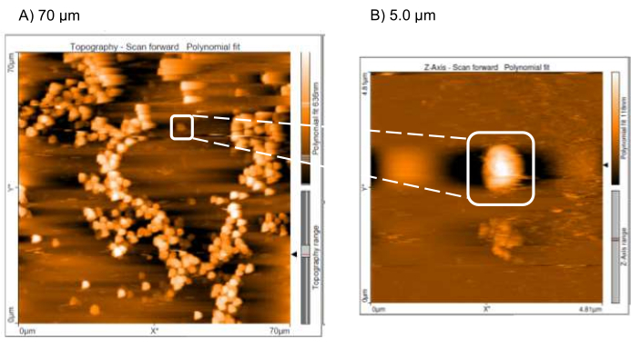
Figure 1: Atomic force microscopy contact mode for Staphylococcus aureus. This figure shows the topographies taken from S. aureus at different scales: (A) 70 µm and (B) 5.0 µm. The mapping areas were chosen to obtain the S. aureus topography; image B clearly shows S. aureus cocci. Please click here to view a larger version of this figure.
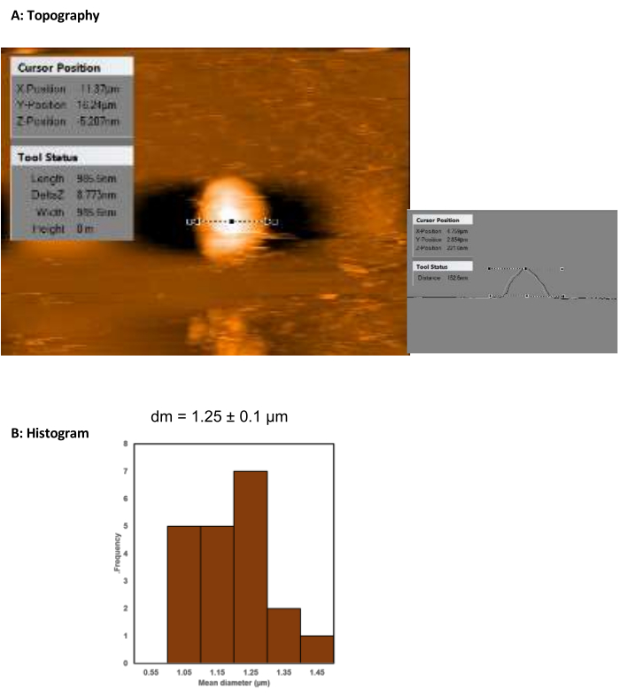
Figure 2: Atomic force microscopy contact mode topographies and histograms of Staphylococcus aureus. The image shows the (A) topography and (B) histogram of the diameters of S. aureus bacteria fixed on a glass support using the heat-fixation procedure. The cell diameter was measured with AFM software; n = 20. The image is a representation of the measurement. Please click here to view a larger version of this figure.
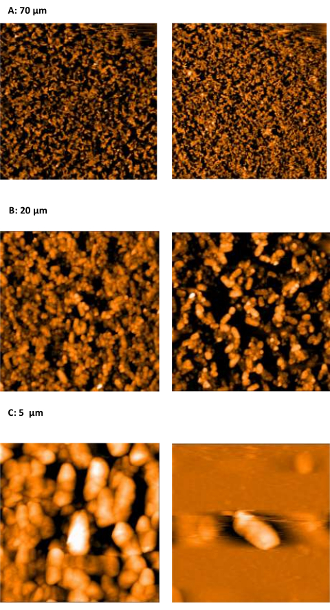
Figure 3: Atomic force microscopy contact mode for Pseudomonas hunanensis 1AP-CY. This figure shows the topographies taken from P. hunanensis 1AP-CY at different scales: (A) 70 µm, (B) 20 µm, and (C) 5.0 µm. The images at different scales show the distribution of the cell population on the slide. To analyze the cell morphology, an area with a lower population density is focused on, as shown in image B, which shows the rod shape of P. hunanensis (the deflection signal is shown in order to enhance the topography image). Please click here to view a larger version of this figure.
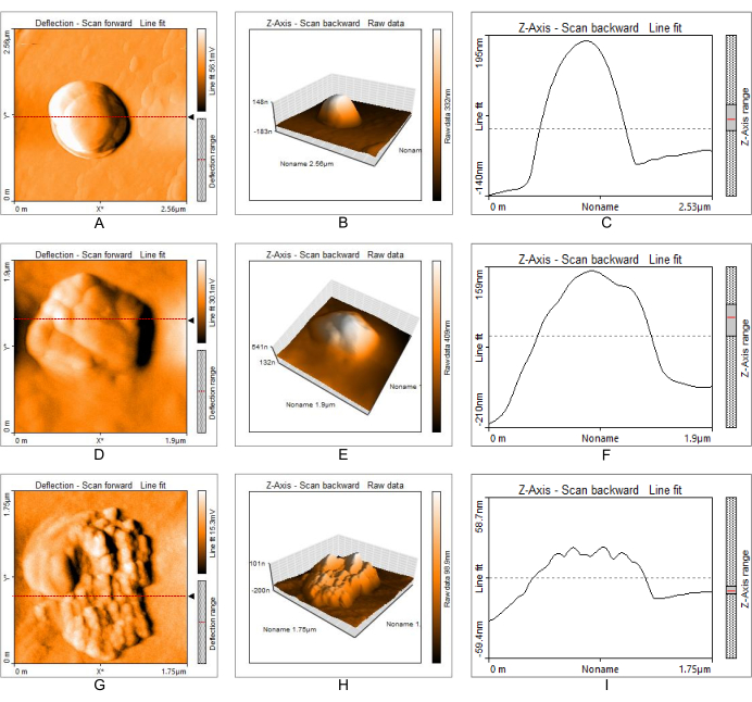
Figure 4. AFM contact mode images obtained for untreated S. aureus (top) and S. aureus exposed to 250 ppm and 500 ppm MgO nanoparticles (middle and bottom) for 24 h. (A,D,G) Topographic images; (B,E,H) three-dimensional images; (C,F,I) cross-sectional images. Structural changes in the bacteria were observed when employing the minimum inhibitory concentration of MgO (250 ppm) and a higher concentration (500 ppm). The deflection signal is shown to enhance the topography image. Figure 4A,D,G are from Muñiz Diaz et al.14. Please click here to view a larger version of this figure.
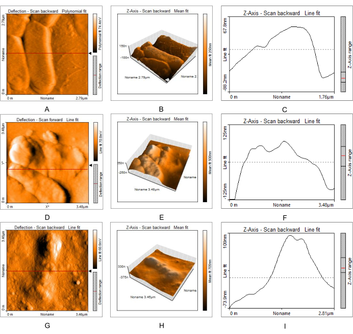
Figure 5: AFM contact mode images obtained for untreated E. coli (top) and E. coli exposed to 500 ppm and 1,000 ppm MgO nanoparticles (middle and bottom) for 24 h. (A,D,G) Topographic images; (B,E,H) three-dimensional images; (C,F,I) cross-sectional images. Structural changes in the bacteria were observed when employing the minimum inhibitory concentration of MgO (500 ppm) and a higher concentration (1,000 ppm). Figure 5A,D,G are from Muñiz Diaz et al.14. Please click here to view a larger version of this figure.
Microscopy is a technique commonly used in biological laboratories that allows for the investigation of the structure, size, morphology, and cellular arrangement of biological samples. To improve this technique, several types of microscopes can be used that differ from each other in terms of their optical or electronic characteristics, which determine the resolution power of the instrument.
In scientific research, the use of microscopy is required for the characterization of bacterial cells; for example, microscopy has demonstrated that NaCl influences the thermal resistance and cell morphology of E. coli strains, Schisandra chinensis extract shows antibacterial effects toward S. aureus, S. aureus develops thermotolerance after sublethal heat shock, and E. coli biomineralizes platinum23,24,25,26,27. Thus, the advantages presented by AFM have allowed its expansion to the research areas of environment and biology.
The atomic force microscope is an instrument used in contact mode to analyze bacteria in situ and in non-contact mode to determine the topography of macro- and nanoscopic materials. The advantage of AFM over other techniques is that it requires fewer samples to obtain topographic images; it is also considered a non-destructive technique, and it does not damage or compromise the structure of the analyzed image. Although we used heat fixation in this protocol, the bacterial cell morphology was not altered. It is important to mention that this technique is commonly used in the microbiology laboratory to study bacterial morphologies. Unlike in plant and animal tissue samples, the heat fixation technique produces changes in proteins and lipids that are manifested in tissue deterioration; in bacteria, the technique causes a slight shrinkage of the cell that does not interfere with the observation of cell shape and identification.
With AFM, sample preparation is easy and does not require any chemical products, as in the case of electron microscopy, where it is essential that the sample to be analyzed is conductive, since the image is produced by the interaction of the electrons emitted by the equipment and the sample. If the sample is not conductive, it must be metalized with a conductive element such as gold by means of the physical vapor deposition technique. Furthermore, the resolution of electron microscopy reaches up to 0.4 nm, depending on the lens and model, whereas AFM in contact mode can reach micrometer, nanometer, or even picometer resolution, which makes it competitive with electron microscopy22. Another advantage of AFM over electron microscopy is that it does not require high vacuum conditions for sample processing20,21. Other advantages of AFM over commonly used techniques are the low cost and short image acquisition time.
With regard to the technique of fixing the bacterial sample to a glass plate, it is important to mention that heat fixation is a technique commonly used in microbiology laboratories. This technique is used so that bacterial samples can be identified by simple or differential staining. At the same time, the shape of the bacteria, commonly cocci or rod, can be seen. A disadvantage of the heat fixation technique is the decrease in the size of the bacteria, but this technique does not compromise the shape of the bacteria.
After fixation, care must be taken to avoid the aging of the sample, which can manifest as a significant decrease in the cell size; hence, it is advisable to analyze the sample within 24 h of fixation. Care should also be taken to ensure that the sample is not exposed to dust; samples should, therefore, be kept in a closed container (e.g., a Petri dish). These conditions are critical as they may alter the AFM images obtained.
It is clear that decreasing the size of the bacteria may affect some research work, as well as that other ways of attaching the bacteria to a support may be required. For example, S. aureus biofilms grown in BM broth on 34 mm plates have been fixed with glyceraldehyde. Furthermore, S. aureus have been immobilized with V-shaped cantilevers treated with poly-l-lysine, while for C. albicans, hyphae have been fixed on glass slides coated with positively charged poly-l-lysine in order to analyze the exchange of adsorbed serum proteins during the adhesion of both to an abiotic surface28,29.
In this work, heat-fixing the samples did not alter the shape or aggregates formed by the bacteria, and the heat-fixing allowed the effect of the nanoparticles to be visualized. Therefore, the AFM topographies could show the shape of the isolated cocci or aggregates of S. aureus and the rods of P. hunanensis (Figure 1 and Figure 3). In addition, we also found that S. aureus and E. coli showed damage in their structures due to the effect of the MgO nanoparticles, whereas the control samples (without nanoparticles) did not show any alteration in their morphology (Figure 4 and Figure 5).
This work demonstrates that the use of the AFM contact mode is a viable alternative for characterizing the surface topology and defects of biological samples, as has been shown with S. aureus, P. hunanensis, and E. coli. Furthermore, the heat fixation technique is not a determining factor that alters the cell surface and prevents analysis. Modestly, we can suggest that heat fixation may be an advantage in the use of AFM. Samples are processed less, and the use of chemical reagents is avoided. Therefore, the methodology is a simple and economical technique. It is worth mentioning that this may change depending on the specific analysis to be performed, as shown for the study of biofilms and bacterial adhesion28,29. As a possible extension of this work, contact mode AFM could be used to analyze the effect of NPs on other medically important bacteria or in research in which cell damage is to be observed.
The authors have nothing to disclose.
Ramiro Muniz-Diaz thanks CONACyT for the scholarship.
AFM EasyScan 2 | NanoSurf | discontinued | Measurement Media |
bacteriological loop | No aplica | not applicable | instrument for bacterial inoculation |
BigDye Terminator v3.1 | ThermoFisher Scientific | 4337455 | Matrix installation kit |
Bioedit | not applicable | version 7.2.5 | Sequence alignment editor |
Cary 60 spectrometer | Agilent Technologies | not applicable | |
ceftriazone | Merck | not applicable | antibiotic |
centrifuge | eppendorf | not applicable | to remove particles that interfere with AFM |
ContAI-G Silicon cantilever | BudgetSensors | ContAl-G-10 | Measurement Media |
eosin and methylene blue agar | Merck | not applicable | bacterial culture medium |
Escherichia coli | American Type Culture Collection | ATCC 25922 | bacterial strain |
GoTaq Flexi DNA Polymerase | Promega | M8295 | PCR of 16S rRNA gene |
microplate | Thermo Scientific | 10558295 | for microdilution analysis |
Müller-Hinton broth | Merck | not applicable | bacterial culture medium |
nutrient agar | Merck | not applicable | bacterial culture medium |
nutritious broth | Merck | not applicable | bacterial culture medium |
Petri dishes | not applicable | not applicable | growth of bacteria |
Pseudomonas hunanensis 9AP | not applicable | not applicable | isolated from the garlic bulb by CNRG |
Sanger sequencing | Macrogen | not applicable | sequencing service |
ScienceDesk Anti-Vibration workstation | ThorLabs | ||
slides | not applicable | not applicable | glass holder for bacterial sample analysis |
Staphylococcus aureus | American Type Culture Collection | ATCC 25923 | bacterial strain |
Thermalcycler | Applied Biosystems | Veriti-4375786 | PCR amplification |
Trypticasein soy agar | BD | BA-256665 | growth media |
ultrasonicator | Cole-Parmer Ultrasonic Processor, 220 VAC | not applicable | for mixing the nanoparticle dilutions |