Summary

The Mouse Isolated Perfused Kidney Technique

Published: November 17, 2016
doi:

Summary

The mouse isolated perfused kidney (MIPK) is a technique for keeping a mouse kidney under ex vivo conditions perfused and functional for 1 hr. The buffers and surgical technique are described in detail.

Abstract

The mouse isolated perfused kidney (MIPK) is a technique for keeping a mouse kidney under ex vivo conditions perfused and functional for 1 hr. This is a prerequisite for studying the physiology of the isolated organ and for many innovative applications that may be possible in the future, including perfusion decellularization for kidney bioengineering or the administration of anti-rejection or genome-editing drugs in high doses to prime the kidney for transplantation. During the time of the perfusion, the kidney can be manipulated, renal function can be assessed, and various pharmaceuticals administered. After the procedure, the kidney can be transplanted or processed for molecular biology, biochemical analysis, or microscopy.

This paper describes the perfusate and the surgical technique needed for the ex vivo perfusion of mouse kidneys. Details of the perfusion apparatus are given and data are presented showing the viability of the kidney's preparation: renal blood flow, vascular resistance, and urine data as functional, transmission electron micrographs of different nephron segments as morphological readouts, and western blots of transport proteins of different nephron segments as molecular readout.

Introduction

The isolated perfusion of organs has been the subject of an ongoing effort among physiologists for many decades1. The technique enables the function of the organ, without systemic influences such as blood pressure, hormones, or nerves, to be studied. Carl Eduard Loebell is considered to be the first to have described the successful perfusion of an isolated kidney, in 18492. Since then, the perfusion apparatus has undergone significant refinement. Frey and Gruber introduced an artificial lung for oxygenation and pulsatile pumps for continuous perfusion2. While early researchers mainly studied the kidneys of large mammals-namely, pigs2 and dogs3-the first report of the use of rat kidneys, by Weiss et al., was a milestone in the study of small-mammal-organ perfusion4. Schurek et al. reported the necessity of adding mammalian erythrocytes to the perfusate if sufficient renal tubular oxygenation was to be achieved5. Critical for long-term experiments was the introduction of continuous dialysis of the buffer by the same research group6. In 2003, Schweda et al. were the first to report a functional mouse isolated perfused kidney (MIPK)7, later refined by Rahgozar et al.18 and Lindell et al.14.

While technically more challenging than the rat isolated perfused kidney, the use of the MIPK bears the advantage of enabling the use of a wide array of genetically altered mice. This paper presents the details of the authors' method for perfusing isolated mouse kidneys for 1 hr. The method allows for the continuous assessment of renal flow rate, vascular resistance, hormone release, blood gas analysis, urine analysis, and the application of drugs. Following the procedure, kidneys could be processed for molecular and biochemical analysis, be fixed for microscopy, or transplanted into a recipient mouse (Figure 1).

Figure 1
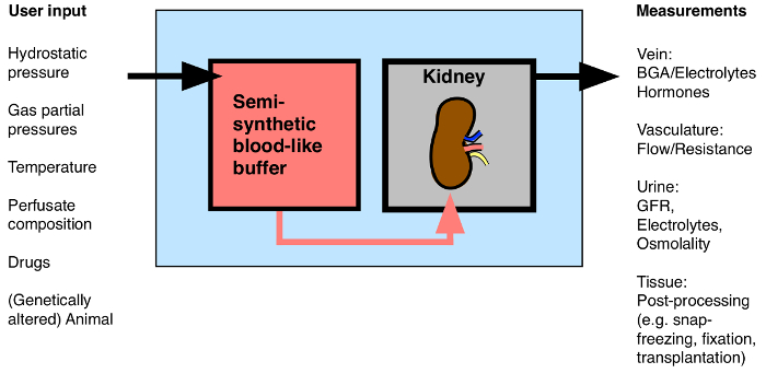
Figure 1: Overview of Possible Input/Output to the Isolated Perfused Kidney. BGA: Blood gas analysis. Please click here to view a larger version of this figure.

This technique likely will receive increasing attention over coming years, as many innovative applications are being discussed with the dawn of prolonged normothermic kidney perfusion prior to transplantation (with or without the application of anti-rejection or genome-editing drugs)8,9, 10, 11, the bioengineering of whole kidneys from decellularized scaffolds12, and the application of high doses of fluorescent dyes for multiphoton imaging13. It is also an ideal model with which to study the role of specific genes during acute kidney injury14.

A step-by-step protocol is given to allow other laboratories to perform isolated mouse kidney perfusion successfully. First, the composition and preparation of the buffer is specified. Then, the surgery is described in detail and the critical steps are shown. Third, data is presented that are representative of a successful preparation: renal blood flow, vascular resistance, glomerular filtration rate , and fractional electrolyte excretion-all as functional measurements of viability-and transmission electron micrographs of the morphology of different nephron segments of perfused kidneys fixed after 1 hr of perfusion.

Protocol

All procedures involving animals described in this manuscript were conducted according to Swiss law and approved by the veterinary administration of the Canton of Zurich, Switzerland. 1. Buffer Preparation Prepare solutions 1 – 4 and the antidiuretic hormone (ADH) solution (Table 1). Prepare the dialysis buffer (Table 1). NOTE: This is the buffer used as the dialysis buffer during the perfusion. Later, the erythrocytes will be dilu…

Representative Results

With the method described, isolated mouse kidneys can remain viable for at least 1 hr. We tested the tissue viability after 1 hr of continuous perfusion with functional (renal blood flow and vascular resistance, blood gas analysis of venous outflow, glomerular filtration rate, urinary fractional Na+ and K+ excretion, and urine osmolality) and morphological (transmission electron microscopy, TEM) methods in four kidneys of wildtype C57Bl/6 mice. Additionally, western …

Discussion

The mouse isolated perfused kidney is a tool for studying kidney function in a controlled environment ex vivo for 1 hr, bridging the gap between in vivo experiments in intact animals, which may be flawed by the impact of numerous systemic factors, and in vitro experiments in isolated nephron segments or cultured cells, which necessarily neglect the impact of intact organ structure on function. There is, to the authors' knowledge, no alternative technique with which to perform this specific …

Disclosures

The authors have nothing to disclose.

Acknowledgements

The authors would like to thank Hans-Joachim Schurek for invaluable scientific advice. The authors would like to thank Monique Carrel and Michèle Heidemeyer for excellent technical assistance, David Penton Ribas and Nourdine Faresse for a critical reading of the manuscript and Carsten Wagner and Jürg Biber for the NaPi-2a antibody. This work was supported by the Swiss National Centre for Competence in Research “Kidney.CH” and by a project grant (310030_143929/1) from the Swiss National Science Foundation.

Materials

Perfusion Circuit:
Moist chamber 834/8 Harvard Apparatus/Hugo Sachs Elektronik GmbH 73-2901
Cannular with basket and side port Harvard Apparatus/Hugo Sachs Elektronik GmbH 73-2947
Thermostat TC120-ST5  Harvard Apparatus/Hugo Sachs Elektronik GmbH 73-4544
ISM 827/230V Roller Pump Reglo Analogue Harvard Apparatus/Hugo Sachs Elektronik GmbH 73-0114
Reservoir jacketed for buffer solution 1L Harvard Apparatus/Hugo Sachs Elektronik GmbH 73-3438
Reservoir jacketed for buffer solution 0.5L Harvard Apparatus/Hugo Sachs Elektronik GmbH 73-3436
Pressure Transducer APT300  Harvard Apparatus/Hugo Sachs Elektronik GmbH 73-3862
TAM-D Plugsys Transducer Harvard Apparatus/Hugo Sachs Elektronik GmbH 73-1793
SCP Plugsys servo controller Harvard Apparatus/Hugo Sachs Elektronik GmbH 73-2806
Windkessel  Harvard Apparatus/Hugo Sachs Elektronik GmbH 73-3717
HSE-USB data acquisition Harvard Apparatus/Hugo Sachs Elektronik GmbH 73-3330
Low-Flux Dialysator Diacap Polysulfone B.Braun 7203525
PE-Tubing for aorta cannulation 1.19mm I.D. x 1.70mm O.D. Scientific Commodities Inc. BB31695-PE/8
Name Company Catalog Number Comments
Buffer reagents:
Aminoplasmal 10% B.Braun 134518064
Sodium pyruvate Sigma-Aldrich P2256-25G
L-Glutamic acid monosodium salt hydrate Sigma-Aldrich G1626-100G
L-(-)-Malic acid sodium salt Sigma-Aldrich M1125-25G
Sodium-L-Lactate Sigma-Aldrich L7022-10G
alpha-Ketoglutaric acid sodium salt Sigma-Aldrich K1875-25G
NaCl Sigma-Aldrich 31434-1KG-R
NaHCO3 Sigma-Aldrich S5761-5KG
KCl Sigma-Aldrich 60130-1KG
Urea Sigma-Aldrich U5378-500G
Creatinine Sigma-Aldrich C4255-10G
Ampicillin Roche 10835242001
MgCl2 * 6H2O Sigma-Aldrich M2393-500G
D-Glucose Sigma-Aldrich G8270-1KG
CaCl2 * 6H2 Riedel-de-Haën 12074
NaH2PO4 Sigma-Aldrich S9638-500G
Na2HPO4 Sigma-Aldrich S0876-500G
Antidiuretic Hormone dDAVP Sigma-Aldrich V2013-1MG
FITC-Inulin Sigma-Aldrich
Filter used for erythrocyte filtration Macherey-Nagel MN 615
BGA Analysis:
ABL 80 flex Radiometer Medical ApS
Electron Microscope:
Philips CM100 TEM FEI

References

  1. Carrel, A., Lindbergh, C. A. The culture of whole organs. Science (New York, N.Y.). 81 (2112), 621-623 (1935).
  2. Skutul, K. . Über Durchströmungsapparate Pflüger’s Archiv. 123 (4-6), 249-273 (1908).
  3. Nizet, A. The isolated perfused kidney: possibilities, limitations and results. Kidney Int. 7 (1), 1-11 (1975).
  4. Weiss, C., Passow, H., Rothstein, A. Autoregulation of flow in isolated rat kidney in the absence of red cells. Am J Physiol. 196 (5), 1115-1118 (1959).
  5. Schurek, H. J., Kriz, W. Morphologic and functional evidence for oxygen deficiency in the isolated perfused rat kidney. Lab Invest. 53 (2), 145-155 (1985).
  6. Stolte, H., Schurek, H. J., Alt, J. M. Glomerular albumin filtration: a comparison of micropuncture studies in the isolated perfused rat kidney with in vivo experimental conditions. Kidney Int. 16 (3), 377-384 (1979).
  7. Schweda, F., Wagner, C., Krämer, B. K., Schnermann, J., Kurtz, A. Preserved macula densa-dependent renin secretion in A1 adenosine receptor knockout mice. AJP Renal Physiol. 284 (4), F770-F777 (2003).
  8. Moers, C., Smits, J. M., et al. Machine perfusion or cold storage in deceased-donor kidney transplantation. N Engl J Med. 360 (1), 7-19 (2009).
  9. Nicholson, M. L., Hosgood, S. A. Renal transplantation after ex vivo normothermic perfusion: the first clinical study. Am J Transplant. 13 (5), 1246-1252 (2013).
  10. Worner, M., Poore, S., Tilkorn, D., Lokmic, Z., Penington, A. J. A low-cost, small volume circuit for autologous blood normothermic perfusion of rabbit organs. Art Org. 38 (4), 352-361 (2014).
  11. Kaths, J. M., Spetzler, V. N., et al. Normothermic Ex Vivo Kidney Perfusion for the Preservation of Kidney Grafts prior to Transplantation. J Vis Exp. (101), e52909 (2015).
  12. Song, J. J., Guyette, J. P., Gilpin, S. E., Gonzalez, G., Vacanti, J. P., Ott, H. C. Regeneration and experimental orthotopic transplantation of a bioengineered kidney. Nature Med. 19 (5), 646-651 (2013).
  13. Hall, A. M., Crawford, C., Unwin, R. J., Duchen, M. R., Peppiatt-Wildman, C. M. Multiphoton imaging of the functioning kidney. JASN. 22 (7), 1297-1304 (2011).
  14. Lindell, S. L., Williams, N., Brusilovsky, I., Mangino, M. J. Mouse IPK: A Powerful Tool to Partially Characterize Renal Reperfusion and Preservation Injury. Open Transplant J. 5, 15-22 (2011).
  15. Wagner, C., De Wit, C., Kurtz, L., Grünberger, C., Kurtz, A., Schweda, F. Connexin40 is essential for the pressure control of renin synthesis and secretion. Circ Res. 100 (4), 556-563 (2007).
  16. Czogalla, J., Vohra, T., Penton, D., Kirschmann, M., Craigie, E., Loffing, J. The mineralocorticoid receptor (MR) regulates ENaC but not NCC in mice with random MR deletion. Pflüger’s Archiv. , (2016).
  17. Poosti, F., et al. Precision-cut kidney slices (PCKS) to study development of renal fibrosis and efficacy of drug targeting ex vivo. Dis Model Mech. 8 (10), 1227-1236 (2015).
  18. Rahgozar, M., Guan, Z., Matthias, A., Gobé, G. C., Endre, Z. H. Angiotensin II facilitates autoregulation in the perfused mouse kidney: An optimized in vitro model for assessment of renal vascular and tubular function. Nephrology. 9 (5), 288-296 (2004).
check_url/cn/54712?article_type=t

Play Video

Cite This Article
Czogalla, J., Schweda, F., Loffing, J. The Mouse Isolated Perfused Kidney Technique. J. Vis. Exp. (117), e54712, doi:10.3791/54712 (2016).

View Video