Summary

Scalable Isolation and Purification of Extracellular Vesicles from Escherichia coli and Other Bacteria

Published: October 13, 2021
doi:

Summary

Bacteria secrete nanometer-sized extracellular vesicles (EVs) carrying bioactive biological molecules. EV research focuses on understanding their biogenesis, role in microbe-microbe and host-microbe interactions and disease, as well as their potential therapeutic applications. A workflow for scalable isolation of EVs from various bacteria is presented to facilitate standardization of EV research.

Abstract

Diverse bacterial species secrete ~20-300 nm extracellular vesicles (EVs), comprised of lipids, proteins, nucleic acids, glycans, and other molecules derived from the parental cells. EVs function as intra- and inter-species communication vectors while also contributing to the interaction between bacteria and host organisms in the context of infection and colonization. Given the multitude of functions attributed to EVs in health and disease, there is a growing interest in isolating EVs for in vitro and in vivo studies. It was hypothesized that the separation of EVs based on physical properties, namely size, would facilitate the isolation of vesicles from diverse bacterial cultures.

The isolation workflow consists of centrifugation, filtration, ultrafiltration, and size-exclusion chromatography (SEC) for the isolation of EVs from bacterial cultures. A pump-driven tangential flow filtration (TFF) step was incorporated to enhance scalability, enabling the isolation of material from liters of starting cell culture. Escherichia coli was used as a model system expressing EV-associated nanoluciferase and non-EV-associated mCherry as reporter proteins. The nanoluciferase was targeted to the EVs by fusing its N-terminus with cytolysin A. Early chromatography fractions containing 20-100 nm EVs with associated cytolysin A – nanoLuc were distinct from the later fractions containing the free proteins. The presence of EV-associated nanoluciferase was confirmed by immunogold labeling and transmission electron microscopy. This EV isolation workflow is applicable to other human gut-associated gram-negative and gram-positive bacterial species. In conclusion, combining centrifugation, filtration, ultrafiltration/TFF, and SEC enables scalable isolation of EVs from diverse bacterial species. Employing a standardized isolation workflow will facilitate comparative studies of microbial EVs across species.

Introduction

Extracellular vesicles (EVs) are nanometer-sized, liposome-like structures comprised of lipids, proteins, glycans, and nucleic acids, secreted by both prokaryotic and eukaryotic cells1. Since the early studies visualizing the release of EVs from gram-negative bacteria2, the number of biological functions attributed to bacterial EVs (20-300 nm in diameter) has constantly been growing in the past decades. Their functions include transferring antibiotic resistance3, biofilm formation4, quorum sensing5, and toxin delivery6. There is also growing interest in the use of bacterial EVs as therapeutics, especially in vaccinology7 and cancer therapy8.

Despite the growing interest in EV research, there are still technical challenges regarding methods of isolation. Specifically, there is a need for isolation methods that are reproducible, scalable, and compatible with diverse EV-producing organisms. To create a unified set of principles for planning and reporting EV isolation and research methods, the International Society for Extracellular Vesicles publishes and updates the MISEV position paper9. Moreover, the EV-TRACK consortium provides an open platform for reporting detailed methodologies for EV isolation used in published manuscripts to enhance transparency10.

In this protocol, previous methodologies used for the isolation of EVs from mammalian cell culture were adapted11,12 to enable the isolation of EVs from bacterial cell culture. We sought to employ methods that enable EV isolation from a variety of microbes, which can be scalable, and balance EV purity and yield (as discussed in the MISEV position paper9). After removing bacterial cells and debris by centrifugation and filtration, the culture medium is concentrated either by centrifugal device ultrafiltration (for a volume of up to ~100 mL) or pump-driven TFF (for larger volumes). EVs are then isolated by SEC using columns optimized for the purification of small EVs.

Figure 1
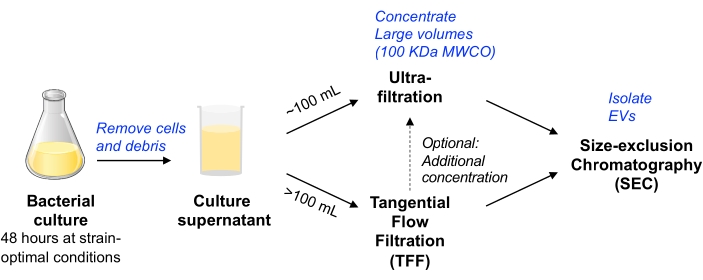
Figure 1: Bacterial EV isolation workflow schematic overview. Abbreviations: EV = extracellular vesicle; TFF = tangential flow filtration; SEC = size exclusion chromatography; MWCO = molecular weight cut-off. Please click here to view a larger version of this figure.

A mouse-commensal strain of Escherichia coli (i.e., E. coli MP113) was used as a model organism and modified to express EV-associated nanoluciferase by fusion to cytolysin A, as previously reported14. The methods used here can process at least up to several liters of bacterial cultures and effectively separate EV-associated from non-EV-associated proteins. Finally, this method can also be used for other gram-positive and gram-negative bacterial species. All relevant data of the reported experiments were submitted to the EV-TRACK knowledgebase (EV-TRACK ID: EV210211)10.

Protocol

NOTE: Ensure that all work involving bacteria and recombinant DNA follows best practices for biosafety containment appropriate for the biosafety hazard level of each strain. Work should be done in accordance with local, national, and international biosafety regulations. 1. Bacterial strains and culturing conditions NOTE: Bacterial strains used in this study were Escherichia coli MP113, Akkermansia mucinophila,…

Representative Results

To assess which SEC chromatography fractions were enriched for EVs, the SEC column was loaded with 2 mL of E. coli MP1-conditioned culture medium that had been concentrated 1,000-fold by TFF, and sequential fractions were collected. Using MRPS, it was found that Fractions 1-6 contained the most EVs (Figure 2A). Subsequent fractions contained very few EVs, comprising instead of EV-free proteins (Figure 2B). EVs were primarily <100 nm in diameter (<st…

Discussion

In the protocol above, a method is described that is scalable and reliably isolates EVs from various gram-negative/positive and aerobic/anaerobic bacteria. It has several potential stopping points throughout the procedure, although it is better to avoid taking longer than 48 h to isolate EVs from conditioned bacterial culture media.

First, it consists of culturing bacteria to generate conditioned bacterial culture medium. It was found that increasing the culture time to at least 48 h and using…

Offenlegungen

The authors have nothing to disclose.

Acknowledgements

The research described above was supported by NIH TL1 TR002549-03 training grant. We thank Drs. John C. Tilton and Zachary Troyer (Case Western Reserve University) for facilitating access to the particle size analyzer instrument; Lew Brown (Spectradyne) for technical assistance with analysis of the particle size distribution data; Dr. David Putnam at Cornell University for providing pClyA-GFP plasmid14; and Dr. Mark Goulian at the University of Pennsylvania for providing us with the E. coli MP113.

Materials

0.5 mL flat cap, thin-walled PCR tubes Thermo Scientific 3430 it is important to use thin-walled PCR tubes to obtain accurate readings with Qubit
16% Paraformaldehyde (formaldehyde) aqueous solution Electron microscopy sciences 15700
250 mL Fiberlite polypropylene centrifuge bottles ThermoFisher 010-1495
500 mL Fiberlite polypropylene centrifuge bottles ThermoFisher 010-1493
65 mm Polypropylene Round-Bottom/Conical Bottle Adapter Beckman Coulter 392077 Allows Vivacell to fit in rotor
Akkermansia mucinophila ATCC BAA-835
Amicon-15 (100 kDa MWCO) MilliporeSigma UFC910024
Avanti J-20 XPI centrifuge Beckman Coulter No longer sold by Beckman. Avanti J-26XP is closest contemporary model.
Bacteroides thetaiotaomicron VPI 5482 ATCC 29148
Bifidobacterium breve NCIMB B8807
Bifidobacterium dentium ATCC 27678
Brain Heart infusion (BHI) broth Himedia M2101 After autoclaving, Both BHI broth and agar were introduced into the anaerobic chamber, supplemented with Menadione (1 µg/L), hematin (1.2 µg/L), and L-Cysteine Hydrochloride (0.05%). They were then incubated for at least 24 h under anaerobic conditions before inoculation with the anaerobic bacterial strains.
C-300 microfluidics cartridge Spectradyne
Chloramphenicol MP Biomedicals ICN19032105
Escherichia coli HST08 (Steller competent cells) Takara 636763
Escherichia coli MP1 Dr. Mark Goulian (gift) commensal bacteria derived from mouse gut
Fiberlite 500 mL to 250 mL adapter ThermoFisher 010-0151-05 used with Fiberlite rotor to enable 250 mL bottles to be used for smaller size of starting bacterial culture
Fiberlite fixed-angle centrifuge rotor ThermoFisher F12-6×500-LEX fits 6 x 500 mL bottles
Formvar Carbon Film 400 Mesh, Copper Electron microscopy sciences FCF-400-CU
Glutaraldehyde (EM-grade, 10% aqeous solution) Electron microscopy sciences 16100
Hematin ChemCruz 207729B Stock solution was made in 0.2 M L-histidine solution as  1.2 mg/mL
Infinite M Nano+ Microplate reader Tecan This equibment was used to measure the mCherry fluorescence
In-Fusion  HD Cloning Plus Takara 638909 For cloning of the PCR fragements into the PCR-lineraized vectors
JS-5.3 AllSpin Swinging-Bucket Rotor Beckman Coulter 368690
Lauria Bertani (LB) broth, Miller Difco 244620
L-Cysteine Hydrochloride J.T. Baker 2071-05 It should be weighed and added directly to the autoclaved BHI media inside the anaerobic chamber
Masterflex Fitting, Polypropylene, Straight, Female Luer to Hose Barb Adapter, 1/8" ID; 25/PK cole-parmer – special HV-30800-08 connection adapters for filtration tubing circuit
Masterflex Fitting, Polypropylene, Straight, Male Luer to Hose Barb Adapter, 1/8" ID; 25/PK cole-parmer – special HV-30800-24 connection adapters for filtration tubing circuit
Masterflex L/S Analog Variable-Speed Console Drive, 20 to 600 rpm Masterflex HV-07555-00
Masterflex L/S Easy-Load Head for Precision Tubing, 4-Roller, PARA Housing, SS Rotor Masterflex EW-07514-10
Masterflex L/S Precision Pump Tubing, PharmaPure, L/S 16; 25 ft Cole Palmer EW-06435-16 low-binding/low-leaching tubing
Menadione (Vitamin K3) MP 102259 Stock solution was made in ethanol as 1 mg/mL
MIDIKROS 41.5CM 100K MPES 0.5MM FLL X FLL 1/PK Repligen D04-E100-05-N TFF device we have used to filter up to 2 L of E. coli culture supernatant
Nano-Glo Luciferase Assay System Promega N1110 This assay kit was used to measure the luminescence of the nluc reporter protein
NanoLuc (Nluc) Luciferase Antibody, clone 965808 R&D Systems MAB10026
nCS1 microfluidics resistive pulse sensing instrument Spectradyne
nCS1 Viewer Spectradyne Analysis software for particle size distribution
OneTaq 2x Master Mix with Standard Buffer NEB M0482 DNA polymerase master mix used to perform the routine PCR reactions for colony checking
Protein LoBind, 2.0 mL, PCR clean tubes Eppendorf 30108450
Q5 High-Fidelity 2x Master Mix NEB M0492 DNA polymerase master mix used to perform the PCR reactions needed for cloning
qEV original, 35 nm Izon maximal loading volume of 0.5 mL
qEV rack Izon for use with the qEV-original SEC columns
qEV-2, 35 nm Izon maximal loading volume of 2 mL
Qubit fluorometer ThermoFisher Item no longer available. Closest available product is Qubit 4.0 Fluorometer (cat. No. Q33238)
Qubit protein assay kit ThermoFisher Q33211 Store kit at room temperature. Standards are stored at 4 °C.
Sorvall Lynx 4000 centrifuge ThermoFisher 75006580
SpectraMax i3x Microplate reader Molecular Devices This equipment was used to measure the nanoluciferase bioluminescence
Stericup Quick-release-GP Sterile Vacuum Filtration system (150, 250, or 500 mL) MilliporeSigma S2GPU01RE
S2GPU02RE
S2GPU05RE
One or multiple filters can be used to accommodate working volumes. In our experience, you can filter twice the volume listed on the product size.
Uranyl acetate Electron microscopy sciences 22400
Vinyl anaerobic chamber Coy Lab
Vivacell 100, 100,000 MWCO PES Sartorius VC1042
Whatman Anotop 10 Plus syringe filters (0.02 micron) MilliporeSigma WHA68093002 to filter MRPS diluent

Referenzen

  1. Yanez-Mo, M., et al. Biological properties of extracellular vesicles and their physiological functions. Journal of Extracellular Vesicles. 4, 27066 (2015).
  2. Chatterjee, S. N., Das, J. Electron microscopic observations on the excretion of cell-wall material by Vibrio cholerae. Journal of General Microbiology. 49 (1), 1-11 (1967).
  3. Ciofu, O., Beveridge, T. J., Kadurugamuwa, J., Walther-Rasmussen, J., Hoiby, N. Chromosomal beta-lactamase is packaged into membrane vesicles and secreted from Pseudomonas aeruginosa. Journal of Antimicrobial Chemotherapy. 45 (1), 9-13 (2000).
  4. Yonezawa, H., et al. Outer membrane vesicles of Helicobacter pylori TK1402 are involved in biofilm formation. BMC Microbiology. 9, 197 (2009).
  5. Mashburn, L. M., Whiteley, M. Membrane vesicles traffic signals and facilitate group activities in a prokaryote. Nature. 437 (7057), 422-425 (2005).
  6. Kato, S., Kowashi, Y., Demuth, D. R. Outer membrane-like vesicles secreted by Actinobacillus actinomycetemcomitans are enriched in leukotoxin. Microbial Pathogenesis. 32 (1), 1-13 (2002).
  7. Petousis-Harris, H., et al. Effectiveness of a group B outer membrane vesicle meningococcal vaccine against gonorrhoea in New Zealand: a retrospective case-control study. Lancet. 390 (10102), 1603-1610 (2017).
  8. Kim, O. Y., et al. Bacterial outer membrane vesicles suppress tumor by interferon-gamma-mediated antitumor response. Nature Communications. 8 (1), 626 (2017).
  9. Thery, C., et al. Minimal information for studies of extracellular vesicles 2018 (MISEV2018): a position statement of the International Society for Extracellular Vesicles and update of the MISEV2014 guidelines. Journal of Extracellular Vesicles. 7 (1), 1535750 (2018).
  10. Consortium, E. -. T., et al. EV-TRACK: transparent reporting and centralizing knowledge in extracellular vesicle research. Nature Methods. 14 (3), 228-232 (2017).
  11. Watson, D. C., et al. Efficient production and enhanced tumor delivery of engineered extracellular vesicles. Biomaterials. 105, 195-205 (2016).
  12. Watson, D. C., et al. Scalable, cGMP-compatible purification of extracellular vesicles carrying bioactive human heterodimeric IL-15/lactadherin complexes. Journal of Extracellular Vesicles. 7 (1), 1442088 (2018).
  13. Lasaro, M., et al. Escherichia coli isolate for studying colonization of the mouse intestine and its application to two-component signaling knockouts. Journal of Bacteriology. 196 (9), 1723-1732 (2014).
  14. Kim, J. Y., et al. Engineered bacterial outer membrane vesicles with enhanced functionality. Journal of Molecular Biology. 380 (1), 51-66 (2008).
  15. Beveridge, T. J. Structures of gram-negative cell walls and their derived membrane vesicles. Journal of Bacteriology. 181 (16), 4725-4733 (1999).
  16. Reimer, S. L., et al. Comparative analysis of outer membrane vesicle isolation methods with an Escherichia coli tolA mutant reveals a hypervesiculating phenotype with outer-inner membrane vesicle content. Frontiers in Microbiology. 12, 628801 (2021).
check_url/de/63155?article_type=t

Play Video

Diesen Artikel zitieren
Watson, D. C., Johnson, S., Santos, A., Yin, M., Bayik, D., Lathia, J. D., Dwidar, M. Scalable Isolation and Purification of Extracellular Vesicles from Escherichia coli and Other Bacteria. J. Vis. Exp. (176), e63155, doi:10.3791/63155 (2021).

View Video