Here, we present a protocol to isolate apoptotic breast cancer cells by fluorescence-activated cell sorting and further detect the transition of breast non-stem cancer cells to breast cancer stem cell-like cells after apoptosis reversal by flow cytometry.
Cancer recurrence has long been studied by oncologists while the underlying mechanisms remain unclear. Recently, we and others found that a phenomenon named apoptosis reversal leads to increased tumorigenicity in various cell models under different stimuli. Previous studies have been focused on tracking this process in vitro and in vivo; however, the isolation of real reversed cells has yet to be achieved, which limits our understanding on the consequences of apoptosis reversal. Here, we take advantage of a Caspase-3/7 Green Detection dye to label cells with activated caspases after apoptotic induction. Cells with positive signals are further sorted out by fluorescence-activated cell sorting (FACS) for recovery. Morphological examination under confocal microscopy helps confirm the apoptotic status before FACS. An increase in tumorigenicity can often be attributed to the elevation in the percentage of cancer stem cell (CSC)-like cells. Also, given the heterogeneity of breast cancer, identifying the origin of these CSC-like cells would be critical to cancer treatment. Thus, we prepare breast non-stem cancer cells before triggering apoptosis, isolating caspase-activated cells and performing the apoptosis reversal procedure. Flow cytometry analysis reveals that breast CSC-like cells re-appear in the reversed group, indicating breast CSC-like cells are transited from breast non-stem cancer cells during apoptosis reversal. In summary, this protocol includes the isolation of apoptotic breast cancer cells and detection of changes in CSC percentage in reversed cells by flow cytometry.
Cancer has been a leading cause of death, causing heavy burden to countries worldwide1. Breast cancer ranks high both in terms of incidence and mortality in female patients among all types of cancer1. Due to the cancer heterogeneity, a combination of drugs is usually used in chemotherapy to achieve cancer cell death2,3,4. However, since common chemotherapeutic drugs often target DNA5,6, protein synthesis7,8 and/or microtubule dynamics9, rapidly growing cells are affected the most while quiescent cells such as cancer stem cell (CSC)s are usually less affected10. CSCs are, therefore, more likely to survive after the treatment, which later leads to drug resistance and cancer relapse10,11. Hence, elimination of CSCs has become an important topic for cancer treatment and study of the origin of CSCs is necessary.
More studies on the phenomenon of apoptosis reversal have been performed in the recent decade12,13,14,15,16,17,18,19. Before the emergence of this concept, it has been widely accepted that cells will irreversibly undergo apoptosis after caspase activation. Caspases are a family of protein enzymes that play key roles in the initiation and execution stages of apoptosis, including the formation of the apoptotic complex and the cleavage of downstream substrates20. Activation of executioner caspases such as caspase 3 or caspase 7 has been considered as the "point of no return" for apoptosis21. However, researchers recently observed that apoptosis reversal occurs both in vitro and in vivo, during which cells can recover from apoptosis even after caspase activation12,13,14,15,16,17,18,19. Moreover, aggressive features such as higher resistance to the original apoptotic inducer and higher invasiveness are detected in the reversed cancer cells15. Hence, it was proposed that the percentage of CSC-like cells would be higher in the reversed population when compared to the untreated cells, eventually contributing to the more malignant features after apoptosis reversal18.
Previously, many efforts have been made to track the apoptosis reversal in vitro and more importantly, in vivo, which greatly help in confirming the universality of this process16,17,19. However, a systemic study on the consequences of reversed cells is lacking due to the unsatisfactory isolation of cells that have genuinely undergone apoptosis reversal. There is a need to acquire pure apoptotic cells and recover them for further study. Thus, we use the traditionally well-accepted marker of executioner caspase activation as the marker of the "point of no return"21 for apoptosis and utilize fluorescence-activated cell sorting (FACS) to discriminate caspase-activated cells stained with Caspase-3/7 Green Detection dye. The dye is covalently linked to a short amino acid sequence, DEVD, which can be recognized and cleaved by active caspases 3/7. The cleavage helps release the dye, which will translocate from the cytosol to the nucleus where it binds to DNA and emits strong fluorescence. This procedure avoids using a bulk cell population in which some cells may not have undergone apoptosis.
CSCs or tumor-initiating cells have been identified in many solid tumors using a single or a combination of several surface marker(s) and very few numbers of these cells are sufficient to form tumors in immunodeficient mice22,23,24,25,26,27. A combination of CD44 and CD24 has been commonly used in breast CSC studies, and CD44+/CD24– cells have been defined as the breast CSCs26,27,28,29,30. Recently, we have performed a series of experiments to confirm the proposed relationship between apoptosis reversal and CSCs and demonstrate that reversed breast cancer cells gained increased tumor-forming ability in vitro and in vivo with an elevated percentage of cells with CSC markers18. Although we could not exclude the possibility that breast CSCs survive better and thus get enriched after apoptosis reversal, importantly, when we isolate non-stem cancer cells and subject them to apoptosis reversal, CSC will emerge in the originally non-stem cancer cell population, suggesting that non-stem cancer cells can contribute to the increase in the percentage of CSCs during apoptosis reversal.
This article aims to demonstrate the transition from breast non-stem cancer cells to breast CSC-like cells after apoptosis reversal and to detect this transition by flow cytometry. The breast non-stem cancer cells are initially prepared by isolating CD44–/CD24+ breast cancer cells by FACS. Then, apoptosis is induced and confirmed by morphological changes under the microscope. Afterwards, apoptotic cells positively-labelled by Caspase 3/7 Green Detection dye are isolated by FACS and further cultured in the absence of apoptotic inducers for apoptosis reversal. The reversed cells are then stained with CSC markers after 7 days of recovery for flow cytometric analysis. Cells with CD44+/CD24– markers re-appear in the reversed population, suggesting that transition from non-stem cancer cells to CSC-like cells has occurred during apoptosis reversal.
Apoptosis reversal has been observed in multiple cancer cell lines as well as normal primary cells treated with different apoptotic stimuli in vitro12,13. This process has also been traced in Drosophila model in vivo16,17,19. Much information regarding the underlying mechanism of cancer relapse in different cancer disease models and the origin of CSCs can be obtained through the use of the technique as described in this manuscript.
1. Preparation of Breast Non-stem Cancer Cells
2. Apoptotic Induction and Detection
3. Isolation of Apoptotic Cells and Apoptosis Reversal Procedure
4. Confirmation of Apoptosis in Caspase-activated Cells
5. Measurement of Breast CSC-like Cells by Flow Cytometry
In order to observe the transition from breast non-stem cancer cells to breast CSC-like cells, a first sorting of CD44–/CD24+ breast cancer cells were needed. For the MCF-7 cell line, which has around 0.15% cells with CSC markers in the original population (Figure 1), this step helped exclude the possibility of CSC enrichment during apoptosis reversal. On the contrary, if there were no cells with CSC markers in the original population, such as for T47D cells (Figure 1), this sorting procedure could be omitted. Indeed, gating affected the definition of the positive and negative of each marker, which would eventually influence the percentage of CSC determined. Therefore, appropriate controls including isotype controls for antibodies of interest, single stained controls for each marker should be carefully chosen and prepared for gate adjustment (Figure 1).
With breast non-stem cancer cells, the apoptosis reversal model could thereafter be established. Typical morphological changes could be observed after adding apoptotic inducers and cells should recover from apoptosis with similar morphology after drug withdrawal (Figure 2). Caspase-3/7 recognizes the amino acid sequence DEVD in the Caspase-3/7 Green Detection dye and the active forms of caspase-3/7 are able to cleave this site41. Originally, the fluorescence of the Caspase-3/7 Green Detection dye in cytosol is weak while if the dye is cleaved and translocated to the nucleus, the fluorescence signal would be amplified after its binding to DNA in the nucleus. This obvious difference could be distinguished by flow cytometry. Hence, those caspase-activated cells could be labelled and sorted out based on their higher fluorescence intensity comparing to those without caspase activation (Figures 3A-3D). During the apoptotic induction process, an appropriate solvent control must be included: solvent-treated cells without caspase activation were collected (Figures 3E and 3F) to exclude the possibility that the FACS procedure or the solvent itself was the cause for transition, if any.
In order to show that those FACS-sorted caspase-activated cells were indeed apoptotic, we co-stained these cells with Annexin V and PI. One of the early-stage changes in apoptotic cells is the translocation of phosphatidylserine from the inner side of plasma membrane to the external surface of the cell13,38,39,40; this externalization of phosphatidylserine could be detected by Annexin V. While this change is not unique to apoptosis, PI can be used for distinguishing apoptosis from necrosis based on the integrity of the cell membrane. Thus, cells with Annexin V binding but without PI staining are regarded as apoptotic cells. Caspase-activated cells in the apoptotic inducer treatment groups were found to be Annexin V positive and PI negative, suggesting that they were apoptotic cells (Figure 4).
After the second sorting based on caspase activation in apoptotic cells, these apoptotic cells were collected and subsequently cultured for recovery. The reversed cells that were alive were able to attach the culture container bottom and continue to proliferate. After 7 days, staining of CD44 and CD24 was performed in the reversed cells while controls were prepared at the same time as before. Compared with the solvent-treated groups (Figure 1), flow cytometric analysis showed that there were events (cells) appearing in the CD44+/CD24– quadrant in the reversed breast non-stem cancer cell population (Figure 1). Since we had already excluded cells with CD44+/CD24– in advance of apoptosis induction and only CD44–/CD24+ breast non-stem cancer cells were chosen, these CD44+/CD24– CSC-like cells could only be transited from breast non-stem cancer cells during apoptosis reversal.
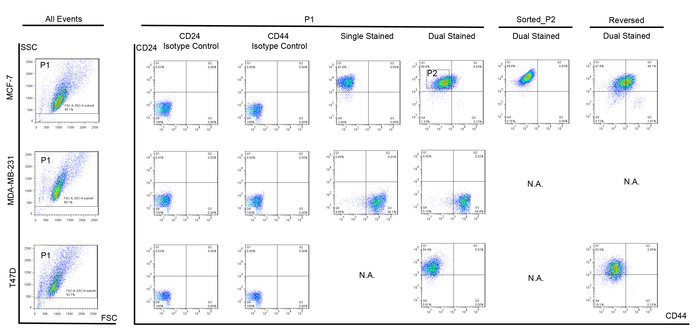
Figure 1: Representative breast CSC marker staining on breast cancer cells in flow cytometry.
MCF-7, MDA-MB-231 and T47D were stained with fluorochrome-conjugated monoclonal antibodies against human CD44 (PerCP-Cy5.5) and CD24 (PE). Cells were first gated by forward scatter (FSC) and side scatter (SSC) (P1) to exclude debris. Isotype controls of CD24 and CD44 were used as negative controls for CD24 and CD44 respectively. MCF-7 cells stained with CD24 was used as positive control for CD24 and MDA-MB-231 cells stained with CD44 was used as positive control for CD44. Non-stem MCF-7 breast cancer cells (P2) were sorted. These sorted cells and T47D cells were subjected to apoptosis reversal procedure. CSC-like (CD44+CD24–) cells appeared after apoptosis reversal. Please click here to view a larger version of this figure.
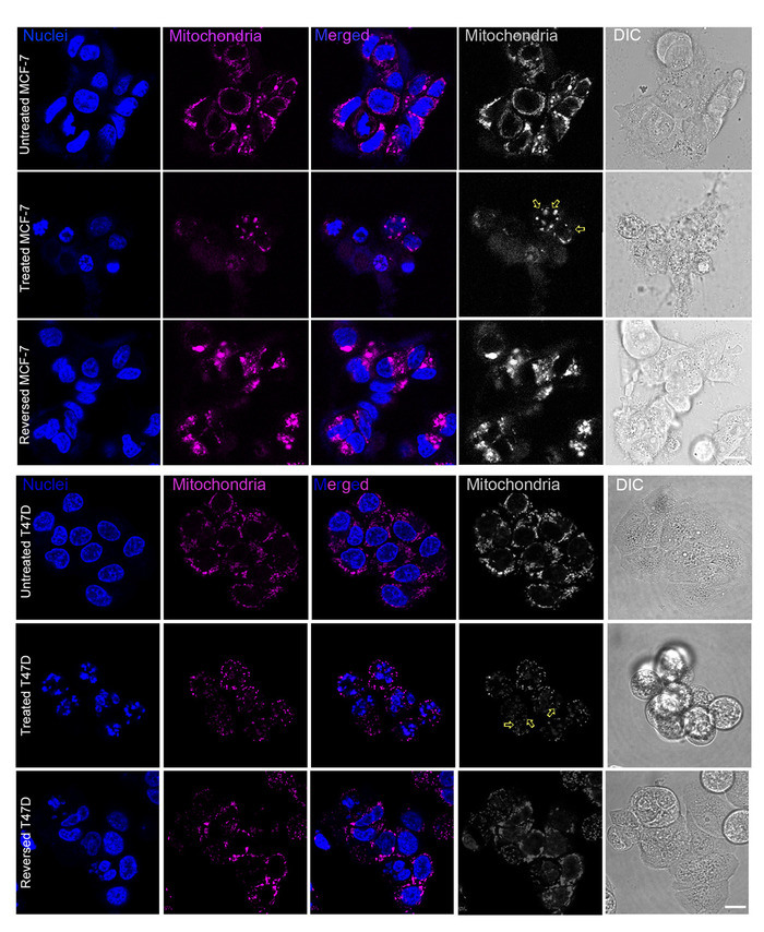
Figure 2: Morphological changes of breast cancer cells under apoptotic stimulus induction.
Breast cancer cells showed typical apoptotic morphological changes including cell shrinkage, membrane blebbing, mitochondria fragmentation (yellow arrows in the monochrome figures) and nuclear condensation. Nuclei (in blue): nuclei of cells stained with Hoechst 33342 were shown in blue color. Mitochondria (in pink): mitochondria of cells stained with Mitotracker Red CMXRos were shown in pink color. Merged: figures merged in dual colors showing both mitochondria and nuclei. Mitochondria (in gray): mitochondria of cells stained with Mitotracker Red CMXRos were shown in monochrome. Differential Interference Contrast (DIC): whole cells. Upper: MCF-7 cells were treated with staurosporine (STS) for 6 h then reversed for 24 h. Lower: T47D cells were treated with paclitaxel for 10 h with reversal for 24 h. Scale bars = 20 μm. Please click here to view a larger version of this figure.
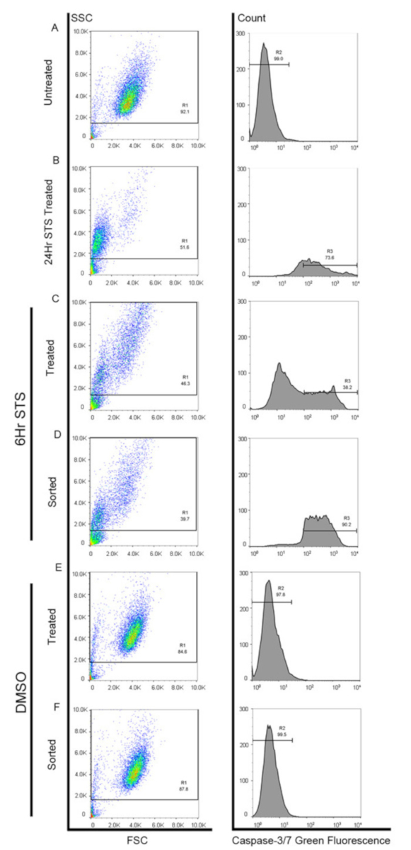
Figure 3: Caspase activation analysis by sorter. (A) Unstained MCF-7 cells without staurosporine (STS) treatment were used as negative control (R2). (B) MCF-7 cells treated with staurosporine (STS) for 24 h. Cells in R3 region were regarded as the caspase-activated cells. (C) MCF-7 cells treated with staurosporine (STS) for 6 h. Cells in R3 region were FACS-sorted. (D) FACS-sorted caspase-activated MCF-7 cells after staurosporine (STS) treatment for 6 h were re-run. (E) DMSO-treated MCF-7 cells. (F) FACS-sorted cells without caspase activation after DMSO treatment for 6 h were re-run. Please click here to view a larger version of this figure.
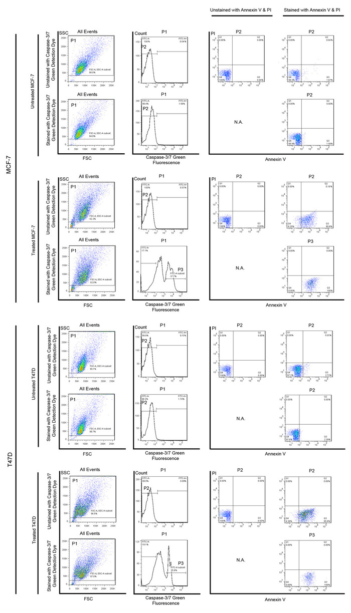
Figure 4: Annexin V and PI staining of caspase-activated cells in flow cytometry. MCF-7 and T47D cells were firstly gated by forward scatter (FSC) and side scatter (SSC) (P1) to exclude debris. Cells that were untreated with apoptotic inducers and without any staining were used as negative controls. For cells treated with apoptotic inducers, caspase-activated cells [i.e., Caspase-3/7 Green positive cells] were selected (P3) to show the fluorescence intensity of Annexin V and PI staining. All Caspase-3/7 Green positive cells were Annexin V positive and PI negative, suggesting that they were apoptotic. Please click here to view a larger version of this figure.
This protocol describes a direct and clear way for detecting the transition of breast non-stem cancer cells into breast CSC-like cells as a result of apoptosis reversal. Confirmation of the CSC properties of these reversed cells could be assisted by using in vitro mammosphere formation assay and in vivo xenograft transplantation in immunodeficient mice18,24,26,27,42,43,44,45. Here, we use two breast cancer cell lines, but this protocol can be further applied in other breast cancer cell lines. Since this phenomenon is apparent in the breast cancer cell line in which fewer number of CD44+/CD24– cells exist originally, it is suggested not to choose cell lines such as MDA-MB-231 in which there are more than 90% of CD44+ cells.
Since gating is important in flow analysis, proper and sufficient controls should be prepared in advance. For sorting, the purity should also be determined before the actual collection of cells. For example, to know the purity of the first sorting, a small portion of cells (such as 10,000 cells) can be collected and re-run on the sorter to check the pattern of CSC markers. If over 90% of these sorted cells are CD44–/CD24+ and are not shown in the CD44+/CD24– region, the collected cells are believed to be relatively pure (Figure 1). If cells appear in the CD44+/CD24– region, the sorting region and/or the sorter should be reset. Sorting should be conducted as sterile as possible either by using a sorter inside culture hood or adding antibiotics in the collection and culture medium for sorted cells.
Cancer cell types other than breast cancer can also be chosen and induced with various apoptotic stimuli to establish the apoptosis reversal model. While caspase activation could be detected as early as 2 h, the sorting time should be carefully selected. Since cells in the population are not synchronized, at one specific time point, each cell may have reached different stages of apoptosis. Meanwhile, cells keep going on to an apoptotic death after caspase activation. Therefore, if the inducers are removed only when all the cells reach caspase activation, some cells may have already reached the stage of DNA fragmentation, making them unable to recover even after inducer removal. It is not suggested to induce apoptosis for too long time to achieve a higher percentage of caspase-activated cells but with the cost of leaving a large number of cells dying after collection. Besides, the incubation time and the dye concentration that labels caspase-activated cells should be optimized whenever a new cell line is used for the first time.
Another concern as of successfully inducing apoptosis reversal in the apoptotic cells is the low recovery rate of the cells (usually less than 10%). The cells have gone through apoptosis plus the sorting procedure; therefore, they require very gentle handling during centrifugation and transfer at the collection and resuspension steps. Also, the choice of plate or dish used for subsequent culture should not be dependent on the number of cells collected but on the expected number of recoveries. Otherwise, cells could be allocated too sparsely to reverse and re-grow. Given that non-stem cancer cells grow at a higher speed and may dominate in the re-constituted cell population46, the detection time should not be too far away from the first recovery day.
This method first isolates breast non-stem cancer cell then applies them for apoptotic induction where a second sorting is done based on caspase activation before cells get recovered. The re-appearance of breast CSC-like cells in the reversed population is detected by flow cytometry. Since this in vitro apoptosis reversal procedure isolates pure apoptotic cancer cells, a number of experiments now can be conducted to understand the consequences of these real reversed cells. Apart from breast cancer cells, other solid tumor types as well as hematologic malignancy that are with known CSC markers can be studied and various apoptotic stimulus can be used. Hence, this method is extendable and applicable to a boarder scope of cancer investigation.
The authors have nothing to disclose.
This work was supported by the Innovative Technology Fund of Innovation Technology Commission: Funding Support from the State Key Laboratory of Agrobiotechnology (CUHK), the Lo Kwee-Seong Biomedical Research Fund and the Lee Hysan Foundation. Y.X. was supported by the postgraduate studentship from the CUHK.
MCF-7 | American Type Culture Collection (ATCC) | HTB-22 | |
MDA-MB-231 | American Type Culture Collection (ATCC) | HTB-26 | |
T47D | American Type Culture Collection (ATCC) | HTB-133 | |
Reagent | |||
0.05% trypsin-EDTA | Invitrogen | 25300054 | |
0.25% trypsin-EDTA | Invitrogen | 25200072 | |
Alexa Fluor 680 annexin V conjugate | Invitrogen | A35109 | |
bovine serum albumin | USB | 9048-46-8 | |
CaCl2 · 2H2O | Sigma-Aldrich | C-5080 | |
CellEvent caspase-3/7 green fluorescent dye | Invitrogen | C10423 | |
dimethyl sulfoxide | Sigma-Aldrich | D2650 | |
Fc block | Miltenyi Biotec | 130-059-901 | |
fetal bovine serum | Invitrogen | 16000044 | heat-inactivated |
HEPES | USB | 16926 | |
Hoechst 33342 | Invitrogen | H3570 | |
L-glutamine | Invitrogen | 25030081 | |
Mitotracker Red CMXRos | Invitrogen | M7512 | |
monoclonal antibodies against human CD24 | BD Biosciences | 555428 | PE Clone:ML5 Lot:5049759 RRID:AB_395822 |
monoclonal antibodies against human CD44 | BD Biosciences | 560531 | PERCP-CY5.5 Clone:G44-26 Lot:7230770 RRID:AB_1727485 |
NaCl | Sigma-Aldrich | 31434 | |
paclitaxel | Sigma-Aldrich | T7402 | |
PE Mouse IgG2a, κ Isotype Control | BD Biosciences | 554648 | Clone:G155-178 (RUO) RRID:AB_395491 |
Penicillin-Streptomycin | Invitrogen | 15070-063 | |
PerCP-Cy5.5 Mouse IgG2b, κ Isotype Control | BD Biosciences | 558304 | Clone:27-35 RRID:AB_647257 |
phosphate buffered saline | Thermo Fisher Scientific | 21600010 | |
propidium iodide | Invitrogen | P1304MP | |
Roswell Park Memorial Institute 1640 medium | Invitrogen | 11835055 | phenol red-free |
sodium azide | Sigma-Aldrich | S2002 | |
staurosporine | Sigma-Aldrich | S4400 | |
Equipment | |||
100 mm culture dish | Greiner Bio-One | 664160 | |
12-well tissue culture plates | Thermo Fisher Scientific | 150628 | |
Cell Strainer 40-μm nylon mesh | BD Biosciences | 08-771-1 | |
FACSuite software bundle v1.0 | BD Biosciences | 651360 | |
FACSVerse | BD Biosciences | 651155 | |
FluoView FV1000 confocal microscope | Olympus | IX81 | 60X objective |
FV10-ASW Viewer software Ver.4.2b | Olympus | – | |
round-bottom polystyrene 12 × 75 mm tubes | BD Biosciences | 352003 | |
S3e sorter | Bio-Rad | 1451006 |