Summary

인간 단핵구 유래 대식세포에서 근접성을 유도한 염증성 카스파제의 시각화

Published: April 06, 2022
doi:

Summary

이 프로토콜은 인간 혈액 샘플로부터 단핵구 유래 대식세포 (MDM)를 얻는 워크 플로우, 세포 생존력과 행동을 손상시키지 않고 염증성 카스파제 BiFC (BiFC) 리포터를 인간 MDM에 효율적으로 도입하는 간단한 방법, 살아있는 세포에서 염증성 카스파제 활성화를 측정하기위한 이미징 기반 접근법을 설명합니다.

Abstract

염증성 카스파제는 카스파제-1, -4, -5, -11, 및 -12를 포함하고, 개시제 카스파제의 하위군에 속한다. Caspase-1은 염증 신호전달의 정확한 조절을 보장하기 위해 요구되며, 염증에 대한 모집 후 근접-유도된 이량체화에 의해 활성화된다. Caspase-1은 단핵구 세포 계보에 풍부하고, 전염증성 사이토카인인 인터루킨(IL)-1β 및 IL-18의 성숙을 활성 분비 분자로 유도한다. 다른 염증성 카스파제, 카스파제-4 및 -5 (및 이들의 뮤린 상동체 카스파제-11)는 피로옵토시스를 유도함으로써 IL-1β 방출을 촉진한다. Caspase Bimolecular Fluorescence Complementation (BiFC)은 카스파제 활성화의 판독으로서 염증성 카스파제 유도 근접성을 측정하는데 사용되는 도구이다. 인플라마좀에 결합하는 영역을 포함하는 카스파제-1, -4, 또는 -5 프로도메인은 카스파아제가 유도된 근접성을 겪을 때 형광성 복합체를 개혁하기 위해 연관되는 황색 형광 단백질 금성(Venus-N[VN] 또는 Venus-C[VC])의 비형광 단편에 융합된다. 이 프로토콜은 핵을 사용하여 이러한 리포터를 일차 인간 단핵구 유래 대식세포 (MDM)에 도입하고, 세포를 치료하여 염증성 카스파제 활성화를 유도하고, 형광 및 공초점 현미경을 사용하여 카스파제 활성화를 측정하는 방법을 설명합니다. 이러한 접근법의 장점은 살아있는 세포에서 염증성 카스파제 활성화 복합체의 성분, 요건 및 국소화를 확인하는데 사용될 수 있다는 것이다. 그러나 세포 생존력과 행동을 손상시키지 않도록 신중한 통제를 고려해야합니다. 이 기술은 인플라마솜 수준에서 동적 카스파제 상호작용을 분석하고 살아있는 MDM 및 인간 혈액 샘플로부터 유래된 단핵구에서 염증성 신호전달 캐스케이드의 심문을 위한 강력한 도구입니다.

Introduction

카스파제는 개시제 카스파제 및 집행자 카스파제로 그룹화될 수 있는 시스테인 아스파르테이트 프로테아제의 패밀리이다. 사형 집행자 카스파제는 카스파제-3, -6 및 -7을 포함한다. 그들은 이량체로서 세포에서 자연적으로 발견되며 아폽토시스1을 실행하기 위해 개시제 카스파제에 의해 절단됩니다. 개시제 카스파제는 인간 카스파제-1, -2, -4, -5, -8, -9, -10 및 -12를 포함한다. 이들은 근접-유도된 이량체화에 의해 활성화되고 자가-단백질 분해 절단 2,3에 의해 안정화되는 비활성 효소원(pro-caspases)으로서 발견된다. 염증성 카스파제는 개시제 카스파제2의 서브세트이고, 인간에서 카스파제-1, -4, -5, 및 -12를 포괄하고, 마우스4,5에서 카스파제-1, -11, 및 -12를 포괄한다. 아폽토시스 역할보다는 염증에서 중심적인 역할을합니다. 이들은 프로인터루킨(IL)-1β 및 프로-IL-18 6,7의 단백질 분해 처리 및 분비를 매개하며, 이는 병원성 침입자 8,9에 반응하여 방출되는 최초의 사이토카인이다. Caspase-1은 활성화 플랫폼으로의 모집시 활성화됩니다. 큰 분자량의 단백질 복합체를 인플람마솜(도 1A)10이라고 불렀다. 카스파제-4, -5 및 -11의 이량체화는 비정준 인플라마솜 경로(11,12)를 통해 이들 플랫폼과 독립적으로 발생한다.

정준 인플라마좀은 인플람마솜 센서 단백질, 어댑터 단백질 ASC(CARD를 함유하는 아폽토시스-관련 얼룩-유사 단백질), 및 이펙터 단백질 카스파제-110으로 구성된 시토졸 다량체 단백질 복합체이다. 가장 잘 연구된 정준 인플라마좀은 피린 도메인 (NLRP), NLRP1 및 NLRP3을 함유하는 NOD 유사 수용체 패밀리, CARD (NLRC), NLRC4, 및 흑색종 2 (AIM2)에 결석한 NLR 패밀리를 함유한다. 그들은 각각 피린 도메인, 카드 또는 두 도메인을 모두 포함합니다. CARD 도메인은 CARD 함유 카스파제와 그들의 업스트림 활성화제 사이의 상호작용을 매개한다. 따라서, N 말단 피린 도메인 (PYD) 및 C 말단 CARD 모티프 13,14로 구성되는 스캐폴드 분자 ASC는 NLRP1 10, NLRP315 및 AIM216 inflammasomes에 대한 카스파제-1의 모집 필요하다.

각 인플람마솜은 뚜렷한 전염증성 자극을 인식하는 고유한 센서 단백질의 이름을 따서 명명되었습니다(그림 1B). 이 경로의 활성화자는 정식 자극이라고합니다. 인플람마좀은 미생물 성분 및 조직 스트레스에 대한 센서로서 작용하고, 염증 카스파제(17)의 활성화를 통해 강력한 염증 반응을 촉발시키기 위해 조립된다. 인플라마솜 어셈블리는 카스파제-1 활성화를 개시하여 그의 주요 기질인 pro-IL-1β 및 pro-IL-18의 성숙 및 분비를 매개한다. 이 프로세스는 두 단계 메커니즘을 통해 발생합니다. 첫째, 프라이밍 자극은 NF-κB 경로의 활성화를 통해 특정 염증성 단백질 및 pro-IL-1β의 발현을 상향조절한다. 둘째, 세포내(정경적) 자극은 프로파스파제-1 6,7의 인플라마솜 조립 및 모집을 유도한다.

Caspase-4 및 caspase-5는 뮤린 카스파제-11 11의 인간 오르토로그이다. 이들은 그람 음성 박테리아 18,19,20의 외막에서 발견되는 분자인 세포내 리포폴리사카라이드(LPS)와 적혈구 용혈(21)의 산물인 세포외 헴에 의해 인플라마솜-비의존성 방식으로 활성화된다. LPS가 이들 단백질의 CARD 모티프에 직접 결합하고 이들의 올리고머화(20)를 유도한다는 것이 제안되었다. 카스파제-4 또는 카스파제-5의 활성화는 기공 형성 단백질 가스더미민 D(GSDMD)18,19의 절단을 통해 피롭토시스라고 불리는 염증성 형태의 세포 사멸을 유도함으로써 IL-1β 방출을 촉진한다. 또한, 카스파제-4 및 GSDMD 매개 열록토시스 사멸로부터 야기되는 칼륨 이온의 유출은 NLRP3 인플라마좀의 활성화 및 카스파제-122,23의 후속 활성화를 유도한다. 따라서, 카스파제-4, -5, 및 -11은특정 자극(11,24)에 반응하여 피옵토시스 및 카스파제-1 활성화를 유도할 수 있는 LPS용 세포내 센서로 간주된다.

Figure 1
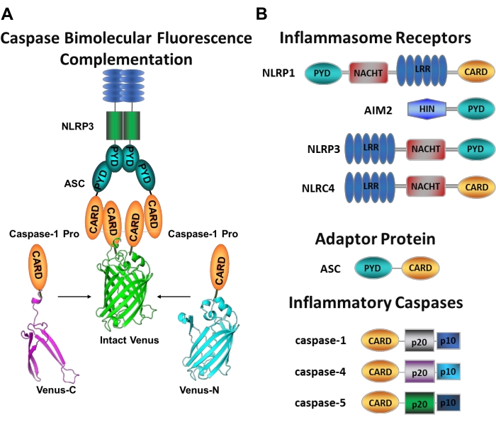
그림 1: 염증성 카스파제 및 카스파제-이분자 형광 보체 (BiFC) 분석. (A) 카스파제-BiFC 시스템을 나타낸 도면으로, 금성의 각 비형광 단편(Venus-C 또는 Venus-N)에 연결된 두 개의 카스파제-1 프로도메인(C1-pro)이 NLRP3 활성화 플랫폼으로 모집되어 금성이 재굴 및 형광을 갖도록 강제한다. 이 복합체는 현미경 하에서 녹색 반점으로 나타나며 개시제 카스파제 활성화의 첫 번째 단계 인 염증성 카스파제 유도 근접성에 대한 판독 역할을합니다. (b) 염증성 카스파아제 및 염증성 카스파제의 도메인 조직을 보여주는 개략도. 이 그림의 더 큰 버전을 보려면 여기를 클릭하십시오.

특정 개시제 카스파제 활성화를 측정하는 것은 어렵고, 이미징 접근법에 의해 그렇게 할 수 있는 방법은 많지 않다. Caspase Bimolecular Fluorescence Complementation (BiFC)은 살아있는 세포에서 직접 염증성 카스파제 활성화를 시각화하는 데 사용할 수 있습니다 (그림 1A)25. 이 기술은 최근 인간 단핵구 유래 대식세포 (MDM)21에서 사용하기 위해 적응되었다. 카스파제 BiFC는 염증성 카스파제 활성화의 첫 번째 단계를 측정하고, 이량체화를 촉진하기 위해 근접성을 유도한다. 광화성 황색 형광 단백질 금성 (Venus-C[VC]) 및 Venus-N[VN])의 비형광 단편에 융합된 CARD 함유 카스파제 프로도메인을 코딩하는 플라스미드의 발현이 사용된다. 두 카스파제 프로도메인이 그들의 활성화 플랫폼으로 모집되거나 유도된 근접성을 겪을 때, 금성의 두 반쪽은 근접하게 가져와 굴착 및 형광을 강요받는다(도 1A,B 참조). 이는 특정 염증성 카스파제 활성화의 실시간 판독을 제공한다.

인간 MDM은 위험 신호 및 병원체 생성물을 식별하는 인플라마솜 유전자와 패턴 인식 수용체를 풍부하게 발현한다. 이것은 염증성 카스파제 경로의 심문을 위한 이상적인 세포 유형을 제공한다. 또한, 이들은 말초 혈액 및 심지어 환자 샘플로부터 유래되어 특정 질병 상태에서의 염증성 카스파제 활성화를 평가할 수 있다. 이 프로토콜은 뉴클레오펙션을 사용하여 BiFC 카스파제 리포터를 MDM에 도입하는 방법, 전기천공 기반 트랜스펙션 방법, 염증성 카스파제 활성화를 유도하기 위해 세포를 치료하는 방법, 현미경 접근 방식을 사용하여 활성 카스파제 복합체를 시각화하는 방법을 설명합니다. 추가적으로, 이 방법론은 이들 복합체의 분자 조성, 아세포 국재화, 동역학, 및 이들 고도로 정렬된 구조의 크기(25,26,27)를 결정하도록 적응될 수 있다.

Protocol

이 프로토콜은 인간 샘플의 조작에 대한 Baylor College of Medicine의 인간 연구 윤리위원회의 지침을 따릅니다. 혈액 샘플은 인간 샘플에 대한 제도적 안전 지침에 따라 처리됩니다. 혈액 샘플은 지역 혈액 은행에서 얻어지며, 구연산 인산염 덱스트로스 (CPD) 용액으로 수집됩니다. 그러나, 나트륨 헤파린, 리튬 헤파린 또는 EDTA와 같은 다른 항응고제로 수집된 혈액은 또한 이 프로토콜28,29</s…

Representative Results

도 2에 도시된 반응식은 인간 MDM을 수득, 형질감염 및 이미지화하는 방법에 대한 개요를 제공한다. 선택된 CD14+ 단핵구를 GM-CSF로 7일 동안 인큐베이션한 후, 분화 기간에 걸쳐 세포 형태가 변화하고(도 3A), 구형 현탁액 세포로부터 스핀드하고 완전히 부착되는 과정(제3일 및 제4일), 마지막으로 완전히 분화되었을 때 세포를 더 많이 퍼뜨린다(제7일째). ?…

Discussion

이 프로토콜은 인간 혈액 샘플로부터 분리된 단핵구로부터 대식세포를 수득하는 워크플로우 및 세포 생존력 및 거동을 손상시키지 않으면서 염증성 카스파제 BiFC 리포터를 인간 MDM에 효율적으로 도입하는 방법을 기술한다.

이 프로토콜은 BiFC 기술(35 )을 활용하여 카스파제 모집 도메인(CARD)에서 염증성 카스파제를 비형광 단백질 금성의 비형광 단편으로 태?…

Divulgaciones

The authors have nothing to disclose.

Acknowledgements

우리는이 기술의 발전에 기여한 LBH의 과거와 현재의 실험실 구성원들에게 감사드립니다. 이 실험실은 NIH / NIDDK T32DK060445 (BEB), NIH / NIDDK F32DK121479 (BEB), NIH / NIGMS R01GM121389 (LBH)가 지원합니다. 도 2는 바이오렌더 소프트웨어를 사용하여 도출하였다.

Materials

48 well tissue culture2:34 plates Genesee Scientific 25-108
10 cm Tissue Culture Dishes VWR 25382-166
2 Mercaptoethanol 1000x Thermo Fisher Scientific 21985023
8 well chambered coverglass with 1.5 HP coverglass Cellvis c8-1.5H-N
AutoMACS columns Miltenyi (Biotec) 130-021-101 For automated separation using AutoMACS Pro Separator only
AutoMACS Pro Separator Miltenyi (Biotec) 130-092-545 For automated separation using AutoMACS Pro Separator only
AutoMACS Pro Washing Solution Miltenyi (Biotec) 130-092-987 For automated separation using AutoMACS Pro Separator only
AutoMACS Rinsing Solution Miltenyi (Biotec) 130-091-222 For automated separation using AutoMACS Pro Separator only
AutoMacs running buffer Miltenyi (Biotec) 130-091-221 For manual or automated separation using QuadroMACS or AutoMACS pro Separator
Axio Observer Z1 motorized inverted microscope equipped with a CSU-X1A 5000 spinning disk unit Zeiss Any confocal microscope equipped with a laser module fitted with laser lines of 568 nm (RFP) and 488 or 512 nm (GFP or YFP) wavelengths can be used
AxioObserver A1, Research Grade Inverted Microscope Zeiss Any epifluorescence microscope with fluorescence filters capable of exciting 568 nm (RFP) and 488 or 512 nm (GFP or YFP) wavelengths can be used
CD14+ MICROBEADS Miltenyi (Biotec) 130-050-201 For manual or automated separation using QuadroMACS or AutoMACS pro Separator
DPBS without calcium chloride and magnesium chloride Sigma D8537-6x500ML
DsRed mito plasmid Clontech 632421 Similar plasmids that can be used as fluorescent reporters can be found on Addgene
Fetal Bovine Serum Thermo Fisher Scientific 10437028
Ficoll-Paque PLUS 6 x 100 mL Sigma GE17-1440-02
GlutaMAX Supplement (100x) Thermo Fisher Scientific 35050079
GM-CSF Thermo Fisher Scientific PHC2011
Hemin BioXtra, from Porcine, ≥96.0% (HPLC) Sigma 51280-1G
HEPES Thermo Fisher Scientific 15630106
Inflammatory caspase BiFC plasmids Available by request from LBH lab
LPS-EB Ultrapure Invivogen TLRL-3PELPS
LS Columns Miltenyi (Biotec) 130-042-401 For manual separation using QuadroMACS Separator only
MACS 15 mL Tube Rack Miltenyi (Biotec) 130-091-052 For manual separation using QuadroMACS Separator only
MACS MultiStand Miltenyi (Biotec) 130-042-303 For manual separation using QuadroMACS Separator only
mCherry plasmid Yungpeng Wang Lab Similar plasmids that can be used as fluorescent reporters can be found on Addgene
Neon Transfection System Thermo Fisher Scientific MPK5000 Includes Neon electroporation device, pipette and pipette station
Neon Transfection System 10 µL Kit Thermo Fisher Scientific MPK1096 Includes resuspension buffer R, resuspension buffer T, electrolytic buffer E, 96 x 10 µL Neon tips and Neon electroporation tubes
Nigericin sodium salt, ready made solution Sigma SML1779-1ML
Penicillin-Streptomycin (10,000 U/mL) Thermo Fisher Scientific 15140122
Poly-D-Lysine Hydrobromide Sigma P7280-5mg
QuadroMACS Separator Miltenyi (Biotec) 130-090-976 For manual separation using QuadroMACS Separator only
qVD-OPh Fisher (ApexBio) 50-101-3172
RPMI 1640 Medium Thermo Fisher Scientific 11875119
Trypsin-EDTA (0.25%), phenol red Thermo Fisher Scientific 25200072
UltraPure 0.5 M EDTA, pH 8.0 Thermo Fisher Scientific 15575020
Zeiss Zen 2.6 (blue edition) software Zeiss Any software used to operate the confocal microscope of choice

Referencias

  1. Boatright, K. M., et al. A unified model for apical caspase activation. Molecular Cell. 11 (2), 529-541 (2003).
  2. Pop, C., Salvesen, G. S. Human caspases: activation, specificity, and regulation. Journal of Biological Chemistry. 284 (33), 21777-21781 (2009).
  3. Boice, A., Bouchier-Hayes, L. Targeting apoptotic caspases in cancer. Biochimica et Biophysica Acta (BBA) – Molecular Cell Research. 1867 (6), 118688 (2020).
  4. Bolívar, B. E., Vogel, T. P., Bouchier-Hayes, L. Inflammatory caspase regulation: maintaining balance between inflammation and cell death in health and disease. The FEBS Journal. 286 (14), 2628-2644 (2019).
  5. Martinon, F., Tschopp, J. Inflammatory caspases: linking an intracellular innate immune system to autoinflammatory diseases. Cell. 117 (5), 561-574 (2004).
  6. Lamkanfi, M., Vishva, M. D. Mechanisms and functions of inflammasomes. Cell. 157 (5), 1013-1022 (2014).
  7. Viganò, E., et al. Human caspase-4 and caspase-5 regulate the one-step noncanonical inflammasome activation in monocytes. Nature Communications. 6, 8761 (2015).
  8. Cerretti, D. P., et al. Molecular cloning of the interleukin-1 beta converting enzyme. Science. 256 (5053), 97 (1992).
  9. van de Veerdonk, F. L., Netea, M. G., Dinarello, C. A., Joosten, L. A. Inflammasome activation and IL-1β and IL-18 processing during infection. Trends in Immunology. 32 (3), 110-116 (2011).
  10. Martinon, F., Burns, K., Tschopp, J. The inflammasome: a molecular platform triggering activation of inflammatory caspases and processing of proIL-beta. Molecular Cell. 10 (2), 417-426 (2002).
  11. Kayagaki, N., et al. Noncanonical inflammasome activation targets caspase-11. Nature. 479 (7371), 117-121 (2011).
  12. Kayagaki, N., et al. Noncanonical inflammasome activation by intracellular LPS independent of TLR4. Science. 341 (6151), 1246-1249 (2013).
  13. Masumoto, J., et al. ASC, a novel 22-kDa protein, aggregates during apoptosis of human promyelocytic leukemia HL-60 cells. The Journal of Biological Chemistry. 274 (48), 33835-33838 (1999).
  14. Bertin, J., DiStefano, P. S. The PYRIN domain: a novel motif found in apoptosis and inflammation proteins. Cell Death and Differentiation. 7 (12), 1273-1274 (2000).
  15. Agostini, L., et al. NALP3 forms an IL-1beta-processing inflammasome with increased activity in Muckle-Wells autoinflammatory disorder. Immunity. 20 (3), 319-325 (2004).
  16. Bürckstümmer, T., et al. An orthogonal proteomic-genomic screen identifies AIM2 as a cytoplasmic DNA sensor for the inflammasome. Nature Immunology. 10 (3), 266-272 (2009).
  17. Latz, E., Xiao, T. S., Stutz, A. Activation and regulation of the inflammasomes. Nature Reviews Immunology. 13 (6), 397-411 (2013).
  18. Kayagaki, N., et al. Caspase-11 cleaves gasdermin D for noncanonical inflammasome signalling. Nature. 526 (7575), 666-671 (2015).
  19. Shi, J., et al. Cleavage of GSDMD by inflammatory caspases determines pyroptotic cell death. Nature. 526 (7575), 660-665 (2015).
  20. Shi, J., et al. Inflammatory caspases are innate immune receptors for intracellular LPS. Nature. 514 (7521), 187-192 (2014).
  21. Bolívar, B. E., et al. Noncanonical Roles of Caspase-4 and Caspase-5 in Heme-Driven IL-1β Release and Cell Death. The Journal of Immunology. 206 (8), 1878-1889 (2021).
  22. Schmid-Burgk, J. L., et al. Caspase-4 mediates noncanonical activation of the NLRP3 inflammasome in human myeloid cells. European Journal of Immunology. 45 (10), 2911-2917 (2015).
  23. Baker, P. J., et al. NLRP3 inflammasome activation downstream of cytoplasmic LPS recognition by both caspase-4 and caspase-5. European Journal of Immunology. 45 (10), 2918-2926 (2015).
  24. Cullen, S. P., Kearney, C. J., Clancy, D. M., Martin, S. J. Diverse activators of the NLRP3 inflammasome promote IL-1β secretion by triggering necrosis. Cell Reports. 11 (10), 1535-1548 (2015).
  25. Sanders, M. G., et al. Single-cell imaging of inflammatory caspase dimerization reveals differential recruitment to inflammasomes. Cell Death & Disease. 6, 1813 (2015).
  26. Charendoff, C. I., Bouchier-Hayes, L. Lighting up the pathways to caspase activation using bimolecular fluorescence complementation. Journal of Visualized Experiments: JoVE. (133), e57316 (2018).
  27. Bouchier-Hayes, L., et al. Characterization of cytoplasmic caspase-2 activation by induced proximity. Molecular Cell. 35 (6), 830-840 (2009).
  28. Riedhammer, C., Halbritter, D., Weissert, R. Peripheral blood mononuclear cells: Isolation, Freezing, thawing, and culture. Methods in Molecular Biology. 1304, 53-61 (2016).
  29. Betsou, F., Gaignaux, A., Ammerlaan, W., Norris, P. J., Stone, M. Biospecimen science of blood for peripheral blood mononuclear cell (PBMC) functional applications. Current Pathobiology Reports. 7 (2), 17-27 (2019).
  30. Perregaux, D., et al. IL-1 beta maturation: evidence that mature cytokine formation can be induced specifically by nigericin. The Journal of Immunology. 149 (4), 1294-1303 (1992).
  31. Cheneval, D., et al. Increased mature interleukin-1β (IL-1β) secretion from THP-1 cells induced by nigericin is a result of activation of p45 IL-1β-converting enzyme processing. Journal of Biological Chemistry. 273 (28), 17846-17851 (1998).
  32. Fernandes-Alnemri, T., et al. The pyroptosome: a supramolecular assembly of ASC dimers mediating inflammatory cell death via caspase-1 activation. Cell Death and Differentiation. 14 (9), 1590-1604 (2007).
  33. Lu, A., et al. Unified polymerization mechanism for the assembly of ASC-dependent inflammasomes. Cell. 156 (6), 1193-1206 (2014).
  34. Muller-Eberhard, U., Javid, J., Liem, H. H., Hanstein, A., Hanna, M. Brief report: Plasma concentrations of hemopexin, haptoglobin and heme in patients with various hemolytic diseases. Blood. 32 (5), 811-815 (1968).
  35. Shyu, Y. J., Liu, H., Deng, X., Hu, C. D. Identification of new fluorescent protein fragments for bimolecular fluorescence complementation analysis under physiological conditions. Biotechniques. 40 (1), 61-66 (2006).
  36. Thornberry, N. A., et al. A novel heterodimeric cysteine protease is required for interleukin-1βprocessing in monocytes. Nature. 356 (6372), 768-774 (1992).
  37. Jensen, K., Anderson, J. A., Glass, E. J. Comparison of small interfering RNA (siRNA) delivery into bovine monocyte-derived macrophages by transfection and electroporation. Veterinary Immunology and Immunopathology. 158 (3-4), 224-232 (2014).
  38. Tada, Y., Sakamoto, M., Fujimura, T. Efficient gene introduction into rice by electroporation and analysis of transgenic plants: use of electroporation buffer lacking chloride ions. Theoretical and Applied Genetics. 80 (4), 475-480 (1990).
  39. Bhowmik, P., et al. Targeted mutagenesis in wheat microspores using CRISPR/Cas9. Scientific Reports. 8 (1), 6502 (2018).
  40. Sansom, D. M., Manzotti, C. N., Zheng, Y. What’s the difference between CD80 and CD86. Trends in Immunology. 24 (6), 314-319 (2003).
  41. Kurokawa, M., Kornbluth, S. Caspases and kinases in a death grip. Cell. 138 (5), 838-854 (2009).

Play Video

Citar este artículo
Bolívar, B. E., Bouchier-Hayes, L. Visualization of Inflammatory Caspases Induced Proximity in Human Monocyte-Derived Macrophages. J. Vis. Exp. (182), e63162, doi:10.3791/63162 (2022).

View Video