1. Setting up the WEC system
- Joint a 0.22 μm membrane filter to the inlet silicon tube within WEC system a).
- Open the valve of a gas cylinder containing O2 (95%) and CO2 (5%). Flow the gas mixture into the drum via the bubbling bottle containing autoclaved water.
- Adjust the flow volume of gas mixture to 50 cc.
- Insert the out let silicon tube into a water bottle and check out the flow of gas mixture.
- Rotate the drum at the speed of 20 rpm/min.
2. Preparation of culture medium
- Thaw rat immediately-centrifuged (IC) serum at 37.0°C b). Add D-glucose (2 mg/ml) into thawed serum in an autoclaved 100 ml beaker. Culture medium should be prepared at the primary culture level (e.g., using a hood).
- Add antibiotic-antimycotic (100X) liquid to the serum with 1: 400 dilution.
- Vaporize remaining isoflurane (due to anesthesia of rats during collecting blood) from the serum for 20 minutes at room temperature (RT) c).
- Sterilize the culture medium using a 0.45 μm membrane filter.
- Pour 3.0 ml of the medium in each culture bottle and seal the top of each bottle with a silicon plug, covered with aluminum foil (see ref. 14) d).
- Prepare three or four disposal Petri dishes containing Tyrode’s saline e).
3. Anesthesia of the rat and isolation of the uterus
- Anesthetize a timed-pregnant rat deeply with inhalational or parenteral anesthetics.
- Clean the abdominal area of the anesthetized pregnant rat with 70% ethanol.
- Pick up the skin with forceps and exteriorize the abdominal wall by cutting the skin with large scissors f).
- Pick up the abdominal wall with a pair of forceps and make U-shaped large longitudinal incisions on the abdominal wall with large scissors up to the thoracic level g).
- Bear away the intestine to the left side with a pair of forceps and expose one end of the uterus connected to the ovary.
- Pick up the end of the uterus with a pair of forceps and cut the position between the uterus and ovary with large scissors.
- Take out the uterus from the pregnant rat by cutting the fat around the uterus until the other end of the uterus.
- Transfer the uterus to a Petri dish containing Tyrode’s saline.
- Euthanize the rat by excessive administration of anesthetics or cervical dislocation after removing the uterus.
4. Removal of embryos from the uterus
- Briefly rinse the uterus in Tyrode’s saline and transfer it to the second Petri dish with a pair of fine forceps h).
- Under a binocular microscope, pick up the uterine wall with a pair of fine forceps and cut the uterine wall at the side opposite to the mesometrium connecting with blood vessels using ophthalmic straight scissors.
- Insert the tip of ophthalmic straight scissors into the space between the uterine wall and decidua. Cut the uterine wall longitudinally along the antimesometrial side.
- Clump the decidua at the mesometrial side and separate conceptuses from the uterus using two pairs of fine forceps i).
- Transfer conceptuses to the third Petri dish with a sterilized glass pipette with a suitable diameter.
5. Dissection of rat embryos
- Insert one tip of the microforceps into the decidua and make a transverse incision on the decidua around the conceptus with two pairs of forceps.
- Insert the tips of the forceps into the decidua at the placental side and make two longitudinal incisions on the decidua to the top.
- Remove the decidua at the placental side by ripping out that with two pairs of forceps. Remove the remaining decidua with two pairs of forceps.
- Pick up Reichert’s membrane, make a horizontal incision on it, and separate the membrane from the conceptus at the anti-placental side.
- Make a small hole on the yolk sac near the head of the embryo with the two pairs of forceps, and cut the yolk sac with a pair of ophthalmic curved scissors. It is important not to damage major blood vessels.
- Pick up the amnion at the site around the head of the embryo with the two pairs of fine forceps, and gently pull out the embryo from the yolk sac by tearing the amnion h). The body of the embryo should be pulled out of the yolk sac to increase the supply of oxygen for the embryo at the midgestation stage.
6. Whole embryo culture
- Check damages on the placenta, yolk sac, and the condition of heart beating and blood circulation.
- Being aware of not stretching the umbilical cord, transfer the embryo to a culture bottle with a sterilized glass pipette. Carry over of Tyrode’s saline to culture bottles should be as little as possible.
- Remove a non-hole silicon plug from the rotator drum. Connect the culture bottle with a plug with a hole to the rotator drum of the WEC system perpendicularly. Be careful not to put force on the drum in unsuitable directions since the rotator drum is a very fine equipment. The bottles containing the dissected embryo are transferred to culture one by one; opening of the front door of the incubator should be minimized to avoid the temperature of the incubator is decreased j).
- After 10 hr of culture, increase the flow volume of gas mixture up to 75 cc and up to 100 cc after 24 hr (Table 1).
- The culture medium should be changed around 24 hr by transferring the cultured embryo into a new culture bottle with freshly prepared medium. Increase the flow volume up to 125 cc after 34 hr and up to 150 cc after 48 hr (Table 1)
- Occasionally check the condition of the cultured embryos by counting heartbeat rate (i.e., 120-150/min is optimal) and by observing blood circulation. If required count the number of somites to judge the growth of embryos in Tyrode’s saline. In normal development, one somite is added in two hours.
- When WEC is finished, stop supply of gas mixture and disconnect the inlet tube and the membrane filter to avoid back flow into the inlet tube from the bubbling bottle.
7. Notes
- The temperature inside the WEC system is constantly kept at 37.0-37.5°C.
- 100% rat IC serum is routinely used in our WEC for rat and mouse embryos, and kept at -20°C before use.
- We purchase rat IC serum, which is collected from rats that are anesthetized with isoflurane.
- Disposable bottles are also available from Ikemoto Rika. Bottles and silicon plugs should be autoclaved.
- Dissection of embryos should be performed at RT because maintaining of embryos at low or high temperature during dissection affect subsequent development in WEC.
- Tools for dissection are sterilized by dry heat.
- The skin and abdominal wall should be cut separately to avoid contamination of hairs into a dish as possible. We change a pair of large scissors and forceps with a new pair of them when we cut the skin and isolate the uterus, respectively.
- Using forceps whose tips are fine is very important to dissect tissues certainly. We manually sharpen the tips of forceps with a stone and machine oil.
- We use one side of the forceps like a knife to avoid excess pressure to embryos.
- When the temperature inside the WEC system decreases by opening the door repeatedly, we continue to turn on the light to keep temperature inside the system.
8. Representative results
Figure 1 shows procedures of dissection of the rat embryo and cultured rat embryos.
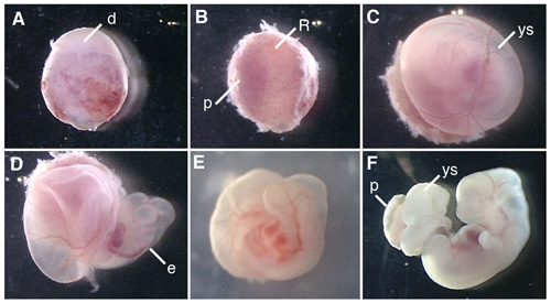
Figure 1. Whole embryo culture of the E12.5 rat embryo. (A) A conceptus dissected from the uterus of the pregnant rat. (B) Removal of the decidua at the placenta side. (C) Removal of Reichert’s membrane. (D) Opening the yolk sac. (E) The rat embryo culturing in the bottle for 6 hr after the beginning of WEC. (F) The rat embryo cultured for 42 hr. d, decidua; R; Reichert’s membrane; p, placenta; ys, yolk sac; e, embryo.
| 0 hr | 12 hr | 24 hr | 36 hr | 48 hr | |
| E12.5 rat embryo | 95% 50 cc | 95% 75 cc (10 hr) |
95% 100 cc | 95% 125 cc (34 hr) | 95% 150 cc |
Table 1. Optimal oxygen condition.
