Primary cilia are extracellular structures associated with the centriole. Primary cilia detection by immunofluorescent staining is a relatively simple procedure that results in extremely high-quality images. In this protocol, fibroblasts expressing primary cilia were fixed, immunostained, and imaged in a fluorescent or confocal microscope.
Primary cilia are dynamically regulated during cell cycle progression, specifically during the G0/G1 phases of the cell cycle, being resorbed prior to mitosis. Primary cilia can be visualized with highly sophisticated methods, including transmission electron microscopy, 3D imaging, or using software for the automatic detection of primary cilia. However, immunofluorescent staining of primary cilia is needed to perform these methods. This publication describes a protocol for the easy detection of primary cilia in vitro by staining acetylated alpha tubulin (axoneme) and gamma tubulin (basal body). This immunofluorescent staining protocol is relatively simple and results in high-quality images. The present protocol describes how four cell lines (C2C12, MEF, NHLF, and skin fibroblasts) expressing primary cilia were fixed, immunostained, and imaged with a fluorescent or confocal microscope.
Primary cilia are sensory, solitary, membrane-bound, nonmotile structures associated with the cell’s mother centriole. Primary cilia are found on most vertebrate cells with the exception of red blood cells, adipocytes1, and hepatocytes2. Primary cilia are formed as an elongated axoneme composed by microtubules, whose main component is α-tubulin. The axoneme grows from the basal body, which is structured from γ-tubulin. The length of the primary cilia varies between 2–10 µm; however, its dimensions can change during glycylation, starvation, hypoxia, cytotoxic stress, or after exposure to ionizing radiation3,4,5,6,7. Usually, cells have only one primary cilium, which is involved in morphogenesis and cell signalling pathways important for cell proliferation and differentiation8,9.
Primary cilia are dynamically regulated during cell cycle progression, specifically during the G0/G1 phases, and resorbed before entering mitosis in a process associated with tubulin deacetylation mediated by HDAC6 (histone deacetylase 6)10. The exact moment of primary cilia resorption depends upon cell type and the expression of genes directly involved in this process, such as Aurora A, Plk1, TcTex-111,12,13. Depending on the cell type, the primary cilia express different types of receptors, ion channels, and active signalling pathways. These include the most important signalling receptors affecting proliferation and survival, EGFR, PDGFR, and FGFR. Also included are some of the signalling pathways that may affect the function of one or more organs, including Hedgehog, Notch, and Wnt. Thanks to these receptors and signalling pathways, the primary cilia also perform a chemosensory function. This function allows primary cilia to detect specific ligands for Notch, hormones, and biologically active substances such as serotonin or somatostatin. Other specific functions exhibited by primary cilia of different lengths include reaction to changes in temperature, gravity, and osmolality14.
Primary cilia can be visualized through various methods, such as live visualization, transmission electron microscopy, 3D imaging, or by software for the automatic detection of primary cilia5,15,16,17. However, these methods are highly specialized and ongoing research needs basic, fast, and easy methods for staining primary cilia in every stage of research. Described is an easy and useful method for the detection of primary cilia in cultured cells.
1. Preparation of culture media, solutions, and dishes
2. Cell culture for immunocytochemistry staining
3. Immunofluorescent staining of primary cilia in vitro
The immunofluorescent staining of primary cilia is a relatively simple procedure that results in high-quality images. In these experiments, fibroblasts expressing primary cilia were fixed, immunostained, and imaged in a fluorescent or confocal microscope following the protocol described above. The primary cilium was detected using acetylated α-tubulin and γ-tubulin. The evaluation of primary cilia can be performed on various levels and any change in this regard can be linked to exposure to ionizing radiation, cell metabolism (e.g., starvation), or chemical treatment (e.g., cytostatics)5,18.
The effect of ionizing radiation on primary cilia has been studied in various cell lines (e.g., the myoblast cell line C2C12), which were irradiated (2, 6, 10, and 20 Gy) and the changes in primary cilia incidence analyzed. According to Filipova at al.4, low irradiation doses do not modify the occurrence of a single primary cilia in C2C12 cells. However, higher doses of ionizing radiation (i.e., 20 Gy) induced the appearance of multiple primary cilia (Figure 1A,B,C). Similarly, when NHLF cells were irradiated at 2 Gy the primary cilia were detected by immunofluorescence (Figure 2).
Metabolic stress is also known to increase the frequency of primary cilia19. In this case, MEF fibroblasts were starved and analyzed for changes in primary cilia incidence (Figure 3).
Immunofluorescence staining revealed that fibroblast cells carried primary cilia after treatment with doxorubin and taxol. Those fibroblasts treated with 120 nM doxorubicin expressed a single primary cilium (Figure 4); higher doses induced the appearance of multiple primary cilia (Figure 5). Treatment with 1.25 nM taxol also resulted in the presence of a single primary cilium (Figure 6). In contrast to the treatment with doxorubicin, multiple cilia were not detected after treatment with higher doses of taxol5.
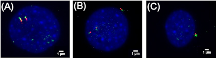
Figure 1: Occurrence of primary cilia in irradiated C2C12 cells. Representative photographs of primary cilia in C2C12 cells. Primary cilia detection was performed by immunofluorescence. The axoneme (arrow) of the primary cilia were assessed with acetylated α-tubulin antibody (red) and the basal body by γ-tubulin antibody (arrow, green). Nuclei were stained with DAPI (blue). (A) and (B) multiple cilia were observed 72 h after irradiation with 20 Gy. (C) Single primary cilia after 72 h irradiation with 20 Gy4. Please click here to view a larger version of this figure.
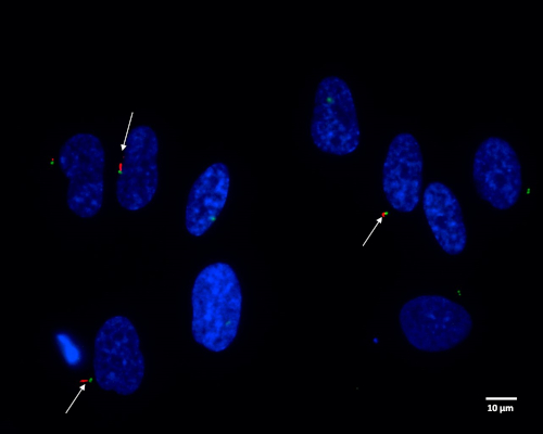
Figure 2: Detection of primary cilia in irradiated NHLF cells. Representative photographs of primary cilia in NHLF cells. Primary cilia (arrow) detection was performed by immunofluorescence. The axonemes of the primary cilia were stained with acetylated α-tubulin antibody (red) and the basal bodies with γ-tubulin antibody (green). Nuclei were stained with DAPI (blue). Single primary cilia 24 hours after irradiation at 2 Gy. Please click here to view a larger version of this figure.
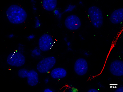
Figure 3: Incidence of primary cilia in the MEF cells after metabolic stress induced by serum starvation. Representative photographs of primary cilia 24 h after serum starvation (0.1% FBS) in MEF cells. Primary cilia (arrow) detection was performed by immunofluorescence. Axonemes were labeled with acetylated α-tubulin antibody (red). Basal bodies were stained with γ-tubulin antibody (green). Nuclei were stained with DAPI (blue). Please click here to view a larger version of this figure.
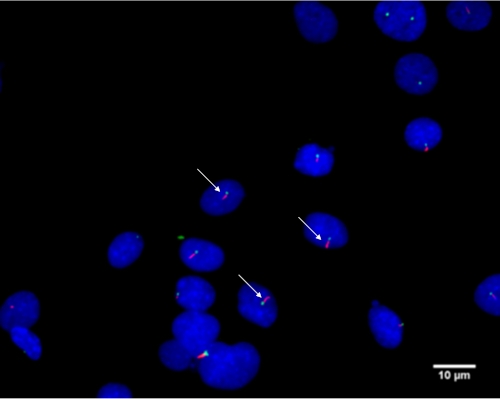
Figure 4: Representative photographs of primary cilia in skin fibroblasts. Primary cilia (arrow) detection was performed by immunofluorescence. Primary cilia were stained with acetylated α-tubulin antibody (red), while the basal bodies were stained with γ-tubulin antibody (green). Nuclei were stained with DAPI (blue). Primary cilia were detected 72 h after treatment with 120 nM doxorubicin5. Please click here to view a larger version of this figure.
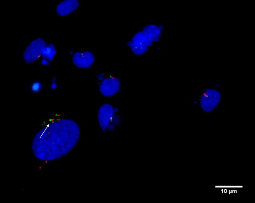
Figure 5: Representative photographs of multiple cilia in skin fibroblasts. Primary cilia (arrow) detection was performed by immunofluorescence. The axonemes were labeled by acetylated α-tubulin antibody (red) and the basal bodies were stained with γ-tubulin antibody (green). Nuclei were stained with DAPI (blue). Multiple cilia were detected 72 h after treatment with 120 nM doxorubicin5. Please click here to view a larger version of this figure.
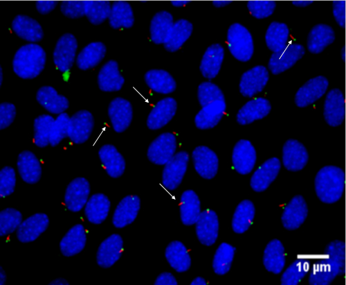
Figure 6: Representative photographs of skin fibroblasts treated with taxol. Primary cilia (arrow) were detected by immunofluorescence. Primary cilia were stained with acetylated α-tubulin antibody (red) and with γ-tubulin antibody (green). Axoneme nuclei were stained with DAPI (blue). Primary cilia were detected 72 h after treatment with 1.25 nM taxol5. Please click here to view a larger version of this figure.
Several authors have described diverse methods for the detection of primary cilia, sometimes also describing various fixation methods that can affect their detection6,20,21,22. Regardless, it is difficult to find a complete and straightforward protocol for detection. The ready availability of such a method would undoubtedly be of great assistance to the study of primary cilia investigation, especially in early stages of research or for a quick and easy method to test the presence of primary cilia in a chosen cell line. Therefore, this protocol is described in as much detail as possible for the detection of primary cilia in vitro after different kinds of treatment.
The present protocol was modified for use on a daily basis20,23,24. For example, 10% formalin was replaced by 4% PFA, whose fresh preparation is recommended due to its short storage life. PFA is a good choice for preserving cell morphology and is especially suited to the visualization of membrane-bound proteins. Organic solvents, such as methanol, have a dehydrating effect on the cell and remove small, soluble molecules and lipids during the fixation process, thus making it unsuitable for use in certain scenarios25. Permeabilization is achieved with 0.5% Triton X-100 in 1x PBS for 15 min. Goat serum in a 1:20 dilution in 1x PBS for 20 min is used as a blocking agent. Both primary antibodies, mouse anti-acetylated tubulin and rabbit anti-γ-tubulin, can be incubated concurrently for 60 min using a 1:800 and 1:300 dilution in 1x PBS, respectively20,21,23,24. In addition, the secondary antibodies, anti-mouse IgG (whole molecule) F(ab′)2 fragment–Cy3 antibody produced in sheep and Alexa Fluor488 AffiniPure F(ab')₂ fragment goat anti-rabbit IgG, were diluted 1:300 in 1x PBS. They were incubated concurrently for 45 min.
It may be necessary to take extra standardization steps should the primary antibodies be incubated overnight. During the development of the protocol it was found that an overnight incubation needs a volume of at least 500–1,000 µL of primary antibodies solution, the 6 well plate must be sealed with parafilm, and storage must be at 4 °C to prevent evaporation.
The most critical steps for the successful staining of primary cilia are: 1) choice of cell line and optimal cell culture practice; 2) use of gelatin coated coverslips; 3) consistent use of fresh 4% paraformaldehyde; 4) incubation of the secondary antibody and DAPI in the dark; 5) performing a gentle flip and placement of the coverslip on top of the mounting media in the slide.
There are no foreseen potential limitations in the future applications of the protocol. Moreover, primary cilia research is becoming more relevant in a variety of fields, and easy, fast, and reliable cilia detection methods are essential. Further, this protocol will facilitate the future study of primary cilia in cell types in which primary cilia have been heretofore undetected.
The authors have nothing to disclose.
This work was supported by the Ministry of Defence of the Czech Republic - Long-term organization development plan Medical Aspects of Weapons of Mass Destruction of the Faculty of Military Health Sciences, University of Defence; the Ministry of Education, Youth and Sport, Czech Republic (Specific Research Project No: SV/ FVZ201703) and PROGRES Q40/06. Thanks also to Daniel Diaz for his kind assistance in English language revision.
6-well plate | TPP | 92406 | Dimensions 128x86x22 mm |
Alexa Fluor488 | Jackson ImmunoResearch | 111-546-047 | AffiniPure F(ab')₂ Fragment Goat Anti-Rabbit IgG |
Anti-Tubulin γ | Sigma-Aldrich | T5192 | Polyclonal Rabbit anti-Mouse IgG2a |
C2C12 | ATCC | CRL-1772 | Myoblast (mouse) |
Cy3 | Sigma-Aldrich | C2181 | Anti-Mouse IgG (whole molecule) F(ab′)2 fragment–Cy3 antibody produced in sheep |
Dapi (4′,6-Diamidino-2-phenylindole dihydrochloride) | Sigma-Aldrich | D9542 | |
Dulbecco´s Modified Eagle´s medium | Thermo Scientific | 11960044 | High glucose, No glutamine, Gibco |
Dulbecco’s Phosphate Buffered Saline | Sigma-Aldrich | D8662 | With MgCl2 and CaCl2, Sterile-filtered, Suitable for cell culture |
Fetal Bovine Serum | Thermo Scientific | 16000044 | Sterile-Filtered, Gibco |
L-Glutamine | Sigma-Aldrich | G7513 | |
MEF | ATCC | SCRC-1039 | Mouse embryonic fibroblast |
Monoclonal Anti-Acetylated Tubulin | Sigma-Aldrich | T7451 | Monoclonal Anti-Acetylated Tubulin antibody produced in mouse |
NHLF | Lonza | CC-2512 | Primary lung fibroblasts (human) |
Normal Goat Serum | Jackson ImmunoResearch | 005-000-121 | |
Paraformaldehyde | Sigma-Aldrich | 158127-500G | Powder |
Penicillin-Streptomycin | Sigma-Aldrich | P0781 | 10,000 units penicillin and 10 mg streptomycin per mL in 0.9% NaCl, Sterile-Filtered |
ProLong Diamond Antifade Mountant | Thermo Scientific | P36961 | |
Skin fibroblasts | Kindly gifted from Charles University, Faculty of Medicine in Hradec Králové. | ||
Square Cover Slips | Thermo Scientific | 22X22-1.5 | Borosilicate glass, 22x22mm, Square |
Triton X-100 | Sigma-Aldrich | 11332481001 | |
Trypsin-EDTA (0.25%) | Thermo Scientific | 25200072 | Sterile-Filtered, Gibco |