Due to the complex anatomy, a consistently reproducible dissection and harvesting protocol for cardiac samples can be challenging to implement. This manuscript presents the key elements of some standard cardiac dissection protocols, highlighting both the gross examination approaches and the sampling sites commonly used for histopathologic examination.
The standard gross examination and sampling are key elements in the reproducibility and success of experimental studies of cardiovascular diseases carried out in large animals. Considering the complex anatomy of the heart, interspecies differences, and the types of compensatory and pathological reactions, consistent protocols are challenging to implement. The utilization of multiple dissection protocols is usually adapted to suit the prosector’s experience, and personal preference continues to be a source of experimental and interobserver variability. Here, the aim is to present the main anatomical features and landmarks, dissection protocols, and histological sampling standards of the heart in some commonly used species (including dogs, pigs, ruminants, and cats) as models of cardiovascular diseases.
Two standard gross examination protocols are presented here. First, the inflow-outflow method, which follows the physiological blood flow direction through the heart and large vessels (frequently used in dogs, ruminants, and pigs), and second, the four-chamber dissection technique (exemplified in cats). Both techniques can be adapted to any species in certain experimental circumstances. The sampling protocols include all the areas of interest (sinoatrial node, ventricles, interventricular septum, atria, valves, and aorta), and if properly carried out, improve both the reproducibility and reliability of experimental studies.
Due to the complex anatomy and early rigor, which can interfere with assessing the cardiac wall thickness, the gross examination of the heart is challenging and prone to several technique-related or interpretation errors. This is amplified by the interspecific morphological variations and by the fact that many clinical, major cardiac pathologies (including early cases of coronary heart diseases, fibrosis, arteritis, and amyloidosis) are not associated with any gross changes, being, in essence, histological pathologies. A standardized dissection and histological sample harvesting protocol can bring consistency between observers and, at the same time, comparability and reproducibility of experimental studies of cardiovascular diseases.
The samples were collected from two dogs (Canis lupus familiaris) (a 3-year-old, male French Bulldog, and an 8-year-old, female mixed-breed), a cat (Felis catus) (a 6-year-old, male European shorthair), a domestic pig (Sus scrofa domesticus) (a 1-year-old, male Large White), and a cow (Bos taurus) (a 2-month-old, female Holstein). Each of the chosen species has particular use as a cardiovascular model for a different disease; for example, dogs are a preferred model for arrhythmia modeling; cats are preferred for hypertrophic cardiomyopathy (HCM), as it is the species with the highest prevalence of HCM; pigs are used as a model for acute myocardial infarction; and ruminants are used as a model for intoxication due to their exposure to toxins that can easily be found on meadows11.
In this manuscript, one necropsy protocol and two dissection protocols of the heart are presented, designed to improve the gross and histological examination of the heart in experimental cardiovascular diseases. The described protocols were developed based on information from veterinary textbooks1,2,4,5,6,7,8,9,10,11,12, journal literature3,13,14, official documents15, and webinars16,17. The samples used in this study were harvested from cadavers submitted to the Pathology Department of the USAMV Cluj-Napoca for routine autopsy diagnostic.
The experimental protocol received bioethical agreement (No. 311 from 2022) and was approved by the Bioethics Committee of the University of Agricultural Sciences and Veterinary Medicine Cluj-Napoca, in compliance with both national (Law No. 43 of 2014) and European (EU Directive No 63 from 2010) legislation. See the Table of Materials for details about all materials and instruments used in this protocol.
1. Necropsy protocol
NOTE: It is recommended that the same necropsy protocol be used for all the species presented due to easier access it is when performed2,12. The following steps represent the necropsy performed in a medium-sized dog. Adapt the steps when performing necropsies on different subjects.
2. Dissection protocol
NOTE: Several necropsy techniques are used for the heart, each with several advantages. For this protocol, two of the most used techniques were chosen: 1) the "inflow-outflow method," which allows a better examination of the valves and the endocardium and is a protocol used for most of the species2,11,12,16, and 2) the "four chambers dissection"/"echocardiographic plane" technique, typically used for cats or small-sized dogs1,17.
3. Sampling protocol for histology16
4. Sampling of coronary arteries3,10,14
5. The "four chambers"/"echocardiographic plane" dissection technique of the heart1,17
NOTE: The four-chamber dissection technique consists of a cut from the base to the apex of the heart to obtain the standard view1. The "four-chamber" technique is best applied to fixed tissues.
6. Photographic documentation
NOTE: Photographic documentation is an optional step in the necropsy examination. However, photography and video recordings are essential to have an "accurate documentation of normal and abnormal anatomy"3.
This protocol was used to visualize anatomical features and collect samples for histological examination of the heart in four different species (dog, cat, pig, and cow). The necropsy protocol was repeated in each of the above-mentioned species but illustrated only in dogs. The necropsy protocol begins with an extensive external examination of the body (Figure 1A) (including the skin, explorable lymph nodes, and exterior mucosa), measuring the overall weight, and scoring the general state of the animal. After the external examination, the necropsy continues with a partial skinning of the body (directed to thoroughly examine the subcutis and superficial muscles) and with the opening of the main body cavities (Figure 1B–D).
The abdominal cavity is typically the first to be examined (Figure 1C), followed by an extensive in situ examination of the peritoneal cavity and abdominal organs, followed by sectioning of the diaphragm, and finally the removal of the right hemithorax (Figure 1D). By this technique, a good in situ examination of the thoracoabdominal organs (including the pericardial cavity and heart) can be achieved, simultaneously allowing a proper assessment of the links of these organs with the local blood vessels, lymphatics, ligaments, and nerves. The examination of the pericardial cavity is achieved by broad sectioning of the parietal pericardium following the longitudinal axis of the heart, followed by the removal of the heart by sectioning the cardiac large vessels close to the lung parenchyma. The external examination of the heart is followed by the sectioning of the cardiac cavities, which allows an extensive inspection and, finally, proper harvesting of the tissue samples for histopathology. Briefly, the procedure and the results of the two cardiac dissection techniques, the "blood flow" (Figure 2, Figure 3, Figure 4, and Figure 5) and the "four chambers" (Figure 10) are presented.
The "blood flow" technique was used in the heart of dogs, swine, and ruminants (Figure 2, Figure 4, and Figure 5). After the heart was removed from the body, the external configuration was observed (Figure 2A, Figure 4A, and Figure 5A). While performing the dissection, one should notice the atrial configuration, the crista terminalis, which is the macroscopic element of identification for the sinoatrial node (Figure 2B, Figure 4B, and Figure 5B). After proceeding with the steps of the protocol (Figure 2B–G, Figure 4B–G, and Figure 5B–G), the dissection is complete, and the harvesting protocol for the histology can be commenced (Figure 2H–J, Figure 4G–J, and Figure 5H–K). Alternatively, the crista terminalis can also be harvested after the heart is fixed (Figure 3) in 10% NBF. Compared with dogs, there is an extra step to be carried out in the heart of the swine-the harvesting of the coronary artery (Figure 4K).
After the harvesting, the samples were fixed in 10% NBF, routinely processed and embedded in paraffin wax, and finally sectioned at 4 µm, and stained by H&E. The histological slides obtained by this technique typically include the following: 1) the epicardium, myocardium, and endocardium of the right atrium and ventricle, the tricuspid valve, and the coronary arteries for histological block 1 (Figure 6); 2) the epicardium, myocardium, and endocardium of the left atrium and ventricle, the mitral valve, the coronary arteries, and the cardiac nerves for histological block 2 (Figure 7); 3) the myocardium, endocardium of the interventricular septum, and epicardium of the right atrium, and the tricuspid valve for histological block 3 (Figure 8); 4) multiple sections from the SA node for histological block 4 (Figure 9); 5) the epicardium, myocardium, and endocardium of the apex of the heart (left and right ventricles and interventricular septum) for histological block 5 (Figure 11A); and 6) the epicardium, myocardium, and endocardium of the base of the heart (left and right atria, ventricles, interventricular septum, atrioventricular valves, and coronary arteries) for histological block 6 (Figure 11B).
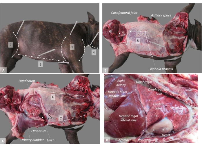
Figure 1: Necropsy technique in a dog. (A) The placement of the subject on the left side; (B) opening of the abdominal cavity; (C) opening of the thoracic cavity; (D) examination of the thoracic cavity. 1-7 indicate the locations and order of the recommended incisions. Please click here to view a larger version of this figure.
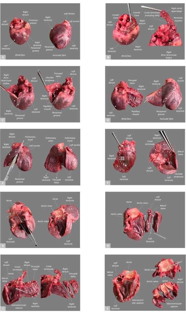
Figure 2: Dissection and sampling protocol in dog's heart. (A) External configuration of the heart; (B) Start from the caudal cava and trim the right atrium, open the right atrium and observe the crista terminalis; (C) trim the right ventricle along the junction with the interventricular septum; display the tricuspid valve; (D) after cutting the chordae tendineae, examine the right ventricle and continue the cut along the paraconal groove; (E) pass the forceps through the pulmonary trunk and use them to guide the cut; examine the pulmonary outflow; (F) 1-open the left atrium, 2-from the base, cut to the apex; examine the mitral valve and the left ventricle; (G) pass the forceps through the aorta and use them to guide the cut; examine the aorta; (H) make two cuts along the left heart and place the middle piece into 10% NBF; (I) make two cuts along the right heart, place the middle piece into 10%NBF, and trim away the crista terminalis; (J) trim away the base of the heart, make two cuts along the piece, and place the middle piece into 10% NBF. Abbreviations: SAN = sinoatrial node; NBF = neutral-buffered formalin. Please click here to view a larger version of this figure.
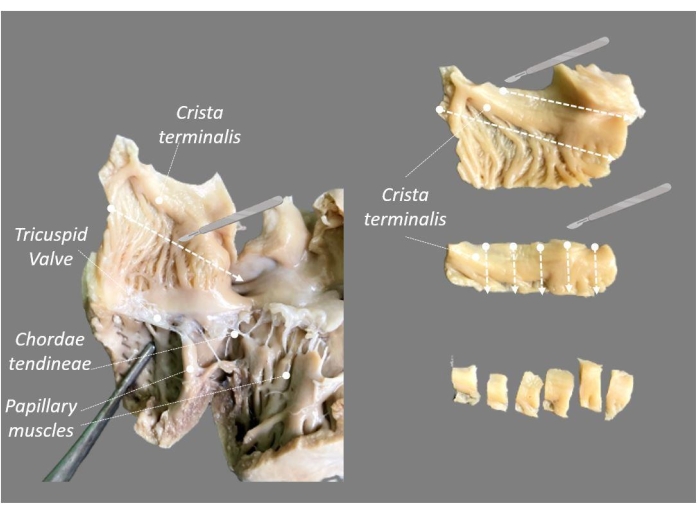
Figure 3: Sampling of the crista terminalis from a formalin-fixed heart. Please click here to view a larger version of this figure.
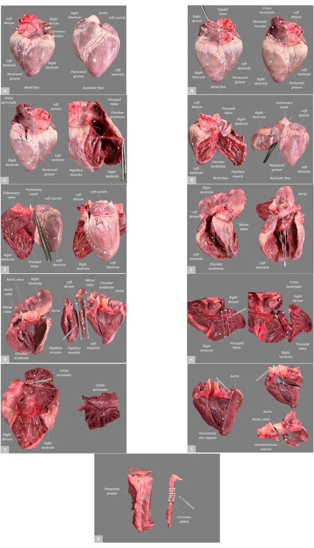
Figure 4: Dissection and sampling protocol in pig's heart. (A) External configuration of the heart; (B) Start from the caudal cava and section the right atrium, examine the right atrium, locate the crista terminalis, and start opening the right ventricle; (C) trim the right ventricle along the junction with the interventricular septum and examine the tricuspid valve; (D) after cutting the chordae tendineae, examine the right ventricle, place the forceps through the pulmonary trunk, and use them to guide the cut; (E) examine the pulmonary outflow; after sectioning the left atrium, cut the left ventricle from base to apex; (F) examine the mitral valve and the left ventricle, and cut the chordae tendineae; place forceps through the aorta and use them to guide the cut; (G) examine the aorta, make two cuts along the left heart, place the middle piece into 10% NBF; (H) make two cuts along the right heart; place the middle piece into 10%NBF; (I) trim away the crista terminalis, place the piece in 10% NBF; (J) trim away the base of the heart, make two cuts along the piece, and place the middle piece into 10% NBF; (K) trim the coronary artery, fix it overnight, and then make multiple transverse cuts at 3 mm intervals. Abbreviation: NBF = neutral-buffered formalin. Please click here to view a larger version of this figure.
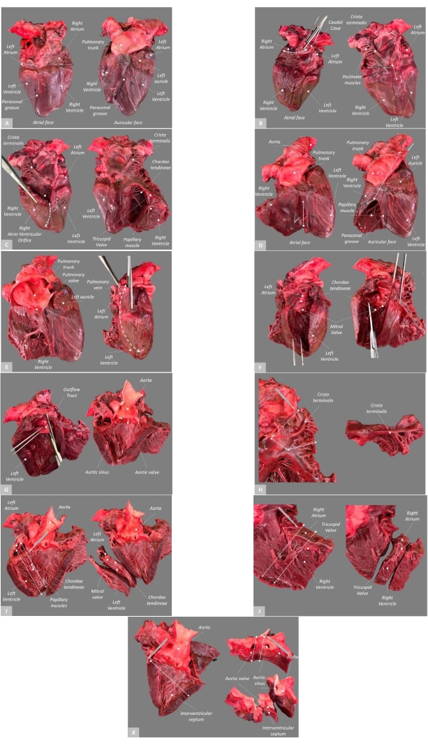
Figure 5: Dissection and sampling protocol in cow's heart. (A) External configuration of the heart; (B) Start from the caudal cava and section the right atrium, examine the right atrium, locate the crista terminalis, and start opening the right ventricle; (C) trim the right ventricle along the junction with the interventricular septum, and examine the tricuspid valve; (D) continue the cut parallel with the paraconal groove, pass the forceps through the pulmonary trunk, and use them to guide the cut; (E) examine the pulmonary outflow, start to cut the pulmonary veins and the left atrium, and then continue to cut the left ventricle from base to apex; (F) examine the mitral valve and the left ventricle, and cut the chordae tendineae; (G) pass the forceps through the aorta, use them to guide the cut, and examine the aorta; (H) trim away the crista terminalis, and place the piece in 10% NBF; (I) make two cuts along the left heart, and place the middle piece in 10% NBF; (J) make two cuts along the right heart, and place the middle piece in 10%NBF; (K) trim away the base of the heart, make two cuts along the piece, and place the middle piece in 10% NBF. Abbreviation: NBF = neutral-buffered formalin. Please click here to view a larger version of this figure.
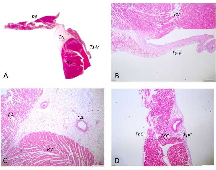
Figure 6: Histological examination of the right ventricle, right atrium, and tricuspid valve in the heart of a dog. H&E staining. (A) Histological sample of the right ventricle, right atrium, and tricuspid valve; (B) right ventricle, right atrium, and tricuspid valve. Scale bars = 500 µm; (C) right ventricle, right atrium, and coronary arteries. Scale bars = 100 µm; (D) right atrium. Scale bars = 500 µm. Abbreviations: H&E = hematoxylin and eosin; RV = right ventricle; RA = right atrium; Ts-V = tricuspid valve; CA = coronary artery; EnC = endocardium; MC = myocardium; EpC = epicardium. Please click here to view a larger version of this figure.
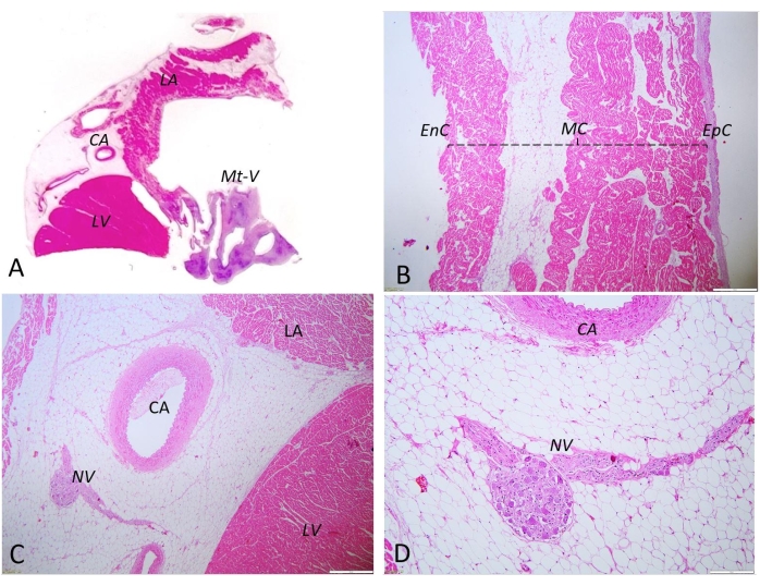
Figure 7: Histological examination of the left ventricle, left atrium, and mitral valve in the heart of a dog. H&E staining. (A) Histological sample of the left ventricle, left atrium, and mitral valve; (B) left atrium. Scale bars = 500 µm; (C) left atrium, left ventricle, coronary artery, and nerve. Scale bars = 100 µm; (D) coronary artery, nerve. Scale bars = 50 µm. Abbreviations: H&E = hematoxylin and eosin; LA = left atrium; LV = left ventricle; Mt-V = mitral valve; CA = coronary artery; EnC = endocardium; MC = myocardium; EpC = epicardium; NV = nerve. Please click here to view a larger version of this figure.
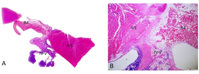
Figure 8: Histological examination of the interventricular septum, the right atrium, and tricuspid valve in the heart of a dog. H&E staining (A) Interventricular septum, right atrium, and tricuspid valve-histological sample; (B) interventricular septum, right atrium, and tricuspid valve. Scale bars = 500 µm. Abbreviations: H&E = hematoxylin and eosin; IVS = interventricular septum; RA = right atrium; Ts-V tricuspid valve. Please click here to view a larger version of this figure.
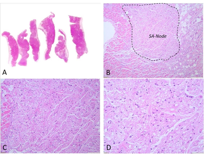
Figure 9: Histological examination of the sinoatrial node in the heart of a dog. H&E staining. (A) Histological sample of the crista terminalis; (B) sinoatrial node 4x magnification. Scale bars = 500 µm; (C) sinoatrial node 20x magnification. Scale bars = 100 µm; (D) sinoatrial node 40x magnification. Scale bars = 50 µm. Abbreviations: H&E = hematoxylin and eosin; SA node = sinoatrial node. Please click here to view a larger version of this figure.
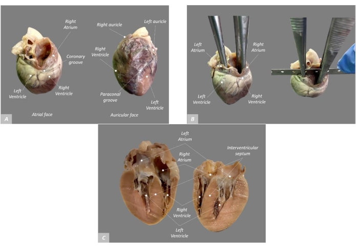
Figure 10: Dissection protocol on a formalin-fixed heart of a cat. (A) External configuration of the heart; (B) pass the forceps through cranial cava and pulmonary veins; the forceps guide the longitudinal section from base to apex; (C) the four-chamber view after the cut. Please click here to view a larger version of this figure.
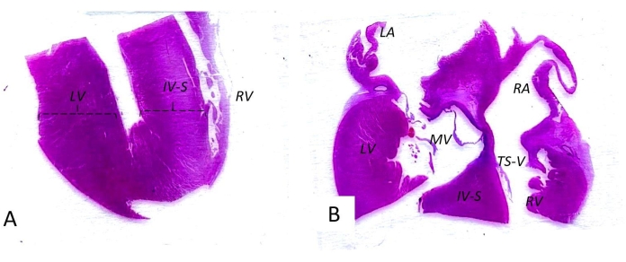
Figure 11: Histological samples of the heart of a cat. H&E staining. (A) Apex of the heart-histological sample; (B) base of the heart-histological sample. Abbreviations: H&E = hematoxylin and eosin; LV = left ventricle; IVS = interventricular septum; RV = right ventricle; LA = left atrium; MV = mitral valve; TS-V = tricuspid valve; RA = right atrium. Please click here to view a larger version of this figure.
Species | Age | Mean %BW | (LV + S)/RV | Citation numbers |
Cat | Newborn | 0.77% | – | 7 |
Cat | Adult | 0.33%-0.46% | 2.94-4.17 | 8, 5, 11 |
Dog | Newborn | 0.47%-0.76% | – | 8, 7 |
Dog | Adult | 0.70%-0.85% | 2.39-5.12 | 8, 6, 11 |
Pig | Adult | 0.32%-0.48% | 2.38-3.84 | 11 |
Sheep | Adult | 0.17%-0.65% | 2.63-4.54 | 11 |
Cow | Adult | 0.30%-0.66% | 2.43-4.00 | 11 |
Table 1: Reference values for normal heart weight/body weight ratio and ventricular ratio in animals. Cardiac measurements for cat, dog, pig, sheep, and cow. Abbreviations: BW = body weight; LV = left ventricle; S = interventricular septum; RV = right ventricle.
When performing the current protocol, several critical steps should be carefully considered for consistent results. In young animals, cardiac measurements are different from those in adults (including ventricular wall thickness), and generally, the heart represents a greater proportion of body weight11,12. The degree of ventricular hypertrophy can be quantified by applying a general weight formula, the ratio between the left ventricle plus septum divided by the free right ventricle weight, which, under normal conditions, should be 2.8-4.0 in mature animals (Table 1). If this ratio exceeds 4.0, it indicates left ventricular hypertrophy, and if it is less than 2.8, it indicates right ventricular hypertrophy4,11,12. To obtain the value of the chamber, the right ventricular free wall must be dissected along the interventricular wall and the coronary groove to be separated from the right atrium. The dissection of the left ventricle from the left atrium is made at the atrioventricular ring4.
The heart is separated from the body by sectioning the large vessels. If the sections of the large vessels are too close to the heart base, several structures can be compromised, mainly the atria and the auricles. To facilitate the process, it is necessary to gently pull the heart ventrally to properly section the large vessels above the base of the heart. A second critical step is recognizing the morphology of the crista terminalis, which is a discrete anatomical structure that requires detailed structural knowledge of the heart. The SA node must be carefully harvested and embedded with special consideration for the arrangement of the tissue samples within the blocks during the histological process.
The modification of the "four-chamber" technique was to change the histological blocking scheme from one block to two. The standard dimensions of broadly used histological cassettes and the adapted histological procedure (including the rotary microtome staining station) are not suitable to fit the entire section of the heart in one block without tissue distortion. Although the "four-chamber" technique eases the examination of the structure of the heart chambers, it limits the examination of the conductive system. This is especially important in cases when cardiac arrhythmia is documented. Further, considering that the "four-chamber" technique requires tissue fixed in NBF, several additional examinations such as bacteriology and molecular biology can be compromised to a certain degree.
When it comes to the heart of a cat or a small-sized dog, an accurate standard echocardiographic plan is obtained on fixed tissue. The four-chamber view is helpful, especially for chamber dimensions, if there are variations in ventricular or atrial sizes1. In addition to the advantages related to the perfect superposition of the gross-trimming planed with those obtained during clinical examination by echocardiography, this technique presents the advantage of including most of the key components of the heart in only two histological blocks.
The "inflow-outflow" method described in pigs was adapted from protocols used in the hearts of humans by adding the coronary artery sampling step. The currently described heart dissection protocols illustrate standard points for sampling. Additionally, when the dissector observes or suspects other lesions besides those covered in the protocol, supplementary samples should be collected from the areas of interest.
While developing the current protocols, other methods were studied, such as the method that involves an initial longitudinal cut of the major cardiac curves12 and the "bread loaf"/short-axis technique9,11. The technique involving a longitudinal cut of the major cardiac curves is a simpler protocol frequently used in biomedical education12. The short-axis sectioning technique ("bread loaf technique") can be performed with one short axis section11 or a series of sections of 1-2 cm thickness from the apex toward the base, leaving the atrioventricular apparatus intact9. This technique is the most suitable if the dissector suspects myocardial pathologies (including inflammatory pathologies and cardiomyopathies) but has the disadvantage of a superficial examination of the valvular components9.
Both these techniques are easily mastered by individuals with a relatively basic knowledge of the heart anatomy but lack robust points for histological sampling and weighting of each cardiac component. After proceeding with these methods, the subsequent harvesting procedure involves laborious work to identify the areas of interest and obtain constant samples. On the hearts dissected using these methods, the separate weighing of the two halves requires effort to be able to calculate the ratio between them. The short-axis method needs additional steps for the examination of the endocardium and all the components located at the base of the heart. Moreover, the harvesting procedure following these methods consists of laborious work to identify the areas of interest and obtain constant samples.
This manuscript presents the key elements of some standard cardiac dissection protocols, highlighting both the gross examination approaches and the sampling sites commonly used for histopathologic examination. Regarding the future applications of the technique, the main purpose of the presented protocol is to offer a robust guideline for cardiac dissection that can be readily used in all the experimental studies that require a detailed cardiac assessment.
The authors have nothing to disclose.
None.
0.9% saline solution | B. BRAUN MELSUNGEN AG | W04479004 | For washing all the blood, and blood clots from the heart. |
10% neutral buffered formalin (NBF) | Q Path | 11699404 | Materials for collecting histopathology samples. |
Bone cutting forceps | HELEN SRL | LS109HV | Sturdy instrument for cutting bone. |
Cutting board | Ambition | 86304 | For an easier manipulation, and cutting the organs. |
Decalcifying solution | Thermo Scientific TBD-2 | 6764004 | 1:1 mixture of 8% formic acid and hydrochloric acid. |
Digital camera | Canon Inc. | PowerShot SX540 HS | For photographic, and videographic documentation. |
Forceps | MKD-Medicale | 15-430 | Dissection instruments. |
Histological cassettes | Q Path | 720-2215 | Materials for collecting histopathology samples. Dimensions: 3 × 2.5 × 0.4 cm |
Knife | TEHNO FOOD COM SERV SRL | D2006/15 | Sharp blade for cutting soft tissue. |
Latex gloves | MKD-Medicale | SANTEX-S | Protection equipment. |
Mask | MKD-Medicale | 21221 | Protection equipment. |
Petri dishes | MKD-Medicale | 0598-1V | Materials for collecting ancillary testing samples. |
Plastic recipients | Corning Gosselin | TP200-02 | Materials for collecting histopathology samples. |
Scale | ESPERANZA | MEEKS008 | For weighing the organs. |
Scale | White Deals | 72 | For weighing the subjects. |
Scalpel | MKD-Medicale | 10322E | Sharp blade for cutting soft tissue. |
Scissors | MKD-Medicale | 13-260 | Dissection instruments. |
Scrub | MKD-Medicale | 410100-52 | Protection equipment. |
Syringes | MKD-Medicale | 10573EU | Materials for collecting ancillary testing samples. |