Cell transplantation represents a strategy for the treatment of retinal degeneration characterized by photoreceptor loss. Here we describe a method for enrichment of transplantable photoreceptors and their subretinal grafting into adult mice.
Vision impairment and blindness due to the loss of the light-sensing cells of the retina, i.e. photoreceptors, represents the main reason for disability in industrialized countries. Replacement of degenerated photoreceptors by cell transplantation represents a possible treatment option in future clinical applications. Indeed, recent preclinical studies demonstrated that immature photoreceptors, isolated from the neonatal mouse retina at postnatal day 4, have the potential to integrate into the adult mouse retina following subretinal transplantation. Donor cells generated a mature photoreceptor morphology including inner and outer segments, a round cell body located at the outer nuclear layer, and synaptic terminals in close proximity to endogenous bipolar cells. Indeed, recent reports demonstrated that donor photoreceptors functionally integrate into the neural circuitry of host mice. For a future clinical application of such cell replacement approach, purified suspensions of the cells of choice have to be generated and placed at the correct position for proper integration into the eye. For the enrichment of photoreceptor precursors, sorting should be based on specific cell surface antigens to avoid genetic reporter modification of donor cells. Here we show magnetic-associated cell sorting (MACS) – enrichment of transplantable rod photoreceptor precursors isolated from the neonatal retina of photoreceptor-specific reporter mice based on the cell surface marker CD73. Incubation with anti-CD73 antibodies followed by micro-bead conjugated secondary antibodies allowed the enrichment of rod photoreceptor precursors by MACS to approximately 90%. In comparison to flow cytometry, MACS has the advantage that it can be easier applied to GMP standards and that high amounts of cells can be sorted in relative short time periods. Injection of enriched cell suspensions into the subretinal space of adult wild-type mice resulted in a 3-fold higher integration rate compared to unsorted cell suspensions.
Vision is one of the prime senses of humans. Impairment of this sense and blindness are one of the main reasons for disability in industrialized countries. The predominant cause for vision impairment or blindness is retinal degeneration, characterized by photoreceptor cell loss, as it can be observed in macular degeneration, retinitis pigmentosa, cone-rod dystrophy, and other conditions. To date, an effective therapy to restore lost vision is not available. In 2006 and 2008 two different labs reported, independent from each other, a successful transplantation of rod photoreceptor precursor cells into adult wild-type mice retinas1,2. Thus, arising the possibility of photoreceptor precursor cell transplantation also into a degenerated retina, to replace degenerated photoreceptors and restore vision. Indeed, it has been demonstrated recently, that such transplanted photoreceptor precursor cells elicit morphological criteria of mature wild-type photoreceptors, such as properly developed outer segments3, synaptic terminals in close proximity to endogenous bipolar cells and a round cell body located in the outer nuclear layer2-4, as well as the ability to integrate functionally into the host neural circuitry5-7. One of the main principles of this strategy is the use of post-natal day 4 (PN 4, PN0 is defined as day of birth) young mice retinas, resulting in a mixture of different cell types for transplantation. On the background of a future therapeutic application, this mixture has to be purified for photoreceptor precursor cells. CD73 has been described as the first cell surface marker specific for young photoreceptors in the retina8-10. Here, we demonstrate a photoreceptor precursor cell purification method based on this cell surface marker and with the use of the magnetic-associated cell sorting (MACS) technique. MACS might have advantages in comparison to fluorescent-activated cell sorting techniques, due to fast sorting times and the easier adjustment to GMP conditions. We could demonstrate a ~90% enrichment and an up to 3-fold higher integration rate when transplanting the enriched population to the subretinal space in adult wild-type retinas. Thus, MACS-based photoreceptor precursor cell enrichment and subretinal transplantation, are reliable and promising techniques for the development of a regenerative therapeutic strategy for the treatment of retinal degeneration.
Ethical use and care of animals statement:
All animal experiments were carried out in strict accordance with European Union and German laws (Tierschutzgesetz) and adhered to the ARVO Statement for the Use of Animals in Ophthalmic and Vision Research. All animal experiments were approved by the animal ethics committee of the TU Dresden and the Landesdirektion Dresden (approval number: 24D-9168.11-1/2008-33).
1. Before Starting Cell Dissociation and Cell Sorting
2. Retina Dissociation
3. Cell Sorting Using Magnetic Associated Cell Sorting (MACS)
4. Transplantation of MAC-sorted Photoreceptor Precursor Cells into the Mouse Retina
In order to assess the ability of rod photoreceptors to integrate into the mouse retina, a mouse reporter line was used, in which GFP is driven by the neural retina leucine zipper (Nrl, Nrl-GFP) promoter11. Nrl is the earliest marker of rod photoreceptors starting its expression at E12.5 throughout adulthood, allowing a specific labeling of donor rod photoreceptor cells.
PN 4 Nrl-GFP pups were decapitated and eyes were enucleated. Retinas were then isolated and dissociated using the method described above. The resulting cell suspension was then sorted using CD73-based MACS (Figures 1 and 2). Following MAC sorting, we analyzed the enrichment achieved during this procedure.
As shown in Figure 3A, the initial cell suspension (Input) contained 30.4% GFP-positive cells, i.e. rod photoreceptors. Following CD73-based MAC-sorting, an enrichment of up to 86.9% in the CD73-positive fraction (CD73+) was observed by flow cytometry. In the CD73-negative fraction (CD73-) only 9.9% of all cells were positively detected for GFP (Figure 3A). The enrichment of Nrl-GFP positive cells following CD73-based MACS is additionally visualized after plating in vitro (Figure 3B).
After MAC-sorting, donor cells in the CD73-positive fraction were spun down and resuspended to a final concentration of 200,000 cells/µl. This cell suspension was further used for transplantation into the subretinal space of wild-type host retinas (Figure 4). Three to four weeks following transplantation, the host retinas were fixed, isolated and sectioned. Several donor cells integrated into the outer nuclear layer of the hosts and acquired the morphology of mature photoreceptors with localization of the cell body in the outer nuclear layer and formation of synaptic spherules and inner segments (Figure 5). Detailed pictures have been published before by our group3,8 showing an outcome comparable with FAC-sorted cells transplanted to host retinae by other groups4,6,10. Consistent in all these studies is, that the transplanted cells stay at the site of injection, defined by the bullous detached host retina, and do not migrate away. Some integrate into the host retina (i.e. outer nuclear layer), and some remain in the subretinal space3. Additionally, it has been shown, that the distribution of donor cells at the site of integration depends on the type of mouse model used5. No acute host/graft rejection signs were detected in transplantations between C57BL/6J mice.
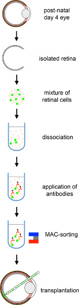
Figure 1. Overall scheme of subretinal transplantation of MACS purified photoreceptor precursors into adult mouse retina. Donor cells from PN 4 Nrl-GFP pups are isolated, followed by CD73-based MAC-sorting and transplanted into the subretinal space of mouse host retinas. Click here to view larger figure.
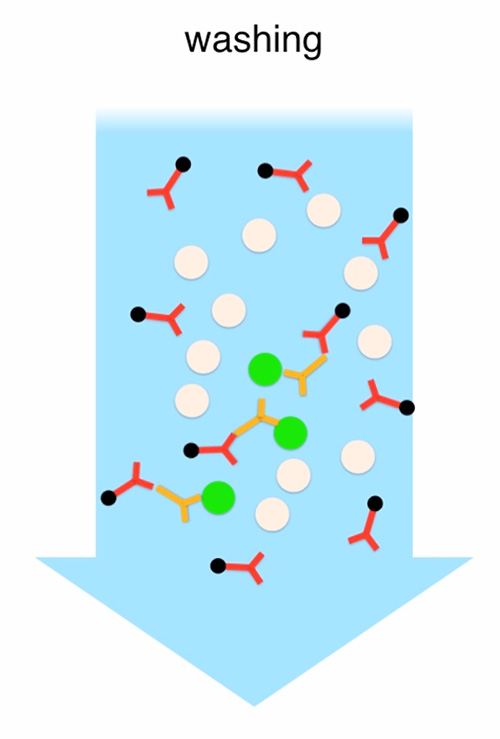
Figure 2. Enrichment of transplantable photoreceptors by MACS. The cell suspension generated from all collected PN4 retinas is incubated with primary rat anti-CD73 antibodies followed by washing and incubation with anti-rat antibodies conjugated with microbeads. Unbound antibodies are washed away. The cell suspension is passed through a LS-column that is connected with a magnetic stand. Cells that are bound by antibodies stay attached to the column while the remaining cells are eluted and collected (CD73-negative fraction). Finally, the LS-column is removed from the magnetic stand and the remaining cells are eluted and collected (CD73-positive fraction). Click here to view full movie.
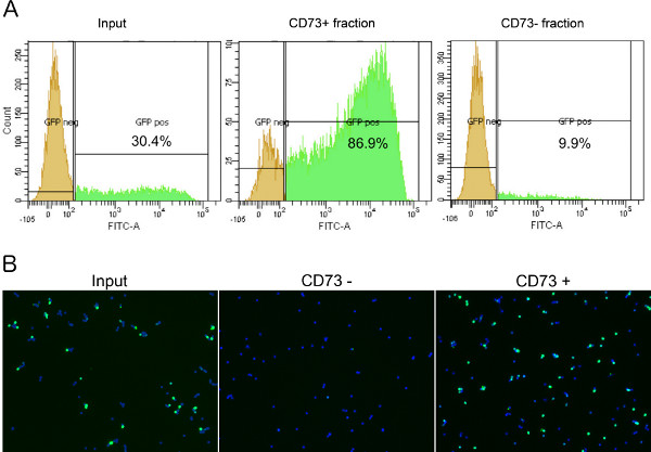
Figure 3. Analysis of Nrl-GFP positive cell enrichment following CD73-based MAC-sorting. (A) Before sorting, the initial population of donor cells contained 30.4% of Nrl-GFP positive cells. Following MAC-sorting, an enrichment of GFP-positive cells could be detected in the CD73+ fraction (86.9%) while the CD73- fraction contained only low amounts of GFP+ cells (9.9%). (B) Representative image demonstrating the result of CD73-based MAC-sorting of PN4 Nrl-GFP cells. 1 million cells from every fraction were plated on laminin-coated coverslips. Cells were fixed 4 hr after plating with 4% PFA for 10 min. Cells were stained with DAPI (4',6-diamidino-2-phenylindole, 1:20,000). A significant enrichment of GFP-positive cells in the CD73+ fraction was observed when compared to the input fraction or the CD73- fraction. Click here to view larger figure.
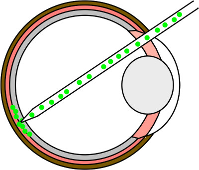
Figure 4. Subretinal injection. Schematic drawing of the transplantation process into the adult mouse retina. The needle is inserted through a hole in the ora serrata, navigated through the vitreal space by avoiding touching the lens and placed at the retina under visual control. By gentle pushing, the needle is punched through the retina and placed in the subretinal space. Eventually, the cell suspension is injected carefully under visual control by evaluating the detachment process.
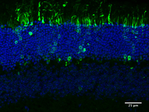
Figure 5. Integration of transplanted photoreceptor cells. Representative picture depicting integrated CD73-based MACS sorted PN4 Nrl-GFP cells following transplantation into the adult mouse retina. Transplanted photoreceptor precursors correctly integrate into the outer nuclear layer (ONL) and generate mature photoreceptor morphology.
Subretinal transplantation of photoreceptor precursor cells represents a reliable tool to achieve integration of these light-sensitive cells into host retinas in significant numbers1,2. This might allow the establishment of a cell therapy for the treatment of retinal degenerative diseases in future6. The donor population of cells, currently isolated from PN 4 retinas, is a mixture of different cell types, from which only the photoreceptor precursor cells integrate after subretinal injection. By using CD73-based MAC-sorting, the proportion of photoreceptors within the donor cell suspension can be increased to ≈ 90% that allows an approximately 3-fold higher integration rate in wild-type host retinas when compared to the unsorted cell population8. In comparison to flow cytometry, MACS has the advantage of a fast sorting procedure and that it can be relatively easy applied to GMP standards. The purification of transplantable photoreceptors is of specific interest in light of recent advantages for the in vitro generation of photoreceptors from pluripotent stem cells12-14. Cultures of differentiated pluripotent stem cells are composed of diverse cell-types making the availability of specific sorting procedures an essential prerequisite for their future use in cell transplantation therapies.
According to the MACS-purification procedure, several points are noteworthy, ensuring a workflow without major disturbances. The higher the number of retinas to be sorted, the more time is needed for their digestion. After digestion, during the sorting procedure, aggregation of cells has to be avoided. This occurs preferably before addition of the secondary antibody and during its incubation. In this case, the aggregate has to be removed immediately with a pipette. During the elution of MACS buffer while collecting the MACS-negative fraction, it is absolutely necessary to not add more than 3 ml to the column. The pressure of the buffer to the column will be too high if more buffer than 3 ml is added. This will disturb the binding of the antibody to the cells and lowers sorting efficiency. When removing the column from the magnetic stand to start collecting the MACS-positive fraction, this step has to be done quickly. The 5 ml of MACS buffer should be added to the column as fast as possible. Also, the plunge should be put and pressed quickly until the entire buffer is through the column. It is not necessary to worry about the applied pressure. The MACS buffer is isotonic and can be varied. It is possible to use FACS buffer, cell culture medium, PBS, EBSS, or HBSS. Sterility of the buffer can be achieved by filtering through a 0.4 µm filter membrane.
After enrichment by MACS, a successful transplantation of donor cells to the subretinal space can be achieved by keeping the following points in mind. Since photoreceptor cells seem to be very sensitive to treatment ex vivo, it is not advisable to keep them on 4 °C longer than necessary. Proceed for transplantation as fast as possible. When inserting the injection needle into the eyeball, it is important to avoid touching the lens as the relatively sharp metal needle might damage the lens resulting in induction of lens-induced uveitis. The last step on the way to the subretinal space, the penetration of the retina, is crucial. Since it cannot visually observed whether the subretinal space is reached it is important to develop a feeling for the right pushing pressure and tissue resistance. To test, if the needle head is placed correctly, a small volume could be applied and the detachment of the retina at the needle head should be carefully observed. If a round, bullous detachment without bleeding is forming, the position is correct and the rest of the solution can be applied carefully. During injection, the needle should remain in its position to avoid further damage to the tissue. The retinal hole created by the injection needle will seal by itself if the needle is retracted slowly. The whole procedure should be done in the minimal amount of time possible to avoid further stress and damage to the animal. During transplantation, the cell suspension tends to clump and block the microliter syringe. If this happens, a dilution of the cell suspension using sterile PBS or HBSS is suggested. Another possibility to overcome this problem is to add 1 µl of DNAse I to the cell suspension, to dissolve DNA clumps from dead cells, which tend to glue living cells together.
This protocol is limited to the magnetic sorting of cells. Therefore, the cells could be sorted for only one marker per sorting round. In comparison to flow cytometry, where cells could be sorted using different markers (including physiological properties as well as different cell surface markers) at the same time, this is a major disadvantage of this technique. Whenever the sorting for different markers in a short time window is needed, flow cytometry might be the better solution. Also, sorting for fluorescent properties of cells, like transgenically expressed GFP or other life cell markers is not possible with this technique. It is currently restricted to the use of cell surface antibodies that can be conjugated with magnetic beads. Nevertheless, this technique could be scaled up based on the technical equipment used.
Since Miltenyi Biotec is currently the only provider of magnetic cell sorting tools, there is no alternative available to date. For cell dissociation, other enzymes than papain (i.e. trypsin) could be used. Of note, our lab achieved the best results with papain, since this seems to be the most gentle dissociation enzyme in our hands. The time window of papain digestion (30-60 min) is variable. In this time window optimal digestion was achieved in our lab. Shorter or longer incubation time might be necessary depending on sample size, sample quality and enzyme used. Individual testing is suggested.
Given that single-step MACS does not reach purity levels as high as flow cytometry, a possible further application of this technique might be negative sorting of unwanted and potential harmful cells. There, the cell fraction positive for a given cell surface marker but not of interest can be sorted out, leaving the fraction of interest isolated in the flow through. This could be done in advance or after a positive sorting step and allows the further discrimination of cells using different cell surface markers as well as the removal of unwanted cells, like feeder layer cells or undifferentiated cells from pluripotent stem cell cultures with their potential tumorgenic risk. Therefore, MACS might be suitable for purification of photoreceptors from ES or iPS cell cultures in possible future therapeutic applications as it represents a gentle, fast and simple treatment for the purification of high amounts of cultured cells. MACS could be easily applied to GMP-conditions since the details of sorting steps, i.e. mechanics, fluids and procedures are reduced compared to flow cytometry. It is noteworthy, that MACS is already routinely used in clinical trials for the enrichment of cells from blood or bone marrow. Interestingly, MACS can be even applied on cells when no suitable cell surface marker is present, by genetically introducing a surface marker to make cells “competent” for subsequent magnetic cell sorting steps15.
In summary, MACS represents a reliable and fast tool to enrich photoreceptor precursor cells using cell surface markers for transplantation studies. Additionally, it is an easily employable method for future clinical applications that require GMP-conditions.
The authors have nothing to disclose.
We like to thank Anand Swaroop for providing Nrl-GFP mice, Jochen Haas for technical support, and Sindy Böhme and Emely Lessmann for animal husbandry.
This work was supported by the Deutsche Forschungsgemeinschaft (DFG): FZT 111 – Center of Regenerative Therapies Dresden, the CRTD Seed Grant Program, the SFB 655, and the ProRetina e.V. foundation, the DIGS-BB Graduate Program Dresden, and the Fundação para a Ciência e Tecnologia (SFRH/BD/60787/2009)
Papain Dissociation System | Worthington Biochemical Corporation | LK003150 | supplied DNase I is not used in the method |
purified rat anti-mouse CD73, clone TY/23 | BD Pharmingen | 550738 | Stock concentration 0.5mg/ml |
Goat Anti-Rat IgG MicroBeads | Miltenyi | 130-048-501 | Total volume of 2ml |
PBS | Gibco | 10010-015 | Used to count the total number of cells |
DNase I | Sigma | D5025-150KU | – |
HBSS | Gibco | 14025050 | Used for dissociation of the retinas |
Trypan blue | Sigma | Fluka93595 | Used to count the total number of cells |
Vidisic | Dr. Mann Pharma / Andreae-Noris Zahn AG | – | – |
Domitor | Pfizer | 76579 | – |
Ketamin 10% | Ratiopharm | 7538843 | – |
Antisedan | Pfizer | 76590 | – |
Phenylephrin 2.5%-Tropicamid 0.5% | University clinics Dresden pharmacy | – | – |
Name of Equipment | Company | Catalog Number | Comments/Description |
Pre-Separation Filters | Miltenyi | 130-041-407 | – |
LS Columns | Miltenyi | 130-042-401 | – |
MACS MultiStand | Miltenyi | 130-042-303 | – |
QuadroMACS Separator | Miltenyi | 130-090-976 | – |
fire polish glass pasteur pipette | Brand | 74777 20 | The pipette’s tips need to be fire-polished and autoclaved. |
MACS 15ml tube rack | Miltenyi | 130-091-052 | – |
Cell count chamber | Carl Roth | T728.1 | – |
Sterile 15ml tubes | Greiner Bio-one | 188271 | – |
Leica M651 MSD | Leica | M651 MSD | can be used instead of Olympus SZX10 |
Olympus SZX10 | Olympus | SZX10 | can be used instead of Leica M651 MSD |
Olympus inverted stereo microscope CKX41 | Olympus | CKX41 | – |
Cell culture hood Thermo Scientific MSC-Advance | Thermo scientific | 51025411 | – |
1.5ml reaction tube | Sarstedt | 727706400 | – |
2ml reaction tube | Sarstedt | 72695 | |
Eppendorf Centrifuge 5702 | VWR (Eppendorf) | 521-0733 | – |
Mouse head holder | myNeurolab | 471030 | – |
BD Microlance 3 30G 1/2” | BD Pharmingen | 304000 | – |
Hamilton microliter syringe 5µl, 75RN | Hamilton | 065-7634-01 | delivered without needles |
Hamilton RN special needle GA34 | Hamilton | 065-207434 | Blunt, 12mm length |
Vannas-Tübingen Spring Scissors – 5mm Blades Straight | Fine Science Tools | 15003-08 | – |
Dumont #7 Forceps – Titanium Biologie | Fine Science Tools | 11272-40 | – |
Diamond pen | Tools-tech | – | – |
15x15mm Cover slips | Sparks | MIC3366 | – |