Summary

Unchelated Gadolinium के लिए एक Aptamer आधारित सेंसर (तृतीय)

Published: January 09, 2017
doi:

Summary

The use of polydeoxynucleotide (44-mer aptamer) molecules for sensing unchelated gadolinium(III) ion in an aqueous solution is described. The presence of the ion is detected via an increase in the fluorescence emission of the sensor.

Abstract

A method for determining the presence of unchelated trivalent gadolinium ion (Gd3+) in aqueous solution is demonstrated. Gd3+ is often present in samples of gadolinium-based contrast agents as a result of incomplete reactions between the ligand and the ion, or as a dissociation product. Since the ion is toxic, its detection is of critical importance. Herein, the design and usage of an aptamer-based sensor (Gd-sensor) for Gd3+ are described. The sensor produces a fluorescence change in response to increasing concentrations of the ion, and has a limit of detection in the nanomolar range (~100 nM with a signal-to-noise ratio of 3). The assay may be run in an aqueous buffer at ambient pH (~7 – 7.4) in a 384-well microplate. The sensor is relatively unreactive toward other physiologically relevant metal ions such as sodium, potassium, and calcium ions, although it is not specific for Gd3+ over other trivalent lanthanides such as europium(III) and terbium(III). Nevertheless, the lanthanides are not commonly found in contrast agents or the biological systems, and the sensor may therefore be used to selectively determine unchelated Gd3+ in aqueous conditions.

Introduction

नैदानिक निदान, जो तकनीक के निहित संवेदनशीलता द्वारा सीमित है में चुंबकीय अनुनाद इमेजिंग (एमआरआई) के बढ़ते महत्व, उपन्यास gadolinium आधारित विपरीत एजेंट (GBCAs) 1 के विकास में अनुसंधान का तेजी से विकास हुआ है। GBCAs अणुओं है कि छवि गुणवत्ता में सुधार करने के लिए प्रशासित रहे हैं, और वे आम तौर पर एक त्रिसंयोजक gadolinium आयन (जी डी 3) एक polydentate ligand के लिए समन्वित की रासायनिक संरचना है। इस complexation unchelated जी.डी. के रूप में 3 + विषैला होता है के महत्व का है; यह गुर्दे की बीमारी या विफलता 2 के साथ कुछ रोगियों में नेफ्रोजेनिक प्रणालीगत फाइब्रोसिस के विकास में फंसाया गया है। नतीजतन, जलीय मुक्त आयन का पता लगाने के GBCAs की सुरक्षा सुनिश्चित करने में महत्वपूर्ण भूमिका निभाई है। GBCA समाधान में unchelated जी.डी. 3 + की उपस्थिति अक्सर ligand और आयन, परिसर की हदबंदी, या displacemen के बीच एक अधूरी प्रतिक्रिया का परिणाम हैअन्य जैविक धातु फैटायनों 3 से टी।

कई तकनीकों वर्तमान में जी.डी. 3 +, बहुमुखी प्रतिभा और प्रयोज्यता 4 के मामले में क्रोमैटोग्राफी और / या स्पेक्ट्रोमेट्री रैंक उच्चतम पर भरोसा उन की उपस्थिति निर्धारित करने के लिए प्रयोग किया जाता है के अलावा। उनकी शक्तियों के बीच उच्च संवेदनशीलता और सटीकता, विभिन्न नमूना matrices, और कई जी.डी. 3 + परिसरों के एक साथ मात्रा का ठहराव (मानव सीरम 5, मूत्र और बालों 6, अपशिष्ट 7, और इसके विपरीत एजेंट योगों 8 सहित) एक सूची का विश्लेषण करने के लिए (क्षमता है अध्ययन के 2013 से पहले Telgmann एट अल।) 4 से एक व्यापक समीक्षा में वर्णित है। केवल दोष यह है कि इन तरीकों में से कई (जैसे उपपादन द्वारा मिलकर प्लाज्मा मास स्पेक्ट्रोमेट्री के रूप में) instrumentations 4 की आवश्यकता है कि कुछ प्रयोगशालाओं के लिए उपयोग नहीं हो सकता है। अनुसंधान और सबूत की अवधारणा के स्तर, एआर पर उपन्यास GBCA खोज के संदर्भ मेंelatively अधिक सुविधाजनक, तेजी से, और लागत प्रभावी स्पेक्ट्रोस्कोपी आधारित पद्धति (जैसे यूवी विज़ अवशोषण या प्रतिदीप्ति के रूप में) एक मूल्यवान विकल्प के रूप में सेवा कर सकते हैं। मन में इन अनुप्रयोगों के साथ, जलीय जी.डी. 3 + के लिए एक फ्लोरोसेंट aptamer आधारित सेंसर विकसित किया गया था 9।

Aptamer (जी.डी.-aptamer) अड्डों में से एक विशिष्ट अनुक्रम कि घातीय संवर्धन (SELEX) 9 से ligands के व्यवस्थित विकास की प्रक्रिया के माध्यम से अलग किया गया था के साथ एक 44 आधार लंबे एकल असहाय डीएनए अणु है। एक फ्लोरोसेंट सेंसर में aptamer अनुकूल करने के लिए, एक fluorophore किनारा, जो फिर एक शमन किनारा (क्यूएस) 13 पूरक कुर्सियां (चित्रा 1) के माध्यम से के साथ संकरित है 5 'टर्मिनस से जुड़ा हुआ है। क्यूएस 3 'टर्मिनस पर एक काले पेय अणु के साथ टैग है। जी.डी. 3 +, सेंसर (जी.डी.-सेंसर) के अभाव में, एक 1 के शामिल में: क्रमश: जी.डी.-aptamer और क्यु के 2 तिल अनुपात, कम से कम प्रतिदीप्ति उत्सर्जन की वजह से टी होगापीने की वस्तु को fluorophore से o ऊर्जा हस्तांतरण। जलीय जी.डी. 3 + के अलावा जी.डी.-aptamer से क्यु विस्थापित, प्रतिदीप्ति उत्सर्जन में वृद्धि में जिसके परिणामस्वरूप।

आकृति 1
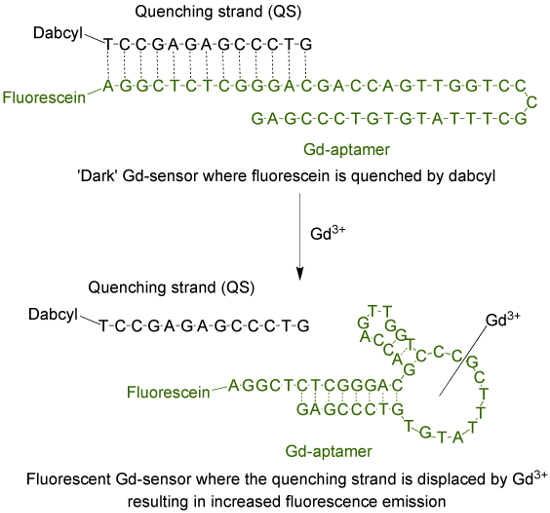
चित्रा 1. सेंसर (जी.डी.-सेंसर) है कि 44 आधार लंबे aptamer (जी.डी.-aptamer) fluorescein (एक fluorophore) और 13 आधार लंबे शमन किनारा (क्यूएस) dabcyl के साथ टैग के साथ टैग (एक अंधेरे पीने की वस्तु) के होते हैं । Unchelated जी.डी. 3 + के अभाव में, सेंसर की रोशनी कम है। जी.डी. 3 + के अलावा के साथ, क्यु के विस्थापन होता है और प्रतिदीप्ति उत्सर्जन में वृद्धि मनाया जाता है। यह आंकड़ा का एक बड़ा संस्करण देखने के लिए यहां क्लिक करें।

वर्तमान में नहीं है, के लिए पता लगाने के लिए एक अधिक इस्तेमाल किया स्पेक्ट्रोस्कोपी आधारित पद्धतिजलीय जी.डी. 3 + हैैं। इस परख अणु xylenol नारंगी, जो आयन 10 के लिए 433 573 एनएम से अधिकतम अवशोषण तरंग दैर्ध्य में बदलाव केलेशन पर से होकर गुजरती है का उपयोग करता है। इन दो absorbance Maxima के अनुपात unchelated जी.डी. 3 + की राशि यों इस्तेमाल किया जा सकता है। aptamer सेंसर, एक विकल्प (भी पूरक हो सकता है) xylenol नारंगी परख करने के लिए है के रूप में दो अलग अलग तरीकों प्रतिक्रिया की स्थिति, लक्ष्य selectivities, मात्रा का ठहराव के रैखिक पर्वतमाला, और पता लगाने के तौर-तरीकों (जैसे पीएच और इस्तेमाल बफर समाधान की संरचना के रूप में) है 9।

Protocol

नोट: आणविक जीव विज्ञान ग्रेड पानी सब बफर और समाधान की तैयारी में प्रयोग किया जाता है। सभी डिस्पोजेबल ट्यूब (microcentrifuge और पीसीआर) और pipet सुझावों DNase- और RNase मुक्त कर रहे हैं। उपयोग करने से पहले सभी रसायनों के लिए स…

Representative Results

Unchelated जी.डी. 3 + की उपस्थिति में जी.डी.-सेंसर समाधान का एक विशिष्ट प्रतिदीप्ति परिवर्तन चित्रा 2 में दिखाया गया है। उत्सर्जन प्रतिदीप्ति गुना परिवर्तन (2A चित्रा) या मनमाना इकाइ?…

Discussion

Aptamer आधारित जी.डी.-सेंसर, प्रतिदीप्ति उत्सर्जन में वृद्धि हुई है कि unchelated जी.डी. 3 + की एकाग्रता मनाया जाता है के लिए आनुपातिक है का उपयोग करना। इस्तेमाल नमूना की मात्रा को कम करने के लिए, परख अच्छी तरह से ?…

Divulgations

The authors have nothing to disclose.

Acknowledgements

We would like to gratefully acknowledge Dr. Milan N Stojanovic from Columbia University, New York, NY for valuable scientific input. This work is supported by funding from the California State University East Bay (CSUEB) and the CSUEB Faculty Support Grant-Individual Researcher. O.E., T.C., and A.L. were supported by the CSUEB Center for Student Research (CSR) Fellowship.

Materials

Gd-aptamer IDTDNA Input sequence and fluorophore modification in the order form A fluorophore with a different emission wavelength may be used. The aptamer may also be ordered from another company.
Quenching strand IDTDNA Input sequence and quencher modification in the order form A different quencher for optimal energy transfer from the fluorophore may be used. The aptamer may also be ordered from another company. 
Molecular biology grade water No specific manufacturer, both DEPC or non-DEPC treated work equally well
Gadolinium(III) chloride anhydrous Strem 936416 Toxic
HEPES Fisher Scientific BP310-500
Magnesium chloride anhydrous MP Biomedicals 0520984480 – 100 g
Sodium Chloride Acros Organics 327300025
Potassium chloride Fisher Scientific P333-500
Sodium hydroxide, pellets Fisher Scientific BP359 Corrosive
Hydrochloric acid Fisher Scientific SA49 Toxic and corrosive
384-well low flange black flat bottom polystyrene NBS plates Corning 3575 Plates which are suitable for fluorescence reading are required. 
Nalgene Rapid-Flow sterile disposable bottle top filter Thermo Scientific  5680020 The bottle top is fitted with  0.2 micron PES membrane
Disposable sterile bottles 250 mL Corning 430281 A larger or smaller bottle may be used
1.5 mL microcentrifuge tubes No specific manufacturer, as long as they are DNAse and RNAse-free
0.2 mL PCR tubes No specific manufacturer, as long as they are DNAse and RNAse-free
Micropipets No specific manufacturer
Pipet tips (non filter) of appropriate sizes No specific manufacturer, as long as they are DNAse and RNAse-free
Name of Equipment
Plate reader Biotek Synergy H1 Plate readers from other manufacturers would work equally well

References

  1. Shen, C., New, E. J. Promising strategies for Gd-based responsive magnetic resonance imaging contrast agents. Curr. Opin. Chem. Biol. 17 (2), 158-166 (2013).
  2. Cheong, B. Y. C., Muthupillai, R. Nephrogenic systemic fibrosis: a concise review for cardiologists. Tex. Heart Inst. J. 37 (5), 508-515 (2010).
  3. Hao, D., Ai, T., Goerner, F., Hu, X., Runge, V. M., Tweedle, M. MRI contrast agents: basic chemistry and safety. J Magn. Reson. Imaging. 36 (5), 1060-1071 (2012).
  4. Telgmann, L., Sperling, M., Karst, U. Determination of gadolinium-based MRI contrast agents in biological and environmental samples: a review. Anal. Chim. Acta. 764, 1-16 (2013).
  5. Frenzel, T., Lengsfeld, P., Schirmer, H., Hütter, J., Weinmann, H. -. J. Stability of gadolinium-based magnetic resonance imaging contrast agents in human serum at 37 degrees C. Invest. Radiol. 43 (12), 817-828 (2008).
  6. Loreti, V., Bettmer, J. Determination of the MRI contrast agent Gd-DTPA by SEC-ICP-MS. Anal. Bioanal. Chem. 379 (7), 1050-1054 (2004).
  7. Telgmann, L., et al. Speciation and isotope dilution analysis of gadolinium-based contrast agents in wastewater. Environ. Sci. Technol. 46 (21), 11929-11936 (2012).
  8. Cleveland, D., et al. Chromatographic methods for the quantification of free and chelated gadolinium species in MRI contrast agent formulations. Anal. Bioanal. Chem. 398 (7), 2987-2995 (2010).
  9. Edogun, O., Nguyen, N. H., Halim, M. Fluorescent single-stranded DNA-based assay for detecting unchelated gadolinium(III) ions in aqueous solution. Anal. Bioanal. Chem. 408 (15), 4121-4131 (2016).
  10. Barge, A., Cravotto, G., Gianolio, E., Fedeli, F. How to determine free Gd and free ligand in solution of Gd chelates. A technical note. Contrast Med. Mol. Imaging. 1 (5), 184-188 (2006).
  11. Johansson, M. K. Choosing reporter-quencher pairs for efficient quenching through formation of intramolecular dimers. Methods Mol. Biol. 335, 17-29 (2006).
  12. Sherry, A. D., Caravan, P., Lenkinski, R. E. A primer on gadolinium chemistry. J. Magn. Reson. Imaging. 30 (6), 1240-1248 (2009).
  13. Shakhverdov, T. A. A cross-relaxation mechanism of fluorescence quenching in complexes of lanthanide ions with organic ligands. Opt. Spectrosc. 95 (4), 571-580 (2003).
  14. Brittain, H. G. Submicrogram determination of lanthanides through quenching of calcein blue fluorescence. Anal. Chem. 59 (8), 1122-1125 (1987).
check_url/fr/55216?article_type=t

Play Video

Citer Cet Article
Edogun, O., Chan, T. Y., Nguyen, N. H., Luu, A., Halim, M. An Aptamer-based Sensor for Unchelated Gadolinium(III). J. Vis. Exp. (119), e55216, doi:10.3791/55216 (2017).

View Video