Summary

从巨 Unilamellar 泡中提取膜纳米管

Published: December 07, 2017
doi:

Summary

许多蛋白质在细胞感觉和导致膜曲度。我们描述了一种从脂泡中提取膜纳米管的方法, 以研究蛋白质或任何曲率-活性分子与弯曲膜的相互作用体外

Abstract

细胞膜的重塑是许多细胞现象的一个组成部分, 如吞、贩运、丝状的形成、。许多不同的蛋白质与弯曲的细胞膜相关联, 因为它们能够感知或诱发膜的曲率。通常, 这些过程涉及大量的蛋白质使它们过于复杂, 无法定量地在细胞中进行研究。我们描述了一个协议, 重建一个弯曲的膜在体外, 模仿一个弯曲的细胞结构, 如吞颈部。一个巨大的 unilamellar 囊 (老爹) 被用作细胞膜的模型, 其内部压力和表面张力受微吸入控制。使用光镊对老大施加点拉力, 就会产生高曲率的纳米管连接到平坦的膜上。该方法传统用于测定脂质膜的基本力学性能, 如弯曲刚度。近年来, 它已经扩展到研究蛋白质与膜曲率的相互作用以及它们对膜的形状和力学的影响方式。结合微、显微注射、光镊和共聚焦显微镜的系统可以同时测量膜的曲率、膜张力和蛋白质的表面密度。从这些测量中可以推断出蛋白质-膜系统的许多重要的力学和形态学特性。此外, 我们还制定了一个在生理盐浓度存在下创建 GUVs 的协议, 以及从标记蛋白和脂类的荧光强度中定量化膜上蛋白质的表面密度的方法。

Introduction

许多细胞过程, 如吞, 贩运, 形成丝状, 感染,, 伴随着细胞膜形状的戏剧性变化1,2。在细胞中, 许多蛋白质通过结合细胞膜和改变它们的形状来参与这些过程。最值得注意的例子是 Bin/Amphiphysin/Rvs (bar) 蛋白质家族的成员, 其中包含一个特征的本质曲线的酒吧域3,4,5,6,7。通常情况下, 它们通过将 BAR 域粘贴到表面上与膜进行交互, 并且在许多情形下, 还将亲螺旋插入双层中。条形域的形状、大小和电荷与亲螺旋的数量一起决定: (1) 膜曲率的方向 (, 它们是否会诱发内陷或突起), 以及 (2) 膜的大小曲率5,8。注意, 这里的正曲率被定义为弯曲膜的凸面,, 对相互作用粒子的凸出, 反之则为负。此外, 对 BAR 蛋白的定量研究表明, 它们对细胞膜的影响取决于一组物理参数: 蛋白质的表面密度、膜张力和膜的形状 (平面管状的vs球形形状)7。根据这些参数, BAR 蛋白质可以: (1) 作为膜曲率传感器, (2) 弯曲膜, 或 (3) 诱导膜断7

由于细胞内膜重塑所涉及的成分数量之多, 研究这些现象的定量方面, 如吞,在体内是极具挑战性的。体外在细胞中模拟弯曲膜的最小成分的重组提供了一种机制, 以获得对膜弯曲蛋白如何操作的机械理解。本文介绍了一种使用微、共焦显微镜和光镊在体外重建膜纳米管的协议。该方法可用于定量地研究蛋白质、脂质或小分子与弯曲膜的相互作用。脂质 GUVs 被用作细胞膜的模型, 其曲率与相互作用的膜弯曲分子的大小相比是微不足道的。他们使用的 electroformation 方法9 , 其中的泡是通过水合脂质膜, 并肿胀成 GUVs 在交流电流 (AC)10。最常见的基板上的 GUVs 是生长的是半导电板镀铟锡氧化物 (ITO) 或铂电线 (Pt 线)11。在这项工作中, GUVs 是在 Pt 线上生长的, 因为这种方法在缓冲12中显示出在使 GUVs 存在的情况下, 比替代品更有效。虽然这里描述了 electroformation 协议以充分的细节来重现它, 但我们将读者介绍给以前的文章, 其中的类似和其他制作 GUVs 的方法已经详细描述了13,14。在我们的手中, electroformation 在 Pt 线上成功地产生了 GUVs 混合的合成脂质或从天然脂提取物的缓冲含有〜100毫米氯化钠。此外, 也有可能在生长过程中封装 GUVs 内的蛋白质。图 1A中显示了一个示例 electroformation 室;它包括两个〜10厘米长的 Pt 线插入到一个持有聚四氟乙烯 (PTFE), 可以密封在双方的玻璃片〜 1-2 厘米分开 (图 1A)。

Figure 1
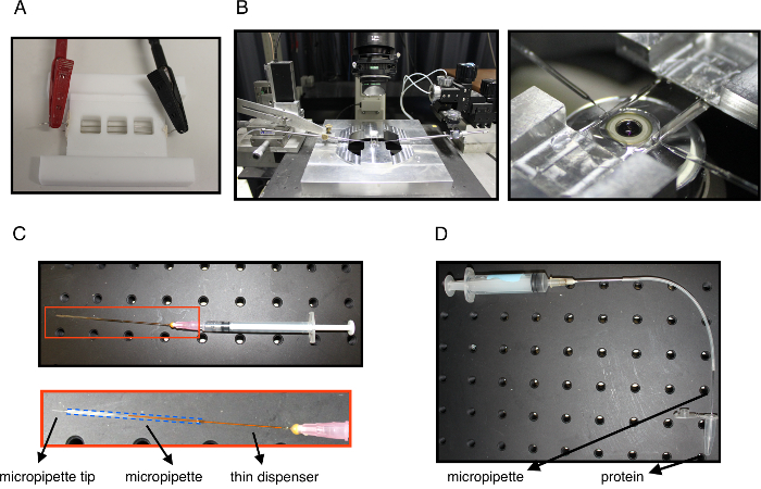
图 1: 实验设置.(A) electroformation 室, 连接在 Pt 线上的电连接器。(B) 左: 实验系统显示显微镜, 实验室以上的目标和两个 micropipettes (左, 右) 连接到动和插入实验室的管拉和蛋白质注射.右: 实验室的一个接近的看法登上了在目标之上显示志向和射入 micropipettes 插入。(C) 注射器装有一个薄薄的分配器插入到微在其后端。底部是微内的饮水机的关闭视图, 蓝色虚线勾勒出微。该系统用于填充微与酪蛋白钝化的玻璃表面, 也可以在需要时回填充矿物油。(D) 一种用于吸µL 蛋白质溶液数量的系统。针连接到注射器和油管连接到注射微。微提示是仔细浸入蛋白溶液和吸气, 以填补微提示。微, 然后返回充满矿物油使用的系统显示在面板 C.请点击这里查看这个数字的更大版本.

一个薄膜纳米管, 范围从7纳米到几百纳米, 可以从一个外部力量从一个老大拉。该方法最初设计用于测量细胞膜和囊泡的弹性特性, 如弯曲刚度15,16。在最近的工作中, 该方法被扩展, 以研究蛋白质与弯曲膜的相互作用, microinjecting 的蛋白质在被拉扯的纳米管附近7,17。其他的方法已经开发的研究膜弯曲蛋白。在一种方法中, 蛋白质是用不同大小的脂质体被拴在钝化表面上孵育的。共焦显微镜是用来测量蛋白质结合作为一个功能的脂质体直径, 这可以指示曲率诱导排序18,19。在另一种方法中, 在微吸气的老爹附近注射蛋白质来测量他们自发诱发小管的能力20,21。本协议中描述的方法非常适合于研究吞中涉及的膜弯曲蛋白, 其中大多数蛋白质通常会遇到与含货物膜套连接的预制膜管。底层平板等离子膜。此外, 在这种方法中, 与栓小脂质体的测定不同, 薄膜纳米管一直与膜连接;因此, 它是在机械平衡与老大, 一个情况下, 预计在体内。因此, 基础膜物理学的应用, 我们可以推断出一个过剩的机械性能从我们的测量22,23,24

为了充分实现这种方法, 必要的设备包括共聚焦显微镜、光镊和一个或两个 micropipettes 连接到水箱 (图 1B)。通过组合所有三, 可以同时测量膜张力、膜曲率、蛋白质的表面密度和管力25。微的愿望是必不可少的, 它是很容易构造的插入一个玻璃微到一个持有人连接到一个水箱, 其中, 通过静水压压力, 控制吸入压力26。微和持有人是由一个微控制, 理想情况下, 在一个方向的压电驱动器的精密运动。为了拉动纳米管, microaspirated 的老大被短暂地粘在微米级的珠子上, 然后拉走了制造纳米管。在这个实现中, 磁珠由光镊持有, 它可以通过遵循已发布的协议27来构造。虽然以精确的力测量为代价, 但可以用不同的方式免除光镊和拉纳米管。如果它是太具挑战性的建立一个光阱或如果力量测量不是必要的, 例如, 如果你简单地想要检查蛋白质的偏爱为弯曲的膜, 管子可以被拉扯用珠吸气在第二微28的尖端。也可以使用引力力29或流3031来拉动管道。此外, 共焦显微镜也不是必要的;然而, 它是比较可取的, 以测量蛋白质的表面密度。它还允许测量管中的脂质的荧光强度的纳米管半径, 从而独立于膜力和张力。从荧光推断管半径是特别重要的, 如果这些数量之间的关系偏离建立良好的方程, 由于存在膜黏附的蛋白质25。重要的是, 人们不能同时省略光阱和共焦显微镜, 因为它不可能测量管曲率。

本协议所述方法用于研究纳米管上各种外周膜蛋白的曲率诱导排序, 主要来自于 BAR 系列25323334.研究还表明, 锥形型跨膜钾通道 KvAP 在弯曲纳米管上的富集方式与棒蛋白35相同。通过优化 GUVs 内蛋白质的封装方法, 研究了负曲率蛋白质的相互作用, 以及36。此外, 该方法已被用来阐明蛋白质支架的形成25,37和研究的机制, 膜断的任何线张力38, 蛋白质动力39, 或由酒吧蛋白质40,41。除了蛋白质, 小分子或离子也能诱发曲率。利用这种方法, 钙离子被证明在无盐条件下诱发正曲率42。有趣的是, 它还表明, 脂质可以接受曲率排序, 虽然只有在分层点43,44附近的组合。总之, 这一方法可以被研究者们所用, 他们有兴趣研究整体膜组件 (例如, 如、脂质或跨膜蛋白) 或外周结合分子 (内外 GUVs) 与圆柱形的弯曲膜, 从机械和定量角度。它也适用于那些有兴趣测量膜本身的机械性能22,23,45

Protocol

1. Electroformation 在 Pt 线上制备 GUVs 清洁 electroformation 室 (参见简介和图 1A) 和带有有机溶剂 (如乙醇或丙酮) 的 Pt 线, 以洗去脂质和水冲洗掉盐。注意: 我们建议用乙醇浸泡过的组织彻底清除残留物, 然后在丙酮、乙醇、水中 sonicating, 每次5分钟。 在氯仿中, 在1毫克/毫升的时候, 准备一个期望的脂质组合。该混合物应包含〜0.05% 摩尔分数的脂质共?…

Representative Results

管拉试验可以提供有关膜的重要机械信息。在没有蛋白质或其他分子与膜曲率耦合的情况下, 膜力和管半径可以与膜张力有关, 将卡纳姆-海尔哈密顿方程应用于从老大50,51 中拉出的管 (公式 1) <p class="jove_content" fo:keep-to…

Discussion

从 GUVs 拉管的方法给膜蛋白系统提供了丰富的信息, 因为它不仅是测量膜的基本力学性能的手段, 而且有助于揭示蛋白质与膜的耦合曲率.正如介绍中所讨论的, 还有其他技术来测量膜弯曲蛋白的影响, 要么通过将亚微米脂质体与钝化表面相连的蛋白质孵化18,19 , 要么通过观察微吸入 GUVs 后小管自发形成的研究20,21</sup…

Divulgations

The authors have nothing to disclose.

Acknowledgements

作者感谢 Benoit Sorre, 达米安 Cuvelier, 皮埃尔 Nassoy, 弗朗索瓦 Quemeneur 和吉尔 Toombes 在该组中建立纳米管方法的重要贡献。莱集团属于 CNRS 财团 CellTiss、Labex CelTisPhyBio (ANR-11-LABX0038) 和巴黎科学文学 (ANR-10-IDEX-0001-02)。EMBO 由长期研究金 (ALTF 1527-2014) 和居里夫人行动 (H2020-MSCA-IF-2014, 项目膜-ezrin-肌动蛋白) 资助。多发性硬化症是西蒙斯研究员协会的初级研究员。

Materials

1,2-Dioleoyl-sn-glycero-3-phosphatidylcholine (DOPC) Avanti 850375 Example lipid used in data for Figure 3
1,2-distearoyl-sn-glycero-3-phosphoethanolamine-N-[biotinyl(polyethyleneglycol)-2000] [DSPE-PEG(2000)-biotin] Avanti 880129 biotinylated lipid
BODIPY-TR-C5-ceramide Molecular Probes (ThermoFisher) D7540 fluorescent lipid
BODIPY- FLC5-hexadecanoyl phosphatidylcholine (HPC*) Molecular Probes (ThermoFisher) fluorescent lipid for protein density calibration
egg L-α-phosphatidylcholine (eggPC) Avanti 840051 used for calibrating the tube radius constant
β-casein from bovine milk (>99%) Sigma Aldrich C6905 used for passivating the micropipette and the experimental chamber
Sucrose Sigma Aldrich S7903
D(+) glucose Sigma Aldrich G7021
NaCl Sigma Aldrich S7653
Tris Sigma Aldrich 10708976001
osmometer Loser n/a
Pt-wires, 0.5 mm diameter, 99.99% pure Goodfellow USA PT005139 used for GUV electroformation
function generator n/a used to create current for GUV electroformation
putty sealant Vitrex (from CML France) CRIT 140013 used to seal the electroformation chamber
bath sonicator n/a useful to clean the electroformation chamber, but not crucial
Nikon TE2000 inverted microscope, eC1 confocal system (Nikon), with two laser lines (λ = 488 nm and 543 nm); optical tweezers induced by a 5 W ytterbium fiber continuous wave laser (λ > 1070 nm; IPG GmBH Germany) an example of a confocal microscopy system equipped with optical tweezers
micromanipulators n/a
borosilicate capillaries (with internal and external radii of 0.78 mm and 1 mm, respectively) Harvard apparatus 30-0036
micropipette puller Sutter Instrument P-2100
microforge Narishige MF-800
piezoelectric actuator Physik Instrumente n/a
polystyrene streptavidin coated beads (diameter 3.2 µm) Spherotech SVP-30-5

References

  1. McMahon, H. T., Gallop, J. L. Membrane curvature and mechanisms of dynamic cell membrane remodelling. Nature. 438 (7068), 590-596 (2005).
  2. Johannes, L., Wunder, C., Bassereau, P. Bending “on the rocks”–a cocktail of biophysical modules to build endocytic pathways. Cold Spring Harb Perspect Biol. 6 (1), (2014).
  3. Dawson, J. C., Legg, J. A., Machesky, L. M. Bar domain proteins: a role in tubulation, scission and actin assembly in clathrin-mediated endocytosis. Trends Cell Biol. 16 (10), 493-498 (2006).
  4. Mim, C., Unger, V. M. Membrane curvature and its generation by BAR proteins. Trends Biochem Sci. 37 (12), 526-533 (2012).
  5. Qualmann, B., Koch, D., Kessels, M. M. Let’s go bananas: revisiting the endocytic BAR code. EMBO J. 30 (17), 3501-3515 (2011).
  6. Rao, Y., Haucke, V. Membrane shaping by the Bin/amphiphysin/Rvs (BAR) domain protein superfamily. Cell Mol Life Sci. 68 (24), 3983-3993 (2011).
  7. Simunovic, M., Voth, G. A., Callan-Jones, A., Bassereau, P. When Physics Takes Over: BAR Proteins and Membrane Curvature. Trends Cell Biol. 25 (12), 780-792 (2015).
  8. Safari, F., Suetsugu, S. The BAR Domain Superfamily Proteins from Subcellular Structures to Human Diseases. Membranes (Basel). 2 (1), 91-117 (2012).
  9. Angelova, M. I., Soléau, S., Méléard, P., Faucon, F., Bothorel, P., Helm, C., Lösche, M., Möhwald, H. . Progress in Colloid and Polymer Science Vol. 89: Trends in Colloid and Interface Science VI. 89, 127-131 (1992).
  10. Meleard, P., Bagatolli, L. A., Pott, T. Giant unilamellar vesicle electroformation from lipid mixtures to native membranes under physiological conditions. Methods Enzymol. 465, 161-176 (2009).
  11. Montes, L. R., Alonso, A., Goni, F. M., Bagatolli, L. A. Giant unilamellar vesicles electroformed from native membranes and organic lipid mixtures under physiological conditions. Biophys J. 93 (10), 3548-3554 (2007).
  12. Mathivet, L., Cribier, S., Devaux, P. F. Shape change and physical properties of giant phospholipid vesicles prepared in the presence of an AC electric field. Biophys J. 70 (3), 1112-1121 (1996).
  13. Collins, M. D., Gordon, S. E. Giant liposome preparation for imaging and patch-clamp electrophysiology. J Vis Exp. (76), (2013).
  14. Garten, M., Aimon, S., Bassereau, P., Toombes, G. E. Reconstitution of a transmembrane protein, the voltage-gated ion channel, KvAP, into giant unilamellar vesicles for microscopy and patch clamp studies. J Vis Exp. (95), e52281 (2015).
  15. Hochmuth, R. M., Evans, E. A. Extensional flow of erythrocyte membrane from cell body to elastic tether. I. Analysis. Biophys J. 39 (1), 71-81 (1982).
  16. Hochmuth, R. M., Wiles, H. C., Evans, E. A., McCown, J. T. Extensional flow of erythrocyte membrane from cell body to elastic tether. II. Experiment. Biophys J. 39 (1), 83-89 (1982).
  17. Bassereau, P., Sorre, B., Levy, A. Bending lipid membranes: experiments after W. Helfrich’s model. Adv Colloid Interface Sci. 208, 47-57 (2014).
  18. Hatzakis, N. S., et al. How curved membranes recruit amphipathic helices and protein anchoring motifs. Nature chemical biology. 5 (11), 835-841 (2009).
  19. Bhatia, V. K., et al. Amphipathic motifs in BAR domains are essential for membrane curvature sensing. EMBO J. 28 (21), 3303-3314 (2009).
  20. Shi, Z., Baumgart, T. Membrane tension and peripheral protein density mediate membrane shape transitions. Nat Commun. 6, 5974 (2015).
  21. Chen, Z., Shi, Z., Baumgart, T. Regulation of membrane-shape transitions induced by I-BAR domains. Biophys J. 109 (2), 298-307 (2015).
  22. Cuvelier, D., Derenyi, I., Bassereau, P., Nassoy, P. Coalescence of membrane tethers: experiments, theory, and applications. Biophys J. 88 (4), 2714-2726 (2005).
  23. Derenyi, I., Julicher, F., Prost, J. Formation and interaction of membrane tubes. Phys Rev Lett. 88 (23), 238101 (2002).
  24. Dimova, R. Recent developments in the field of bending rigidity measurements on membranes. Adv Colloid Interface Sci. 208, 225-234 (2014).
  25. Sorre, B., et al. Nature of curvature coupling of amphiphysin with membranes depends on its bound density. Proc Natl Acad Sci U S A. 109 (1), 173-178 (2012).
  26. Evans, E., Rawicz, W. Entropy-driven tension and bending elasticity in condensed-fluid membranes. Phys Rev Lett. 64 (17), 2094-2097 (1990).
  27. Neuman, K. C., Nagy, A. Single-molecule force spectroscopy: optical tweezers, magnetic tweezers and atomic force microscopy. Nat Methods. 5 (6), 491-505 (2008).
  28. Capraro, B. R., Yoon, Y., Cho, W., Baumgart, T. Curvature sensing by the epsin N-terminal homology domain measured on cylindrical lipid membrane tethers. J Am Chem Soc. 132 (4), 1200-1201 (2010).
  29. Bo, L., Waugh, R. E. Determination of bilayer membrane bending stiffness by tether formation from giant, thin-walled vesicles. Biophys J. 55 (3), 509-517 (1989).
  30. Rossier, O., et al. Giant Vesicles under Flows: Extrusion and Retraction of Tubes. Langmuir. 19 (3), 575-584 (2003).
  31. Borghi, N., Rossier, O., Brochard-Wyart, F. Hydrodynamic extrusion of tubes from giant vesicles. EPL (Europhysics Letters). 64 (6), 837 (2003).
  32. Zhu, C., Das, S. L., Baumgart, T. Nonlinear sorting, curvature generation, and crowding of endophilin N-BAR on tubular membranes. Biophys J. 102 (8), 1837-1845 (2012).
  33. Ramesh, P., et al. FBAR syndapin 1 recognizes and stabilizes highly curved tubular membranes in a concentration dependent manner. Sci Rep. 3, 1565 (2013).
  34. Knorr, R. L., et al. Membrane morphology is actively transformed by covalent binding of the protein Atg8 to PE-lipids. PLoS One. 9 (12), e115357 (2014).
  35. Aimon, S., et al. Membrane shape modulates transmembrane protein distribution. Dev Cell. 28 (2), 212-218 (2014).
  36. Prévost, C., et al. IRSp53 senses negative membrane curvature and phase separates along membrane tubules. Nat Commun. , (2015).
  37. Simunovic, M., et al. How curvature-generating proteins build scaffolds on membrane nanotubes. Proc Natl Acad Sci U S A. 113 (40), 11226-11231 (2016).
  38. Allain, J. M., Storm, C., Roux, A., Ben Amar, M., Joanny, J. F. Fission of a multiphase membrane tube. Phys Rev Lett. 93 (15), 158104 (2004).
  39. Morlot, S., et al. Membrane shape at the edge of the dynamin helix sets location and duration of the fission reaction. Cell. 151 (3), 619-629 (2012).
  40. Renard, H. F., et al. Endophilin-A2 functions in membrane scission in clathrin-independent endocytosis. Nature. 517 (7535), 493-496 (2015).
  41. Simunovic, M., et al. Friction Mediates Scission of Tubular Membranes Scaffolded by BAR Proteins. Cell. 170 (1), 172-184 (2017).
  42. Simunovic, M., Lee, K. Y., Bassereau, P. Celebrating Soft Matter’s 10th anniversary: screening of the calcium-induced spontaneous curvature of lipid membranes. Soft Matter. 11 (25), 5030-5036 (2015).
  43. Sorre, B., et al. Curvature-driven lipid sorting needs proximity to a demixing point and is aided by proteins. Proc Natl Acad Sci U S A. 106 (14), 5622-5626 (2009).
  44. Heinrich, M., Tian, A., Esposito, C., Baumgart, T. Dynamic sorting of lipids and proteins in membrane tubes with a moving phase boundary. Proc Natl Acad Sci U S A. 107 (16), 7208-7213 (2010).
  45. Dasgupta, R., Dimova, R. Inward and outward membrane tubes pulled from giant vesicles. Journal of Physics D: Applied Physics. 47 (28), 282001 (2014).
  46. Vitkova, V., Genova, J., Mitov, M. D., Bivas, I. Sugars in the aqueous phase change the mechanical properties of lipid mono- and bilayers. Molecular Crystals and Liquid Crystals. 449, 95-106 (2006).
  47. Bouvrais, H. . Mechanical properties of giant vesicle membranes investigated by flickering technique. , (2011).
  48. Koster, G., Cacciuto, A., Derenyi, I., Frenkel, D., Dogterom, M. Force barriers for membrane tube formation. Phys Rev Lett. 94 (6), 068101 (2005).
  49. Kwok, R., Evans, E. Thermoelasticity of large lecithin bilayer vesicles. Biophys J. 35 (3), 637-652 (1981).
  50. Helfrich, W. Elastic properties of lipid bilayers: theory and possible experiments. Z Naturforsch C. 28 (11), 693-703 (1973).
  51. Canham, P. B. The minimum energy of bending as a possible explanation of the biconcave shape of the human red blood cell. J Theor Biol. 26 (1), 61-81 (1970).
  52. Marcerou, J. P., Prost, J., Gruler, H. Elastic Model of Protein-Protein Interaction. Nuovo Cimento Della Societa Italiana Di Fisica D-Condensed Matter Atomic Molecular and Chemical Physics Fluids Plasmas Biophysics. 3 (1), 204-210 (1984).
  53. Leibler, S. Curvature Instability in Membranes. J Phys-Paris. 47 (3), 507-516 (1986).
check_url/fr/56086?article_type=t

Play Video

Citer Cet Article
Prévost, C., Tsai, F., Bassereau, P., Simunovic, M. Pulling Membrane Nanotubes from Giant Unilamellar Vesicles. J. Vis. Exp. (130), e56086, doi:10.3791/56086 (2017).

View Video