Intravenous injection of cancer cells is often used in metastasis research, but the metastatic tumor burden can be difficult to analyze. Herein, we demonstrate a tail-vein injection model of metastasis and include a novel approach to analyze the resulting metastatic lung tumor burden.
Metastasis, the primary cause of morbidity and mortality for most cancer patients, can be challenging to model preclinically in mice. Few spontaneous metastasis models are available. Thus, the experimental metastasis model involving tail-vein injection of suitable cell lines is a mainstay of metastasis research. When cancer cells are injected into the lateral tail-vein, the lung is their preferred site of colonization. A potential limitation of this technique is the accurate quantification of the metastatic lung tumor burden. While some investigators count macrometastases of a pre-defined size and/or include micrometastases following sectioning of tissue, others determine the area of metastatic lesions relative to normal tissue area. Both of these quantification methods can be exceedingly difficult when the metastatic burden is high. Herein, we demonstrate an intravenous injection model of lung metastasis followed by an advanced method for quantifying metastatic tumor burden using image analysis software. This process allows for investigation of multiple end-point parameters, including average metastasis size, total number of metastases, and total metastasis area, to provide a comprehensive analysis. Furthermore, this method has been reviewed by a veterinary pathologist board-certified by the American College of Veterinary Pathologists (SEK) to ensure accuracy.
Despite being a highly complex and inefficient process1, metastasis is a significant contributor to the morbidity and mortality of cancer patients2. In fact, most cancer-related deaths are attributed to metastatic spread of disease3,4. In order for tumor cells to successfully metastasize, they must detach from the primary site, invade through adjoining stroma, intravasate into blood circulation or lymphatics, travel to the capillary bed of a secondary site, extravasate into the secondary tissue, and proliferate or grow to form metastatic lesions5. The use of mouse models has been critical to furthering the understanding of the molecular mechanisms responsible for metastatic seeding and growth6,7. Herein, we focus on breast cancer metastasis, for which both genetically modified mouse models as well as methods of transplantation are often used – each with their own set of advantages and limitations.
Genetically engineered mammary tumor models make use of mammary gland specific promoters, including MMTV-LTR (mouse mammary tumor virus long terminal repeat) and WAP (Whey Acidic Protein), to drive expression of transgenes in the mammary epithelium8. Oncogenes including polyoma middle T antigen (PyMT), ErbB2/Neu, c-Myc, Wnt-1, and simian virus 40 (SV40) have been expressed in this manner9,10,11,12,13, and while these genetic models are useful for studying primary tumor initiation and progression, few readily metastasize to distant organs. Furthermore, these genetic mouse models are often more time and cost prohibitive than spontaneous or experimental metastasis models. Given the limitation of most genetically engineered mammary tumor models to study metastasis, transplantation techniques have become attractive methods to study this complex process. This includes orthotopic, tail-vein, intracardiac, and intracranial injection of suitable cell lines.
Although several breast cancer cell lines readily metastasize following orthotopic injection into the mammary fat pad14,15, the consistency and reproducibility of metastatic tumor burden can be a challenge, and the duration of such studies can be on the order of several months. For evaluating lung metastasis, in particular, intravenous injection into the tail-vein is often a more reproducible and time-effective method with metastatic spread typically occurring within the span of a few weeks. However, since the intravenous injection model bypasses initial steps of the metastatic cascade, care must be taken in interpreting the results of these studies. In this demonstration, we show tail-vein injection of mammary tumor cells along with an accurate and comprehensive method of analysis.
Even though the research community has made significant progress in understanding the complex process of breast cancer metastasis, it is estimated that over 150,000 women currently have metastatic breast cancer16. Of those with stage IV breast cancer, >36% of patients have lung metastasis17; however, the site-specific pattern and incidence of metastases can vary based on molecular subtype18,19,20,21. Patients with breast cancer-associated lung metastases have a median survival of only 21 months highlighting the need to identify effective treatments and novel biomarkers for this disease17. The use of experimental metastasis models, including the intravenous injection of tumor cells, will continue to advance our knowledge of this important clinical challenge. When combined with digital imaging pathology and the method of metastatic lung tumor burden analysis described within this protocol, tail-vein injections are a valuable tool for breast cancer metastasis research.
Animal use followed University Laboratory Animal Resources (ULAR) regulations under the OSU Institutional Animal Care and Use Committee (IACUC)–approved protocol 2007A0120-R4 (PI: Dr. Gina Sizemore).
1. Tail-vein injection of breast cancer cells
2. Lung tissue fixation and analysis of metastatic lung tumor burden
If using unlabeled cells for tail-vein injection, it may be difficult to confirm lung colonization until (1) the time of necropsy if macrometastases can be observed or (2) following histological analysis if microscopic metastases exist. With extensive metastatic lung tumor burden, mice will have labored breathing. As with any tumor study, mice should be carefully monitored throughout the study duration. The use of labeled cells is an easy way to confirm successful tail-vein injection; hence the use of luciferase-tagged MDA-MB-231 cells in the demonstration. However, in vivo imaging is not always possible or necessary depending on the experimental design and other factors. Figure 1A shows bioluminescence signal in the thoracic space less than 2 hours after tail-vein injection of luciferase-tagged MDA-MB-231 cells as confirmation of accurate injection. For this experiment, photon counts in the thoracic region increase over time and a strong bioluminescence signal is present at day 24 post-injection (Figure 1B and C; note the change in scale bar). At the time of necropsy, many macroscopic lung lesions were observed in these mice (Figure 1D).
After proper tissue processing and staining, H&E-stained lung sections can be scanned or imaged. Metastatic lung tumor burden quantification can be effectively achieved using image analysis software and a custom algorithm. Using the customized algorithm, whole lung tissue is segmented by different tissue features (Figure 2A and B). By segmenting the lung tissue in this manner, the software can quantify the various parameters listed in Table 1. This analysis has been performed on lung tissue from mice injected with MDA-MB-231 cells followed by treatment with a drug designed to block metastatic colonization or a vehicle control (DMSO). The raw data from this analysis are shown in Table 2. Furthermore, Figure 3A shows representative H&E images of MDA-MB-231 lung metastases from either DMSO or drug-treated mice. While a difference in metastatic tumor burden between these treatment groups may have easily been overlooked as the total number of lung nodules is no different, a comprehensive analysis of all parameters indicates a significant difference in the percent net lung metastasis area (Figure 3B,C). This underscores the need for a comprehensive and thorough approach to analyze metastatic lung tumor burden such as the method described herein.
For the data presented in Figure 3, all statistical analyses were conducted using GraphPad Prism 7. Data were considered normally distributed upon passing any of the following standard normality tests: D’Agostino-Pearson omnibus, Shapiro-Wilk, and Kolmogorov-Smirnov. Comparison between the vehicle and drug-treated groups (Figure 3) was done by unpaired two-tailed Student’s t-test. Statistical significance was established at P ≤ 0.05.
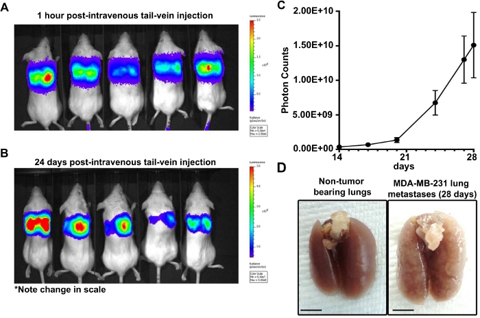
Figure 1: In vivo bioluminescence confirmation of successful tail-vein injection.
(A) Representative bioluminescence signal in mice 1 hour after tail-vein injection of luciferase-tagged MDA-MB-231 cells. (B) Representative bioluminescence signal in the same set of mice as (A) 24 days after tail-vein injection of luciferase-tagged MDA-MB-231 cells. [Note the change in scale bar between (A) and (B)]. (C) Quantification of photon counts over time in MDA-MB-231 tail-vein injected mice. Error bars represent mean ± SEM. (n = 8 mice) (D) Representative non-tumor bearing lung tissue (right) and MDA-MB-231 macrometastases in the lungs (left) at time of necropsy. Scale bars = 50 mm. Please click here to view a larger version of this figure.
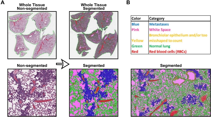
Figure 2: Tissue segmentation using Visiopharm software.
(A) Representative snips of unsegmented and segmented tissue mark-ups using the customized software algorithm. (B) Legend for all tissue categories segmented using software. Please click here to view a larger version of this figure.
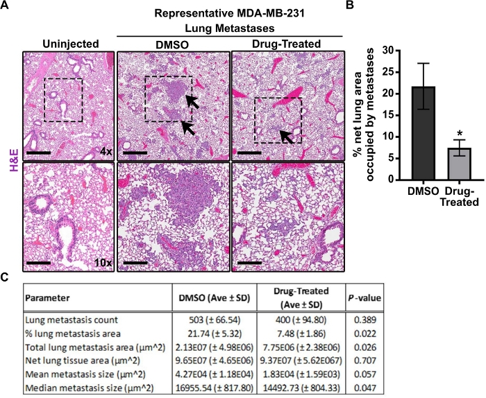
Figure 3: Representative metastatic lung tumor burden analysis of H&E-stained tissues.
(A) Representative H&E staining of lung tissue from uninjected mice and control (DMSO) and drug-treated mice following tail-vein injection of MDA-MB-231 cells. Representative tumor metastases are indicated with arrows. Scale bars = 500 µm for 4x magnification and 200 µm for 10x magnification. (B) Graph of percent net lung metastasis area of control and drug-treated mice. Error bars represent mean ± SD. (*) P = 0.022 by Student’s t-test. (C) Table summarizing the metastatic lung tumor burden analysis (n = 9 DMSO; n = 9 drug-treated). After checking for normal distribution of data, all P-values in the table were determined by unpaired, two-tailed Student’s t-test. Please click here to view a larger version of this figure.
Parameter | Description | |
Total Tissue Area (µm2) | Area in square microns inclusive of all tumor metastases, normal lung and areas of red blood cells. | |
Metastasis Count | Total number of metastases within the lung tissue. | |
Metastasis Area Percentage (µm2) | Total metastasis area divided by net tissue area x 100. | |
Total Tissue + White Space Area (µm2) | Area in square microns inclusive of all tissue and white space. | |
Net Tissue Area (µm2) | Tissue area in square microns (mets and normal lung) without white space and red blood cells. | |
Total Metastasis Area (µm2) | Total metastasis area in square microns as segmented by the Decision Forest algorithim. | |
Mean metastasis Area (µm2) | Mean (average) area in square microns of metastases within each image. | |
Median Metastasis Area (µm2) | Median metastasis area in square microns. An equal number of metastases falls below this value and an equal number of metastases are greater than the median value. |
Table 1: Parameters measured with software. List of parameters along with a description of each measurement that is computed using the custom algorithm.
Slide | Metastasis Count | Metastasis Area Percentage (µm2) | Total Tissue + White Space Area (µm2) | Total White Space (µm2) | Net Tissue Area (µm2) | Total Metastasis Area (µm2) | Red Blood Cells Area (µm2) | Mean Metastasis Area (µm2) | Median Metastasis Area (µm2) |
171 Lung Slide 1 | 435 | 8.90 | 185698000 | 83201800 | 92031400 | 8189250 | 10464800 | 18825.86 | 14748.73 |
171 Lung Slide 2 | 323 | 8.37 | 185698000 | 83201800 | 92054740 | 7708990 | 10441460 | 23866.84 | 14748.73 |
172 Lung Slide 1 | 151 | 2.73 | 181546000 | 89509904 | 81571296 | 2225220 | 10464800 | 14736.56 | 12486.37 |
172 Lung Slide 2 | 142 | 2.60 | 170708000 | 81735504 | 80558196 | 2093040 | 8414300 | 14739.72 | 12119.62 |
173 Lung Slide 1 | 634 | 11.60 | 234104992 | 102153000 | 115606692 | 13406800 | 16345300 | 21146.37 | 15472.22 |
173 Lung Slide 2 | 667 | 12.70 | 223180992 | 86778600 | 122374592 | 15542700 | 14027800 | 23302.40 | 16531.00 |
174 Lung Slide 1 | 40 | 0.55 | 192452992 | 80340896 | 87591096 | 485121 | 24521000 | 12128.03 | 10484.05 |
174 Lung Slide 2 | 34 | 0.51 | 183918000 | 71287904 | 91242796 | 464830 | 21387300 | 13671.47 | 11181.81 |
175 Lung Slide 1 | 780 | 23.93 | 179544992 | 44799200 | 126995782 | 30388600 | 7750010 | 38959.74 | 19307.76 |
175 Lung Slide 2 | 1001 | 12.58 | 169191536 | 43425608 | 120610754 | 15169100 | 5155174 | 15153.95 | 19703.08 |
188 Lung Slide 1 | 569 | 13.20 | 162290000 | 54210000 | 98486310 | 12997300 | 9593690 | 22842.36 | 14463.91 |
188 Lung Slide 2 | 271 | 5.15 | 157146000 | 54250800 | 91996500 | 4738100 | 10898700 | 17483.76 | 12657.83 |
189 Lung Slide 1 | 74 | 1.70 | 185292992 | 95700800 | 77779392 | 1318820 | 11812800 | 17821.89 | 14551.08 |
189 Lung Slide 2 | 74 | 1.76 | 182272992 | 95700800 | 74759392 | 1318820 | 11812800 | 17821.89 | 14551.08 |
816 Lung Slide 1 | 246 | 5.65 | 185876000 | 87568896 | 81916204 | 4631050 | 16390900 | 18825.41 | 14371.99 |
816 Lung Slide 2 | 565 | 6.05 | 183220000 | 76954304 | 90305396 | 5462670 | 15960300 | 9668.44 | 14244.82 |
876 Lung Slide 1 | 468 | 10.36 | 208308000 | 99300096 | 100947064 | 10454500 | 8060840 | 22338.68 | 16011.37 |
876 Lung Slide 2 | 528 | 11.74 | 199750896 | 81642568 | 110450391 | 12963400 | 7657937 | 24551.89 | 16699.13 |
877 Lung Slide 1 | 732 | 17.98 | 219340992 | 99918600 | 107869992 | 19397100 | 11552400 | 26498.77 | 18137.52 |
877 Lung Slide 2 | 605 | 14.64 | 207925504 | 88539712 | 108168329 | 15839700 | 11217463 | 26181.32 | 18014.64 |
878 Lung Slide 1 | 377 | 10.05 | 178534000 | 85610896 | 81931104 | 8232340 | 10992000 | 21836.45 | 16671.03 |
878 Lung Slide 2 | 376 | 9.88 | 170544000 | 75337904 | 86108406 | 8511710 | 9097690 | 22637.53 | 16754.38 |
879 Lung Slide 1 | 205 | 5.22 | 167556000 | 89999000 | 68123630 | 3553860 | 9433370 | 17335.90 | 13845.69 |
879 Lung Slide 2 | 213 | 4.64 | 167931008 | 80789400 | 78489588 | 3638720 | 8652020 | 17083.19 | 14058.12 |
881 Lung Slide 1 | 1122 | 38.81 | 218880000 | 79713504 | 130893816 | 50802300 | 8272680 | 45278.34 | 22044.99 |
881 Lung Slide 2 | 628 | 21.67 | 184200992 | 74502600 | 99122692 | 21475200 | 10575700 | 34196.18 | 19857.40 |
882 Lung Slide 1 | 678 | 24.05 | 194476992 | 83941904 | 98484788 | 23684500 | 12050300 | 34932.89 | 20748.06 |
882 Lung Slide 2 | 645 | 21.93 | 185537040 | 75790040 | 101412430 | 22241700 | 8334570 | 34483.26 | 20325.11 |
883 Lung Slide 1 | 429 | 10.79 | 179400992 | 84955696 | 84699866 | 9138800 | 9745430 | 21302.56 | 15080.23 |
883 Lung Slide 2 | 342 | 85.30 | 175220992 | 76210896 | 90472386 | 77170200 | 8537710 | 225643.86 | 17078.26 |
884 Lung Slide 1 | 359 | 6.42 | 206751008 | 87752600 | 103825008 | 6669710 | 15173400 | 18578.58 | 14333.41 |
884 Lung Slide 2 | 480 | 9.12 | 200990000 | 77052496 | 111060804 | 10125700 | 12876700 | 21095.21 | 15679.88 |
885 Lung Slide 1 | 332 | 7.79 | 191398000 | 92896304 | 84752596 | 6605490 | 13749100 | 19896.05 | 14500.11 |
885 Lung Slide 2 | 537 | 81.02 | 187475008 | 85938000 | 89378408 | 72411104 | 12158600 | 134843.77 | 15360.29 |
886 Lung Slide 1 | 305 | 7.93 | 158435008 | 80433296 | 76541662 | 6068720 | 1460050 | 19897.44 | 14500.11 |
886 Lung Slide 2 | 898 | 8.84 | 155460000 | 70808600 | 83457470 | 7380490 | 1193930 | 8218.81 | 14744.92 |
Table 2: Representative table of results. Table of results for each parameter of the algorithm from a cohort of mice tail-vein injected with MDA-MB-231 cells.
As researchers continue to use intravenous injection of tumor cells as an experimental model for metastasis, standard practices to analyze the resulting metastatic tumor burden are lacking. In some cases, significant differences in metastatic tumor burden upon manipulation of particular cell lines and/or use of chemical compounds can be observed macroscopically. However, in other instances, subtle differences in metastatic seeding and growth may be overlooked or misinterpreted without thorough pathological analysis. This study advances previously published tail-vein injection protocols by including a comprehensive method of metastatic lung tumor burden analysis. Importantly, this method of digital pathology analysis can also be applied to the quantification of lung metastatic tumor burden following orthotopic injection of tumor cells which are capable of spontaneous metastasis as well as other experimental metastasis models (i.e. intracardiac, etc.) and patient-derived xenograft (PDX) models. The use of digital imaging and software algorithm development by veterinary pathologists ensures the reproducibility, accuracy, and thoroughness of this approach to analyze metastatic lung tumor burden25.
Thoughtful decision of cell lines, cell concentration, and endpoints based on either previously published studies or careful experimental optimization is absolutely necessary. Given that metastatic seeding and colonization are highly dependent on interactions with various immune cell populations26,27, the use of immune-competent mice is ideal, albeit not always feasible. For the same reason, the interpretation of experimental metastasis studies using athymic or NSG mice, which lack key immune cell components, should be taken with caution. There are several mouse mammary tumor cell lines, including the MVT1 cells used in this study, that have been derived from the FVB/N mouse strain22,28,29. Other syngeneic models exist as well. In regard to cell concentration, injection of a large number of cells may greatly accelerate and enhance metastatic lung tumor burden. However, if the lungs are overwhelmed, it may be difficult to distinguish individual metastatic foci and emboli are more likely to occur. Also, the tail-vein injection procedure requires ample practice and training before safely and/or routinely performing injections. Many institutions will offer technical training and may provide mice for practice purposes. Proper placement of the needle and a smooth injection should indicate success; however, for training/practice purposes, Evans Blue dye can be used to help determine successful injection (1% in sterile PBS). The extremities of the mouse will turn blue shortly after injection, but the animal should be euthanized afterwards.
Additionally, the importance of standard necropsy and tissue sampling techniques to control and prevent slide artifacts that may impair slide scanning and analysis by the image analysis software cannot be stressed enough. Inflation of the lungs at time of necropsy is a critical step in maintaining tissue integrity and improves subsequent H&E staining as well as final analysis. For consistency with resolution and reproducibility, it is recommended that all slides in a study set are scanned with the same objective. In this study, all slides were scanned at 40x to ensure accuracy of algorithm settings and proper identification of tumor metastases when applied to analyzed fields. For each slide, the same lung lobes were consistently scanned and analyzed for each mouse. It is also strongly recommended that a pathologist review tissue markups for accuracy of the applied algorithm and that the same algorithm is applied to every slide in a study.
The presented protocol can be modified according to experimental design, user preferences, and desired outcome measurements. One such modification includes the use of an anesthesia induction chamber rather than conventional restrainer device for a conscious animal. In terms of animal health and wellness, neither approach is superior to the other and each has its own set of advantages as well as limitations30. Also, for black or brown mice, a light source or heating device may be needed in order to visualize the tail veins. Infrared lamps or a warm water bath can be used to dilate the veins. However, temperature should be carefully monitored. Furthermore, there are illuminated restraint devices available as well as other commercial versions of rodent restraint devices. Some investigators prefer Luer-Lok syringes for injections. We find it more difficult to eliminate air bubbles with Luer-Lok syringes, but it is a matter of preference. The viability of cells is an important consideration for the tail-vein injection procedure, and therefore, accurate cell counts as well as maintaining cells on ice prior to injection are necessary steps. If comparing lung seeding and colonization of manipulated cell lines, it is critical to determine any differences in cell size and viability prior to injection as these may complicate the interpretation of results. Cell death and/or damage may occur when using a narrow gauge needle; however, it is not recommended that a needle larger than 25 G be used as it may cause pain and discomfort to the animal.
As a way to validate that lung lesions are formed by injected tumor cells, immunostaining can be done on tissue sections. If using human cell lines, human-specific antibodies can be used to discern metastatic lesions. Alternatively, if using tagged cell lines (e.g., GFP), corresponding antibodies can be used. Also, many breast cancer cell lines are positive for epithelial markers (i.e., cytokeratins, E-cadherin, and EpCAM), but prior knowledge of expression is essential. However, the lung epithelium lining the airways will also be positive for these markers and thus, structure must also be considered. There may be cases in which primary lung tumor development must be ruled out. To do so, immunohistochemical staining for Thyroid Transcription Factor-1 (TTF1) can be used as a marker of primary lung adenocarcinoma. TTF1 staining should be evaluated by a certified pathologist.
Herein, a custom algorithm was written using a Decision Forest classification algorithm because the established lung metastasis algorithm could not be fine-tuned for accurate detection of metastases that varied in size. This customized algorithm enables complex measurements, allows for accurate segmentation of metastases by size, and supports a size cutoff so that small misshaped areas and normal structures are not misinterpreted and can therefore be included in the final data set. We anticipate that this algorithm will be applicable to most in vivo lung metastasis studies, but users may need to adjust settings within the software to fit their individual study needs. However, this algorithm serves as a platform for investigators wishing to analyze lung metastatic burden in a similar manner. There are many different options for image analysis platforms whereby access or availability, cost and training, as well as experience level may dictate the best platform to utilize36. The range of options include free platforms such as QuPath and more expensive, but sophisticated platforms, such as Visiopharm. It is advised that one consults with an image analysis pathology core and pathologist when deciding which platform may be available and best utilized for a particular research project.
Spontaneous mouse mammary tumor models (e.g., MMTV-PyMT) or orthotopic mammary fat pad injection methods represent the most physiologically relevant model for studying lung metastasis. A serious drawback to the tail-vein injection model is that it does not recapitulate the full metastatic cascade and is therefore limited to the study of tumor cell extravasation and secondary organ colonization. However, this experimental metastasis model is relevant for breast cancer research as lung metastases formed following tail-vein injection have genomic profiles similar to metastatic lesions that develop after orthotopic implantation of the same cells31. In order to establish a lung metastasis model, a large number of cells are often injected intravenously which may not accurately represent the process of metastasis as it pertains to seeding, immune reaction, and dormancy. Also, based on the circulatory pathway, pulmonary metastases are most common with tail-vein injection32. With most breast cancer cell lines, published reports indicate a relatively low incidence of bone, liver, or brain metastases following tail-vein injection7. Alternative experimental metastasis methods such as intracardiac, intratibial, portal vein and intracarotid injections are more appropriate for examining metastases to other sites33,34,35,36,37. Again, spontaneous mammary tumor models or orthotopic fat pad injection methods that recapitulate all steps of the metastatic cascade are preferred. Issues with consistent metastatic tumor burden, duration of study, and numbers of animals required for such studies are a downside. However, the method of digital pathology analysis presented here in can be applied to lung metastases formed through any spontaneous or experimental metastasis model.
The method of analysis also yields certain limitations such as subjectivity in algorithm creation. Even though whole slide imaging allows for digital analysis on an entire tissue section and on all lung lobes of a single mouse, it is limited to a two-dimensional analysis of a 3D tissue. Stereology is becoming a common practice that obtains 3D information for image analysis and can account for factors such as tissue shrinkage that occurs during tissue processing38. Stereology, however, has its own limitations such as tissue, resource, and time constraints.
Given the number of cancer patients affected by metastatic spread of their disease, the tail-vein injection method to study metastasis will continue to be a useful tool in terms of understanding the complicated biology of metastatic spread and in determining the pre-clinical efficacy of novel therapeutics. In vivo mouse models of metastasis, particularly those using immune-competent animals, are becoming even more important for cancer research given the widespread interest in immunotherapy29. Also, experimental metastasis models are critical in terms of investigating metastasis suppressor genes (i.e., those that suppress the metastatic potential of cancer cells without affecting primary tumor growth), and therefore, continue to be a valuable research tool.
Digital imaging and slide analysis have rapidly become a mainstay in diagnostic and experimental mouse modeling39. Using the type of approach described herein to analyze lung metastatic tumor burden will allow for high throughput analyses in a more comprehensive and accurate manner. Furthermore, digital imaging pathology provides an avenue for more collaborative research projects involving pathologists that specialize in areas such as mouse models of breast cancer metastasis. As multiplex tissue imaging methods and 3D imaging technologies (as mentioned above) continue to be developed, digital imaging pathology, sophisticated software programs for image analysis, and the expertise of pathologists will certainly be necessary for advancing metastasis research.
The authors have nothing to disclose.
Representative data was funded through the National Cancer Institute (K22CA218549 to S.T.S). In addition to their assistance in developing the comprehensive analysis method reported herein, we thank The Ohio State University Comprehensive Cancer Center Comparative Pathology and Mouse Phenotyping Shared Resource (Director – Krista La Perle, DVM, PhD) for histology and immunohistochemistry services and the Pathology Imaging Core for algorithm development and analysis.
alcohol prep pads | Fisher Scientific | 22-363-750 | for cleaning tail prior to injection |
dissection scissors | Fisher Scientific | 08-951-5 | for mouse dissection and lung tissue inflation |
DMEM with L-Glutamine, 4.5g/L Glucose and Sodium Pyruvate | Fisher Scientific | MT10013CV | cell culture media base for MDA-MB-231 and MVT1 cell lines |
Dulbecco's Phosphate-Buffered Salt Solution 1x | Fisher Scientific | MT21030CV | used for resuspending tumor cells for injection |
ethanol (70 % solution) | OSU | used to minimize mouse's fur during dissection; use caution – flammable | |
Evan's blue dye | Millipore Sigma | E2129 | used at 1 % in sterile PBS for practice with tail-vein injection method; use caution – dangerous reagent |
Fetal Bovine Serum | Millipore Sigma | F4135 | cell culture media additive; used at 10% in DMEM |
forceps | Fisher Scientific | 10-270 | for dissection and lung tissue inflation |
FVB/NJ mice | The Jackson Laboratory | 001800 | syngeneic mouse strain for MVT1 cells |
hemacytometer (Bright-Line) | Millipore Sigma | Z359629 | for use in cell culture to obtain cell counts |
insulin syringe (28 G) | Fisher Scientific | 14-829-1B | for tail-vein injections (BD 329424) |
MDA-MB-231 cells | ATCC | human breast cancer cell line | |
MVT1 cells | mouse mammary tumor cells | ||
needles (26 G) | Fisher Scientific | 14-826-15 | used to inflate the mouse's lungs |
neutral buffered formalin (10%) | Fisher Scientific | 245685 | used as a tissue fixative and to inflate lung tissue; use caution – dangerous reagent |
NOD.Cg-Prkdcscid Il2rgtm1Wjl/SzJ (NSG) mice | The Jackson Laboratory | 005557 | maintained by OSUCCC Target Validation Shared Resource |
Penicillin Streptomycin 100x | ThermoFisher | 15140163 | cell culture media additive |
sterile gauze | Fisher Scientific | NC9379092 | for applying pressue to mouse's tail if bleeding occurs |
syringe (5 mL) | Fisher Scientific | 14-955-458 | used to inflate mouse lung tissue |
tail-vein restrainer | Braintree Scientific, Inc. | TV-150 STD | used to restrain mouse for tail-vein injections |
Trypan blue (0.4 %) | ThermoFisher | 15250061 | used in cell culture to assess viability |
Trypsin-EDTA 0.25 % | ThermoFisher | 25200-114 | used in cell culture to detach tumor cells from plate |