Summary

재조합 G 단백질 결합 수용체의 고처리량 스크리닝을 위한 "이중 첨가" 칼슘 형광 분석

Published: December 02, 2022
doi:

Summary

이 작업에서는 재조합 G 단백질 결합 수용체(GPCR)에서 소분자 라이브러리를 스크리닝하기 위한 384웰 플레이트에 대한 고처리량 세포 내 칼슘 형광 분석에 대해 설명합니다. 표적, 소열 진드기, Rhipicephalus microplus 의 키닌 수용체는 CHO-K1 세포에서 발현됩니다. 이 분석은 하나의 “이중 첨가” 분석에서 동일한 세포를 사용하여 작용제와 길항제를 식별합니다.

Abstract

G 단백질 결합 수용체 (GPCR)는 수용체의 가장 큰 수퍼 패밀리를 나타내며 수많은 인간 약물의 표적입니다. GPCR에 대한 무작위 소분자 라이브러리의 고처리량 스크리닝(HTS)은 제약 산업에서 표적별 약물 발견을 위해 사용됩니다. 이 연구에서는 치명적인 인간 및 수의학 병원체 벡터의 생리학적 연구를 위한 프로브로서 무척추동물 특이적 신경펩티드 GPCR의 새로운 소분자 리간드를 식별하기 위해 HTS를 사용했습니다.

무척추 동물 특이 적 키닌 수용체는 이뇨, 수유 및 소화를 포함하여 무척추 동물의 많은 중요한 생리적 과정을 조절하기 때문에 표적으로 선택되었습니다. 또한, 많은 무척추 동물 GPCR의 약리학은 특성이 좋지 않거나 전혀 특성화되지 않습니다. 따라서 다른 후생 동물, 특히 인간의 관련 GPCR과 관련하여 이러한 수용체 그룹의 차별적 약리학은 수퍼 패밀리로서 GPCR의 구조-활성 관계에 지식을 추가합니다. HTS 분석은 소열 진드기 또는 남부 소 진드기 인 Rhipicephalus microplus에서 키닌 수용체의 리간드를 발견하기 위해 384 웰 플레이트의 세포에 대해 개발되었습니다. 틱 키닌 수용체는 CHO-K1 세포에서 안정적으로 발현되었다.

키닌 수용체는 내인성 키닌 신경 펩티드 또는 다른 소분자 작용제에 의해 활성화 될 때 칼슘 저장소에서 세포질로의Ca 2+ 방출을 유발합니다. “이중 첨가” 접근법과 결합된 이 칼슘 형광 분석은 동일한 분석 플레이트에서 기능성 작용제와 길항제 “적중” 분자를 검출할 수 있습니다. 각각의 분석은 320개의 무작위 소분자의 어레이를 운반하는 약물 플레이트를 사용하여 수행되었다. 0.7의 신뢰할 수 있는 Z’ 계수가 얻어졌고, HTS가 2μM 최종 농도일 때 3개의 작용제 및 2개의 길항제 히트 분자가 확인되었습니다. 여기에 보고된 칼슘 형광 분석은Ca2+ 신호전달 캐스케이드를 활성화하는 다른 GPCR을 스크리닝하도록 조정할 수 있습니다.

Introduction

효모에서 인간으로 존재하는 G 단백질 결합 수용체 (GPCR)는 많은 유기체에서 수용체의 가장 큰 수퍼 패밀리를 나타냅니다1. 그들은 동물의 거의 모든 생물학적 과정을 조절하는 데 중요한 역할을합니다. 절지동물의 게놈에는 50-200개의 GPCR이 있으며, 이는 그들이 가장 큰 막 수용체 수퍼패밀리2를 대표한다는 것을 의미합니다. 이들은 서열 유사성과 기능에 따라 6개의 주요 클래스인 AF로 분류됩니다3. GPCR은 호르몬, 신경 펩티드, 생체 아민, 글루타메이트, 양성자, 지방 당 단백질 및 광자4와 같은 다양한 세포 외 신호를 전달합니다. GPCR은 이종 삼량체 G 단백질 (Gα, Gβ 및 Gγ)에 결합하여 다운 스트림 신호를 전송합니다. Gαs 또는 Gαi/o 단백질에 결합된 GPCRs는 각각 아데닐릴 사이클라제를 활성화 또는 억제함으로써 세포내 3′,5′-사이클릭 아데노신 모노포스페이트 (cAMP) 수준을 증가 또는 감소시킨다. q/11에 결합된 GPCR은 포스포리파아제 C(PLC)-이노시톨-1,4,5-삼인산(IP3) 경로를 활성화하여 소포체 칼슘 저장소에서 칼슘 방출을 유도합니다. Gα12/13에 결합 된 GPCR은 RhoGTPase 뉴클레오티드 교환 인자 5,6을 활성화시킵니다. GPCR은 인간 약물의 50 % 이상과 살비제 인 amitraz4의 표적입니다. GPCR은 이러한 다양한 신호를 전달하기 때문에 무척추 동물 고유의 생리 기능을 방해하는 새로운 살충제 개발을위한 유망한 표적입니다.

HTS의 목표는 수용체 기능을 조절할 수있는 히트 분자를 확인하는 것입니다. HTS에는 분석 개발, 소형화 및 자동화가 포함됩니다7. 절지동물 신경펩티드 GPCR은 발달, 탈피 및 엑디시스, 배설, 에너지 동원및 생식과 같은 대부분의 생리적 기능에 관여합니다4. 절지 동물 및 후생 동물의 대부분의 신경 펩티드 GPCR은 검게 다리 진드기 Ixodes 견갑골의 근억제 펩티드 및 SIFamide 수용체와 같은 칼슘 신호 전달 캐스케이드 2,6,8,9,10을 통해 신호를 보냅니다. 그들의 리간드는 뒷부 운동성 분석에서 길항성이며, SIF는 수축을 유도하고 MIP는11,12를 억제합니다. 황열병 모기의 NPY 유사 수용체 인 Aedes aegypti13을 찾는 암컷 숙주를 조절합니다. 애쿼린 칼슘 생물발광 분석법14와 같은 다른 대안적인 칼슘 동원 분석과 비교하여, 칼슘 형광 분석은 수행하게 용이하고, 다른 재조합 칼슘 검출 단백질의 형질감염을 필요로 하지 않으며, 비용 효율적이다. 칼슘 형광 분석은 애쿼린 칼슘 생물발광 분석14,15에서 수득된 빠른 운동 신호에 비해 연장된 신호를 생성한다.

여기 예에서, 소열병 진드기의 키닌 수용체인 Rhipicephalus microplus를 CHO-K1 세포주에서 재조합적으로 발현시키고 칼슘 형광 분석에 사용했습니다. R. microplus에서 발견되는 키닌 수용체 유전자는 하나뿐입니다. 수용체는 Gq 단백질 의존성 신호전달 경로를 통해 신호를 보내고 칼슘 저장소로부터 세포 내 공간16으로Ca2+의 유출을 유발합니다. 이 과정은 칼슘 이온과 결합할 때 형광 신호를 유도하는 형광단으로 감지하고 정량화할 수 있습니다(그림 1).

키닌 수용체는 클래스 A 로돕신 유사 수용체에 속하는 무척추 동물 특이 적 GPCR입니다. Kinin은 연체 동물, 갑각류, 곤충 및 Acari 4,17,18에 존재하는 고대 신호 전달 신경 펩티드입니다. 딱정벌레 (딱정벌레)는 키닌 신호 시스템이 부족합니다. 모기 Aedes aegypti에는 3 개의 aedeskinins와 결합하는 키닌 수용체가 하나만 있으며, Drosophila melanogaster에는 독특한 리간드 19,20,21로 drosokinin과 함께 하나의 키닌 수용체가 있습니다. 척추 동물에는 상동 키닌 또는 키닌 수용체가 없습니다. 진드기에서 키닌의 정확한 기능은 알려져 있지 않지만, 키닌 수용체 RNAi- 침묵 된 R. microplus의 암컷은 생식 적합성이 현저히 감소 된 것으로 나타났습니다22. Kinins는 곤충의 다방성 펩타이드입니다. 초파리 멜라노가스터에서, 그들은 중추 및 말초 신경 조절 시스템23, 전-엑디시스(pre-ecdysis)24, 섭식25, 대사(26), 수면 활동 패턴(26, 27) 및 유충 운동28 모두에 관여한다. Kinins는 모기 A. aegypti 29,30,31 에서 뒷장 수축, 이뇨 및 먹이를 조절합니다. 키닌 펩티드는 보존된 C-말단 펜타펩티드 Phe-X1-X2-Trp-Gly-NH2를 가지며, 이는 생물학적 활성(32)에 필요한 최소 서열이다. 절지 동물 특이성, 내인성 리간드의 작은 크기로 인해 소분자 간섭에 민감하며 곤충의 다발성 기능은 키닌 수용체를 해충 방제를위한 유망한 표적으로 만듭니다4.

“이중-첨가” 분석(도 2)은 동일한 HTS 분석(15)에서 작용제 또는 길항제의 식별을 허용한다. 이는 약물 발견33을 위해 제약 산업에서 일반적으로 사용되는 “이중 첨가” 분석으로부터 채택된다. 간단히 말해서, 세포 플레이트에 약물을 먼저 추가하면 용매 제어의 적용에 비해 더 높은 형광 신호가 검출될 때 화학 라이브러리에서 잠재적 작용제를 식별할 수 있습니다. 이러한 소분자와 함께 5분 동안 배양한 후 알려진 작용제(키닌 펩타이드)를 모든 웰에 적용합니다. 약물 플레이트로부터 길항제를 무작위로 받은 웰은 첫 번째 첨가에서 용매를 수용한 대조군 웰에 비해 작용제 첨가 시 더 낮은 형광 신호를 표시합니다. 그런 다음 이 분석을 통해 동일한 세포를 가진 잠재적 작용제 및 길항제를 식별할 수 있습니다. 표준 HTS 프로젝트에서 이러한 적중 분자는 용량 반응 분석 및 여기에 표시되지 않은 추가 생물학적 활성 분석을 통해 추가로 검증됩니다.

Figure 1
그림 1: 칼슘 형광 분석 메커니즘의 그림. Gq 단백질은 세포 내 칼슘 신호 전달 경로를 유발합니다. 키닌 수용체(G 단백질 커플링 수용체)는 CHO-K1 세포에서 재조합적으로 발현되었다. 작용제 리간드가 수용체에 결합할 때, 키닌 수용체와 연관된 Gq 단백질은 PLC를 활성화시키고, 이는PIP2 분자의IP3 및 DAG로의 전환을 촉매한다. 그런 다음 IP 3는 소포체 표면의 IP3R에 결합하여Ca2+가 세포질로 방출되고, 여기서Ca 2+ 이온은 형광단에 결합하여 형광 신호를 유도합니다. 형광 신호는 490nm에서 여기하여 얻을 수 있고 514nm에서 검출할 수 있습니다. 약어 : GPCR = G 단백질 결합 수용체; PLC = 포스포리파아제 C; PIP2 = 포스파티딜이노시톨 4,5-비스포스페이트; IP3 = 이노시톨 트리포스페이트; DAG = 디아실글리세롤; IP3 R = IP3 수용체. BioRender.com 로 만들었습니다. 이 그림의 더 큰 버전을 보려면 여기를 클릭하십시오.

Figure 2
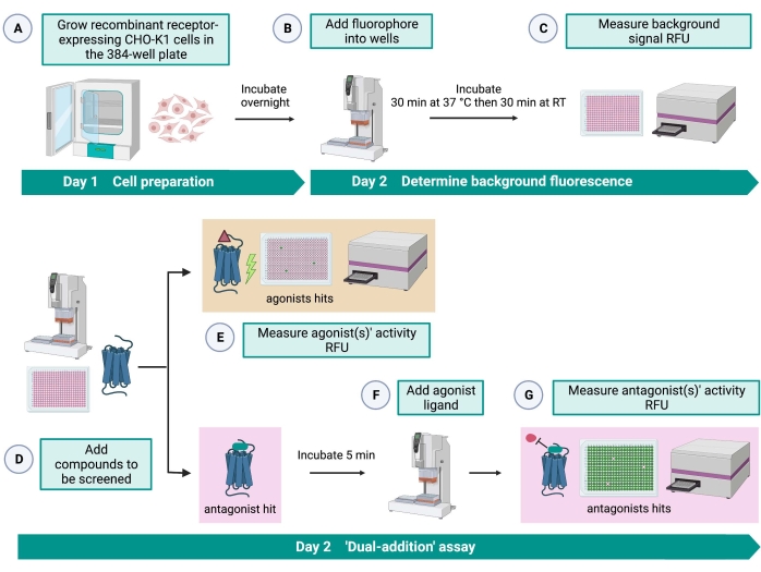
그림 2: CHO-K1 세포에서 발현되는 G 단백질 결합 수용체에서 소분자의 고처리량 스크리닝을 위한 워크플로. (A) 키닌 수용체를 안정적으로 발현하는 재조합 CHO-K1 세포를 액체 처리 시스템(25μL/웰)을 사용하여 384웰 플레이트(10,000세포/웰)에 첨가하고 가습된 CO2 인큐베이터에서12-16시간 동안 배양하였다. (B ) 형광 염료(25μL/웰)를 함유하는 분석 완충액을 액체 처리 시스템을 사용하여 세포 플레이트에 첨가하였다. 플레이트를 37°C에서 30분 동안 인큐베이션하고, RT에서 또 다른 30분 동안 평형화시켰다. (C) 각 웰에서 세포의 배경 형광 신호를 플레이트 리더로 측정하였다. (D) 384웰 라이브러리 플레이트와 블랭크 용매(모두 0.5μL/웰)의 약물 용액을 액체 처리 시스템을 사용하여 세포 분석 플레이트에 첨가했습니다. (e) 세포 칼슘 형광 반응은 약물 용액의 첨가 직후에 플레이트 리더로 측정되었다; 평균 형광 신호보다 높은 형광 신호를 유도하는 화합물은 작용제 히트로 선택되었습니다. GPCR을 차단하는 길항제 히트(아래 아이콘)는 단계 G 동안 펩티드 작용제의 첨가 후에 밝혀졌다. (f) 동일한 분석 플레이트에서, 세포를 스크리닝 화합물로 5분간 배양한 후, 진드기 키닌 수용체의 내인성 작용제 펩티드 Rhimi-K-1 (QFSPWGamide)을 각 웰(1 μM)에 첨가하였다. (g) 작용제 펩티드 첨가 후의 세포 형광 반응은 즉시 플레이트 리더로 측정하였다. 형광 신호를 억제하는 화합물(들)을 길항제 히트(들)로서 선택하였다. 약어 : GPCR = G 단백질 결합 수용체; RT = 실온; RFU = 상대 형광 단위. BioRender.com 로 만들었습니다. 이 그림의 더 큰 버전을 보려면 여기를 클릭하십시오.

Protocol

1. 세포 유지 관리 참고: R. 마이크로플러스의 키닌 수용체를 안정적으로 발현하는 CHO-K1 세포주(BMLK3)는 Holmes et al.16에 의해 개발되었습니다. 세포주의 발달에 대한 세부사항은 다른 곳에 제시되어 있다14. 다음 모든 단계는 클래스 II 생물 안전 캐비닛의 멸균 조건에서 수행됩니다. 표적 수용체의 발현을 보?…

Representative Results

320개의 무작위 소분자로 구성된 사내 약물 플레이트(SAC2-34-6170)를 예로 이 HTS 분석을 시연하는 데 사용했습니다. HTS는 Z’ 계수가 0.7인 우수한 분석 품질을 가졌습니다(표 1). 이 Z’ 인자는 시험된 화합물34와 무관한 분석 품질을 반영한다. 0.5 이상의 Z’ 계수는 양성 대조군과 음성 대조군의 RFU 사이의 양호한 분석 신호 동적 범위를 나타냅니다. Z’ 계수가 0…

Discussion

HTS의 목표는 대량의 소분자를 스크리닝하여 적중 분자를 식별하는 것입니다. 따라서 이 예제의 결과는 기존 HTS 실험의 작은 부분만을 나타냅니다. 또한, 확인된 적중 분자는 동일한 재조합 세포주 및 빈 벡터만 운반하는 CHO-K1 세포주에 대한 용량 의존적 분석과 같은 다운스트림 분석에서 검증되어야 하며, 이는 소분자를 저장하기 위해 동시에 수행할 수 있습니다. 세포독성 분석은 제2 첨가에서의…

Divulgations

The authors have nothing to disclose.

Acknowledgements

이 작업은 USDA-NIFA-AFRI 동물 건강 및 웰빙 상 (수상 번호 2022-67015-36336, PVP [프로젝트 디렉터])과 텍사스 A & M AgriLife 연구 곤충 벡터 질병 보조금 프로그램 (FY’22-23)의 경쟁 기금에서 P.V.P.로 지원되었습니다. 농업 생명 과학 대학 TAMU의 A.W.E.S.O.M.E. 교수 그룹은 원고 편집에 도움을 준 것으로 인정 받았습니다. 보충 표 S2 에는 텍사스 A&M 대학과 텍사스 A&M AgriLife Research의 James Sacchettini 박사 실험실에서 얻은 사내 무작위 소분자 라이브러리의 데이터가 포함되어 있습니다.

Materials

0.25% trypsin-EDTA Gibco Invitrogen 15050-065 with phenol red
0.4% trypan blue MilliporeSigma T8154 liquid, sterile
1.5 mL microcentrifuge tubes Thermo Fisher AM12400 RNase-free Microfuge Tubes
5 mL serological pipette Corning 29443-045 Corning Costar Stripette individually wrapped 
10 mL serological pipette Corning 29443-047 Corning Costar Stripette individually wrapped 
15 mL conical tubes Falcon 352196 sterile
20 µL filter tips USA Scientifc Inc. P1121 sterile, barrier
25 mL serological pipette Corning 29443-049 Corning Costar Stripette individually wrapped 
50 mL conical tubes Corning 430828 graduated, sterile
150 mL auto-friendly reservior Integra Bioscience 6317 sterile, individually wrapped for cell seeding in day 1
150 mL auto-friendly reservior Integra Bioscience 6318 sterile, stacked, for loading dye in day 2
384/ 12.5 µL low retention tips Integra Bioscience 6405 long, sterile filter
384/ 12.5 µL tips Integra Bioscience 6404 long, sterile filter
384-well plate Greiner 781091 CELLSTAR, clear polystyrene, µClear, Black/Flat
Aluminum plate seals Axygen Scientific PCR-AS-200 polyester-based
Aluminum foil wrap Walmart
Biosafty cabinet II NuAire NU-540-300
Cell counter Nexcelom AutoT4
cell counting slides Nexcelom SD-100 20 µL chamber
CO2 humidified incubator Thermo Fisher Forma Series II
Desk Lamp SunvaleeyTEK RS1000B
Dimethyl sulfoxide MilliporeSigma 276855 anhydous, >99.9%
Drug plate Corning 3680
Dulbecco's phosphate-buffered saline Corning 21-031-CV DPBS, 1x without calcium amd magnesium
Ethanol Koptec 2000
F-12K Nutrient Mixture  Corning 45000-354 (Kaighn's Mod.) with L-glutamine
Fetal bovine serum Equitech-Bio SFBU30
Fluorescent calcium assay kit ENZO Lifescience ENZ-51017 10×96 tests
G418 sulfate Gibco Invitrogen 10131-027 Geneticin selective antibiotic 50 mg/mL
Hank's buffer MilliporeSigma 55037C HBSS modified, with calcium, with magnesium, without phenol read
HEPES buffer Gibco Invitrogen 15630-080 1 Molar
HTS data storage plateform CDD vault  https://www.collaborativedrug.com/
Liquid handling system Integra Bioscience Viaflo 384/12.5 µL
Plate centrifuge Thermo Fisher Sorvall ST8
Plate reader BMG technology Clariostar
Poly-D-lysine MilliporeSigma P6407
Rhimi-K-1 agonist peptide Genscript custom order QFSPWGamide
T-75 flask Falcon 353136

References

  1. Hanlon, C. D., Andrew, D. J. Outside-in signaling – A brief review of GPCR signaling with a focus on the Drosophila GPCR family. Journal of Cell Science. 128 (19), 3533-3542 (2015).
  2. Liu, N., Li, T., Wang, Y., Liu, S. G-protein coupled receptors (GPCRs) in insects-A potential target for new insecticide development. Molecules. 26 (10), 2993 (2021).
  3. Pierce, K. L., Premont, R. T., Lefkowitz, R. J. Seven-transmembrane receptors. Nature Reviews Molecular Cell Biology. 3, 639-650 (2002).
  4. Pietrantonio, P. V., Xiong, C., Nachman, R. J., Shen, Y. G protein-coupled receptors in arthropod vectors: Omics and pharmacological approaches to elucidate ligand-receptor interactions and novel organismal functions. Current Opinion in Insect Science. 29, 12-20 (2018).
  5. Hilger, D., Masureel, M., Kobilka, B. K. Structure and dynamics of GPCR signaling complexes. Nature Structural & Molecular Biology. 25 (1), 4-12 (2018).
  6. Liu, N., Wang, Y., Li, T., Feng, X. G-protein coupled receptors (GPCRs): Signaling pathways, characterization, and functions in insect physiology and toxicology. International Journal of Molecular Sciences. 22 (10), 5260 (2021).
  7. Hansen, K. B., Bräuner-Osborne, H., Leifert, W. FLIPR® assays of intracellular calcium in GPCR drug discovery. G Protein-Coupled Receptors in Drug Discovery. , (2009).
  8. Bauknecht, P., Jekely, G. Large-scale combinatorial deorphanization of Platynereis neuropeptide GPCRs. Cell Reports. 12 (4), 684-693 (2015).
  9. Frooninckx, L., et al. Neuropeptide GPCRs in C. elegans. Frontiers in Endocrinology. 3, 167 (2012).
  10. Caers, J., et al. More than two decades of research on insect neuropeptide GPCRs: An overview. Frontiers in Endocrinology. 3, 151 (2012).
  11. Šimo, L., Koči, J., Park, Y. Receptors for the neuropeptides, myoinhibitory peptide and SIFamide, in control of the salivary glands of the blacklegged tick Ixodes scapularis. Insect Biochemistry and Molecular Biology. 43 (4), 376-387 (2013).
  12. Šimo, L., Park, Y. Neuropeptidergic control of the hindgut in the black-legged tick Ixodes scapularis. International Journal for Parasitology. 44 (11), 819-826 (2014).
  13. Liesch, J., Bellani, L. L., Vosshall, L. B. Functional and genetic characterization of neuropeptide Y-like receptors in Aedes aegypti. PLoS Neglected Tropical Diseases. 7 (10), 2486 (2013).
  14. Lu, H. L., Kersch, C. N., Taneja-Bageshwar, S., Pietrantonio, P. V. A calcium bioluminescence assay for functional analysis of mosquito (Aedes aegypti) and tick (Rhipicephalus microplus) G protein-coupled receptors. Journal of Visualized Experiments. (50), e2732 (2011).
  15. Xiong, C., Baker, D., Pietrantonio, P. V. The cattle fever tick, Rhipicephalus microplus, as a model for forward pharmacology to elucidate kinin GPCR function in the Acari. Frontiers in Physiology. 10, 1008 (2019).
  16. Holmes, S. P., Barhoumi, R., Nachman, R. J., Pietrantonio, P. V. Functional analysis of a G protein-coupled receptor from the Southern cattle tick Boophilus microplus (Acari: Ixodidae) identifies it as the first arthropod myokinin receptor. Insect Molecular Biology. 12 (1), 27-38 (2003).
  17. Cox, K. J., et al. Cloning, characterization, and expression of a G-protein-coupled receptor from Lymnaea stagnalis and identification of a leucokinin-like peptide, PSFHSWSamide, as its endogenous ligand. Journal of Neuroscience. 17 (4), 1197-1205 (1997).
  18. Dircksen, H., Kastin, A. J. Chapter 32 – Crustacean bioactive peptides. Handbook of Biologically Active Peptides (Second Edition). , 209-221 (2013).
  19. Halberg, K. A., Terhzaz, S., Cabrero, P., Davies, S. A., Dow, J. A. Tracing the evolutionary origins of insect renal function. Nature Communications. 6, 6800 (2015).
  20. Pietrantonio, P. V., Jagge, C., Taneja-Bageshwar, S., Nachman, R. J., Barhoumi, R. The mosquito Aedes aegypti (L.) leucokinin receptor is a multiligand receptor for the three Aedes kinins. Insect Molecular Biology. 14 (1), 55-67 (2005).
  21. Radford, J. C., Davies, S. A., Dow, J. A. Systematic G-protein-coupled receptor analysis in Drosophila melanogaster identifies a leucokinin receptor with novel roles. Journal of Biological Chemistry. 277 (41), 38810-38817 (2002).
  22. Brock, C. M., et al. The leucokinin-like peptide receptor from the cattle fever tick, Rhipicephalus microplus, is localized in the midgut periphery and receptor silencing with validated double-stranded RNAs causes a reproductive fitness cost. International Journal for Parasitology. 49 (3-4), 287-299 (2019).
  23. Nässel, D. R. Leucokinin and associated neuropeptides regulate multiple aspects of physiology and behavior in Drosophila. International Journal of Molecular Sciences. 22 (4), 1940 (2021).
  24. Kim, Y. -. J., et al. Central peptidergic ensembles associated with organization of an innate behavior. Proceedings of the National Academy of Sciences of the United States of America. 103 (38), 14211-14216 (2006).
  25. Al-Anzi, B., et al. The leucokinin pathway and its neurons regulate meal size in Drosophila. Current Biology. 20 (11), 969-978 (2010).
  26. Yurgel, M. E., et al. A single pair of leucokinin neurons are modulated by feeding state and regulate sleep-metabolism interactions. PLoS Biology. 17 (2), 2006409 (2019).
  27. Nässel, D. R., Zandawala, M. Recent advances in neuropeptide signaling in Drosophila, from genes to physiology and behavior. Progress in Neurobiology. 179, 101607 (2019).
  28. Okusawa, S., Kohsaka, H., Nose, A. Serotonin and downstream leucokinin neurons modulate larval turning behavior in Drosophila. Journal of Neuroscience. 34 (7), 2544-2558 (2014).
  29. Kersch, C. N., Pietrantonio, P. V. Mosquito Aedes aegypti (L.) leucokinin receptor is critical for in vivo fluid excretion post blood feeding. FEBS letters. 585 (22), 3507-3512 (2011).
  30. Kwon, H., et al. Leucokinin mimetic elicits aversive behavior in mosquito Aedes aegypti (L.) and inhibits the sugar taste neuron. Proceedings of the National Academy of Sciences of the United States of America. 113 (25), 6880-6885 (2016).
  31. Xiong, C., Baker, D., Pietrantonio, P. V. A random small molecule library screen identifies novel antagonists of the kinin receptor from the cattle fever tick, Rhipicephalus microplus (Acari: Ixodidae). Pest Management Science. 77 (5), 2238-2251 (2021).
  32. Torfs, P., et al. The kinin peptide family in invertebrates. Annals of the New York Academy of Sciences. 897 (1), 361-373 (1999).
  33. Ma, Q., Ye, L., Liu, H., Shi, Y., Zhou, N. An overview of Ca2+ mobilization assays in GPCR drug discovery. Expert Opinion on Drug Discovery. 12 (5), 511-523 (2017).
  34. Zhang, J. -. H., Chung, T. D., Oldenburg, K. R. A simple statistical parameter for use in evaluation and validation of high throughput screening assays. Journal of Biomolecular Screening. 4 (2), 67-73 (1999).
  35. Zhang, R., Xie, X. Tools for GPCR drug discovery. Acta Pharmacologica Sinica. 33 (3), 372-384 (2012).
  36. Offermanns, S., Simon, M. I. Gα15 and Gα16 couple a wide variety of receptors to phospholipase C. Journal of Biological Chemistry. 270 (25), 15175-15180 (1995).
  37. Murgia, M. V., et al. High-content phenotypic screening identifies novel chemistries that disrupt mosquito activity and development. Pesticide Biochemistry and Physiology. 182, 105037 (2022).
  38. Lismont, E., et al. Can BRET-based biosensors be used to characterize G-protein mediated signaling pathways of an insect GPCR, the Schistocerca gregaria CRF-related diuretic hormone receptor. Insect Biochemistry and Molecular Biology. 122, 103392 (2020).
check_url/fr/64505?article_type=t

Play Video

Citer Cet Article
Xiong, C., Baker, D., Pietrantonio, P. A “Dual-Addition” Calcium Fluorescence Assay for the High-Throughput Screening of Recombinant G Protein-Coupled Receptors. J. Vis. Exp. (190), e64505, doi:10.3791/64505 (2022).

View Video