JoVE Science Education
Biomedical Engineering
Un abonnement à JoVE est nécessaire pour voir ce contenu.  Connectez-vous ou commencez votre essai gratuit.
JoVE Science Education Biomedical Engineering
Tensile Strength of Resorbable Biomaterials
  • 00:07Vue d'ensemble
  • 01:10Principles of Resorbable Biomaterials
  • 03:08Sample Preparation
  • 04:45Control Sample Test
  • 05:51Strength Loss Profile
  • 06:59Résultats
  • 08:20Applications
  • 09:41Summary

リバーブルバイオマテリアルの引張強度

English

Diviser

Vue d'ensemble

出典:ペイマン・シャーベイギ・ルードポシュティとシナ・シャーバズモハマディ、バイオメディカル工学部、コネチカット大学、ストールズ、コネチカット州

4000年以上にわたり、縫合糸は医学的介入として使用されてきました。最も初期の記録は、リネンが選択の生体材料であったを示しています。現在も使用されているCatgutは、紀元前150年頃に剣闘士を治療するために使用されたと伝えられています。今日では、縫合糸に使用されている材料が数多くあります。縫合糸は、その組成(天然または合成)および吸収(非リバーブルまたは再吸収性)によって分類される。

再吸収可能な(または吸収可能な)縫合糸は、酵素分解またはポリマー鎖内の特定のグループとの水の相互作用によって引き起こされるプログラムされた分解のいずれかを介して体内で分解する。これらの縫合糸は、多くの場合、ポリグリコール酸、ポリジオキサノール、ポリカプロラクトンなどの合成材料、またはシルクなどの天然バイオマテリアルから作成されます。それらは通常、一般的な外科のような特定の内部処置のために使用される。吸収可能な縫合糸は、治癒のために十分な長さのために一緒に傷を保持しますが、その後、彼らは最終的に体によって崩壊します。一方、非リバーブル縫合糸は劣化せず、抽出する必要があります。それらは通常ポリプロピレン、ナイロンおよびステンレス鋼に由来する。これらの縫合糸は、通常、整形外科や心臓手術のために実装され、後日それらを除去するために医療専門家を必要とします。

ここでは、2種類の再吸収性縫合糸の引張強度を、人体内の異なるpH環境に対応する中性、酸性、アルカリ性溶液に曝露した後に試験される。テストは2つの部分で構成されます。まず、制御サンプルを準備し、引張試験を介して分析されます。その後、サンプルは、数週間にわたって様々なpHの溶液に連続的に曝露した後に試験されます。

Principles

Procédure

1. サンプル調製 以下の情報を含む 6 つのラベルを作成し、そのラベルをスクリュートップ試験管に取り付けます。 日付: 月と日 サンプルタイプ:ポリグリコネートまたはポリジオキサネ 溶液の種類:酸性(A)、アルカリ性(B)、または中性(N)溶液は、2〜14の間のpHの範囲である。 縫合糸の包装を開き、縫合糸を取り外します。針を切り取り、シャー?…

Résultats

Over the course of five weeks, all treated specimens were tested and analyzed. From the overall trials, the average tensile strengths were calculated using Equation 1:

Equation 1   (1)

The standard deviations of all the forces at failure with respect to suture type and solution environment were also calculated. Finally, the percent tensile strength retained was determined using average tensile strength. Below are the graphs showing representative results.

Equation 2    (2)

The average strength-loss profile for the polyglyconate sutures across all pH ranges were around 81%, 76%, 66%, and 54% for the first four weeks, respectively. During the first four weeks of the experiment, this profile is nearly identical to the manufacturer claims for these sutures. It is also evident that the original polyglyconate profile degrades at a slightly faster rate than the experimental in vitro sutures. This is attributed to the fact that the manufacturer performed in vivo tests, where factors such as enzymatic degradation were present. The presence of biological enzymes can greatly increase the rate of degradation and reabsorption of biomaterials. In vivo testing subjects the specimen to different stresses and biochemical interactions that in vitro procedures lack. In vivo testing is generally preferred over in vitro testing because it allows for the overall effects of an experiment on a living subject to be observed.

Figure 4
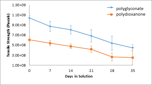
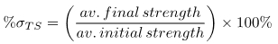
Figure 4: Acidic Solution: Suture Tensile Strength.

Figure 5
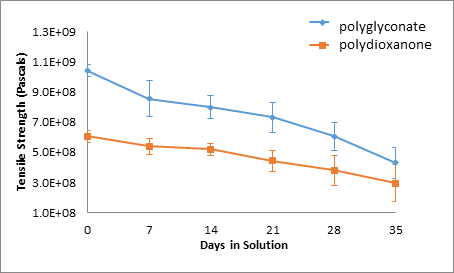
Figure 5: Neutral Solution, Suture Tensile Strength.

Figure 6
Figure 6: Alkaline Solution, Suture Tensile Strength.

Figure 7
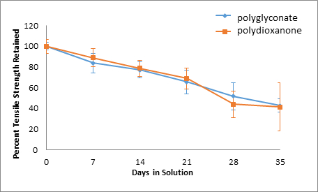
Figure 7: Acid Solution, Percent Tensile Strength Retained.

Figure 8
Figure 8: Neutral Solution, Percent Tensile Strength Retained.

Figure 9
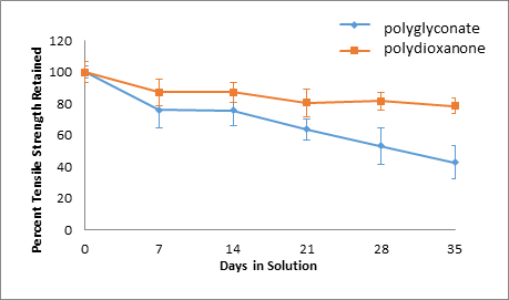
Figure 9: Basic Solution, Percent Tensile Strength Retained.

Control 7 days 14 days
Average pH Average pH Average pH
N/A Acid Neutral Base Acid Neutral Base
5 6 8 4 6 9
Force (N) Force (N) Force (N)
93.63 83.67 85.67 78.40 74.63 83.53 78.40
102.07 98.53 93.50 82.77 71.73 77.30 80.83
101.43 78.13 81.03 86.77 75.08 81.73 80.33
97.80 79.50 75.73 82.40 76.50 74.67 81.17
86.43 79.93 81.63 75.33 67.00 87.10 94.80
94.23 96.80 98.07 89.27 91.43 87.47
21 days 28 days 35 days
Average pH Average pH Average pH
Acid Neutral Base Acid Neutral Base Acid Neutral Base
4 6 9 4 6 8 4 6 8
Force (N) Force (N) Force (N)
56.53 58.70 85.97 51.53 58.57 73.22 36.37 38.77 74.67
60.73 65.33 75.80 49.70 51.43 72.20 24.20 34.83 67.70
58.27 63.53 69.23 56.87 72.20 83.20 36.30 42.37 73.27
64.93 66.83 81.60 40.63 28.40 72.90 21.60 36.83 74.63
68.57 63.90 81.90 29.70 58.70 80.93 42.00 40.97 75.67
75.20 76.17 61.63 20.83 69.47 83.33 31.37 45.33 81.77
85.63 94.17 85.00 36.37 78.13 76.73 87.53 90.77 81.83
60.33 75.83 80.47 52.33 66.67 85.83

Table 1: Overall 5-Week Polydioxanone Suture Data, Forces at Failure

Control 7 days 14 days
Average pH Average pH Average pH
N/A Acid Neutral Base Acid Neutral Base
4 6 9 4 6 9
Force (N) Force (N) Force (N)
170.80 131.37 147.03 146.23 122.07 117.87 135.17
170.93 147.70 142.60 152.63 129.30 132.13 129.87
167.70 134.00 153.80 120.13 107.93 113.13 101.57
162.37 112.90 102.87 111.07 139.63 120.47 111.20
156.70 153.20 124.63 103.80 123.80 131.47 129.57
152.87 145.90 123.33 143.57 146.13 144.57
21 days 28 days 35 days
Average pH Average pH Average pH
Acid Neutral Base Acid Neutral Base Acid Neutral Base
4 6 8 4 6 8 4 5 7
Force (N) Force (N) Force (N)
110.63 109.13 115.27 93.67 93.40 74.57 50.43 54.03 44.80
115.10 113.13 87.90 75.40 100.50 77.93 82.47 78.67 78.70
120.50 128.93 116.37 111.43 108.00 109.73 80.47 42.83 80.20
114.03 116.43 101.03 84.23 87.17 80.10 69.40 81.13 77.10
118.83 110.93 107.43 51.47 66.90 81.60 68.70 81.50 46.97
78.33 87.90 115.57 59.87 93.77 61.07 76.87 82.73 82.53
131.20 141.07 107.83 105.60 111.73 112.21 68.00 57.27 86.23
80.47 122.70 91.67 103.67 110.10 105.67

Table 2: Overall 5-Week Polyglyconate Suture Data, Forces at Failure

Over time, the tensile strengths of all suture specimens decreased. In addition, for polydioxanone sutures, an acidic environment was the most damaging as only 41.46% of the original tensile strength was retained, whereas 78.58% and 48.95% of the original tensile strength was retained for polydioxanone sutures in alkaline and neutral solutions, respectively. On the other hand, the strength retention percentages over time for polyglyconate sutures across different pH solutions were all similar. The greatest decrease in tensile strength for the polyglyconate sutures was observed in a neutral environment, where only 41.22% of the original strength was retained. In acidic and alkaline environments, 42.79% and 42.81% of original tensile strength was retained for the polyglyconate sutures, respectively.

If the sutures were incubated at a higher temperature, they would have degraded faster due to the increased inherent energy found within the system. This would allow for more spontaneous depolymerization into monomers to occur. In other words, as the temperature increases, the tensile strength is negatively affected. In addition, if the sutures were held at constant stress, chances of decay would also increase. This would be due to creep deformation; stretching the sutures creates weaker locales that are prime for absorption. If the sutures were to be tied into knots, a similar scenario would occur.

Applications and Summary

In this experiment, the tensile strength of sutures in different pH environments were evaluated. Over five weeks, the tensile strengths of two different types of sutures were explored after exposure to acidic, alkaline, and neutral solutions. The results overwhelmingly indicate that bioabsorbable sutures will degrade over time in any pH environment.

Although the polyglyconate sutures degrade at a faster rate, the remain stronger compared to the polydioxanone sutures. The experimental results also show that in prolonged time frames, polydioxanone sutures retain more of their strength than the polyglyconate sutures, as the faster degradation rate of polyglyocnate sutures becomes more evident. Nonetheless, since the experiment was performed in vitro, no substantial conclusions can be drawn for the effectiveness of either the polyglyconate or polydioxanone sutures in a more active biochemical model. Enzymatic degradation is a critical aspect that must be considered. Regardless, both sutures are viable candidates for surgical procedures. This study confirms the importance of this type of research.

Resorbable sutures provide temporary wound support, allowing the wound to heal well enough to withstand normal forces. Generally, resorbable sutures are used for internal procedures, so additional surgical procedures would be unnecessary for suture removal. Upon disintegration, little to no traces of the suture remain. Resorbable sutures are also used in patients that cannot return for a suture removal procedure. On the other hand, non-resorbable sutures are commonly used in epidermal would closure, where the sutures can be easily removed after a certain amount of time. Additionally, non-resorbable sutures are often used in stressful internal environments as well, when resorbable sutures are incapable of providing enough wound support. Internal structures such as the heart, which consistently withstands various pressures and movements, require non-resorbable sutures. Other applications of non-resorbable sutures include orthopedic surgeries and sternal closure in cardiac surgeries. Since resorbable sutures are used in internal and more critical parts of the body, it is important to test their strength and analyze the product quality.

Materials List

Name Company Catalog Number Comments
Equipment
Suture
Ruler
Scissors
Calipers
Tweezers
Scale
Tinius Olsen Tester
Oven
Sample Container
Beakers
Pipette
Pipette fillers
Pipette tube
Glassware
Chemicals
De-ionized water
Hydrochloric Acid (HCL)
Sodium Hydroxide (NaOH)

References

  1. Wise, Donald L., et al. Encyclopedic Handbook of Biomaterials and Bioengineering. Marcel Dekker, Inc., New York. 1995. 567-569.
  2. Dattilo, P.P., King, M.W., Cassill, N.L., et al. Medical Textiles: Application of an Absorbable Barbed Bi-directional Surgical Suture. J. Text. & App., Tech. & Mgmt. 2002, 2, 1

Transcription

Sutures have been used for thousands of years for medical intervention, with the earliest materials being linen or cat gut.

The sutures used today are now classified by two different categories, first by composition, either natural or synthetic materials, and by absorption, either non-resorbable or resorbable. Resorbable materials degrade in the body primarily through programmed degradation caused by the interaction of water with specific chemical groups in the polymer chain. Thus, these materials are used to hold a wound together for a timeframe long enough for healing without the need for removal.

In this video, we will discuss the mechanisms behind resorbable material degradation and demonstrate how to evaluate the change in strength of the materials over time as they are exposed to different environments.

Resorbable materials primarily degrade in the body by oxidative, hydrolytic, and enzymatic degradation. Materials may undergo oxidation in vivo as the body reacts to the foreign object and releases oxidative species to attack it. The oxidative effect on polymers can cause chain scission and contribute to degradation. In hydrolytic degradation, water attacks susceptible bonds in the polymer to generate oligomers and finally monomers.

Polyesters like polydioxanone are commonly utilized as resorbable materials because the ester group is easily degraded via hydrolysis. After the material is implanted, it begins to absorb water. Hydrolytic scission then begins wherever the material is in contact with water. Hydrophilic materials absorb more water, and therefore degrade more rapidly throughout. However, hydrophobic materials absorb water more slowly and tend to degrade from the outside in.

Enzymes in the body catalyze various reactions and thus, catalyze the hydrolytic degradation of materials as well. The hydrolysis reaction is catalyzed by enzymes called hydrolases which can increase the rate of hydrolytic degradation by as much as 10 times. As the material degrades, the mechanical properties of the material change as well.

Let’s take a look at how to analyze the change in strength of resorbable materials over time due to hydrolytic degradation in acidic, neutral, and alkaline environments.

For this experiment, obtain two types of resorbable sutures. Here, we use polyglyconate and polydioxanone.

Prepare six screw cap sample tubes each labeled with the date, sample type, and solution that the sample will be placed in. There should be one acidic, one alkaline, and one neutral solution for each sample type. Here, we show one of each sample. However, you should prepare three samples of each suture type for every time point.

Next, open the suture packaging and remove the suture. Cut the needle off the suture and dispose of it in the sharps container. Cut each suture into three pieces approximately 10 to 12 inches long. Make note of the physical characteristics of the suture. Use a caliper to measure the diameter of each suture and note the initial dimension.

Finally, weigh each suture, record the weight, and place one suture in each sample tube. Fill the neutral sample tubes with enough deionized water so that the suture is fully submerged, and cap the tube. Then, fill the acidic tubes with dilute hydrochloric acid and fill the alkaline sample tubes with dilute sodium hydroxide solution. Finally, place all six sample tubes in a rack in an incubator at 37 degrees Celsius.

Now let’s take a look at how to determine the strength of the sutures using tensile testing. The tensile test loads a sample by stretching it until failure, enabling the determination of material strength.

First, test fresh sutures that have not been incubating in test solutions. Place the suture in the fixture of the instrument and secure it in place. The control sample should be the same length as the instrument which is approximately 10 to 12 inches. Next, zero the instrument and record the displacement speed setting. Ensure that peak hold is displayed on the control panel. Then initiate tension on the suture. The force and displacement will start to change on the instrument. Load the suture until failure. Then, turn off the instrument and record the peak force from the display panel.

Now let’s measure the tensile strength of the samples that had been exposed to solutions at varying pH.

After the specified amount of time, remove the samples from the oven. Measure the pH of the solution in each tube using pH paper. After the pH of all solutions have been measured, remove the suture to be tested and rinse it with deionized water. Make note of the physical characteristics of the material.

Pat the sample dry with a paper towel, then weigh it and record the new mass. Next, place the specimen in the grips of the tensile tester and lock it into place. Zero the instrument and make sure the displacement speed is the same as used for the control sample. Also check that peak hold is displayed. Now, load the specimen until failure. Record the peak force from the display. Repeat the tensile test for each sample over the course of the time study.

Now let’s see how to analyze the data to determine the strength of the samples.

First, calculate the average tensile stress of each sample by dividing the peak force by the cross-sectional area of the suture. Then, calculate the percent tensile strength retained by the suture after incubation using the formula shown. A plot of tensile strength over time for each sample shows that the strength of both types of sutures decreased over time in acidic, neutral, and alkaline solutions.

The polydioxanone structures degraded more in the acidic solution, with only 41% of the original tensile strength retained after five weeks, while 49 and 78% of strength was retained for the neutral and alkaline solutions, respectively. The polyglyconate sutures degraded similarly in all three solutions retaining around 42% of strength in acidic, neutral, and alkaline solutions after five weeks. The results are expected as the materials both possess ester bonds that are susceptible to hydrolytic scission, which is enhanced at high and low pH.

Now let’s take a look at where resorbable materials are used in the biomedical engineering field.

Resorbable materials like the sutures tested in this video are most commonly used in surgical procedures to enable the healing of surgical sites while eliminating the need for suture removal. However, resorbable materials also play a role in tissue engineering as the scaffold for engineered tissue. Resorbable tissue scaffolds provide the initial three-dimensional structure for tissue, but degrade slowly as the cells grow and create their own structural material. Eventually, the initial scaffold is no longer needed and the engineered tissue more closely resembles native tissue.

Bone grafting involves replacing missing or damaged bone in order to help large fractures heal. In this study, researchers created a defect in the skull by drilling a five-millimeter hole. The bone fragment was detached and the bone graft attached to the bone using fibrin glue. Although donor bone is often used, resorbable materials present an alternative enabling the graft to degrade away as native bone grows.

You’ve just watched JoVE’s introduction to resorbable materials. You should now understand how these materials degrade in vivo and in vitro, how to test for strength changes due to degradation, and some applications of these materials in the biomedical engineering field. Thanks for watching!

Tags

Cite This
JoVE Science Education Database. JoVE Science Education. Tensile Strength of Resorbable Biomaterials. JoVE, Cambridge, MA, (2023).