We present a protocol to obtain cell-derived matrices rich in extracellular matrix proteins, using macromolecular crowders (MMC). In addition, we present a protocol which incorporates MMC in 3D organotypic skin co-culture generation, which reduces culture time while maintaining maturity of construct.
The glycoprotein family of collagens represents the main structural proteins in the human body, and are key components of biomaterials used in modern tissue engineering. A technical bottleneck is the deposition of collagen in vitro, as it is notoriously slow, resulting in sub-optimal formation of connective tissue and subsequent tissue cohesion, particularly in skin models. Here, we describe a method which involves the addition of differentially-sized sucrose co-polymers to skin cultures to generate macromolecular crowding (MMC), which results in a dramatic enhancement of collagen deposition. Particularly, dermal fibroblasts deposited a significant amount of collagen I/IV/VII and fibronectin under MMC in comparison to controls.
The protocol also describes a method to decellularize crowded cell layers, exposing significant amounts of extracellular matrix (ECM) which were retained on the culture surface as evidenced by immunocytochemistry. Total matrix mass and distribution pattern was studied using interference reflection microscopy. Interestingly, fibroblasts, keratinocytes and co-cultures produced cell-derived matrices (CDM) of varying composition and morphology. CDM could be used as "bio-scaffolds" for secondary cell seeding, where the current use of coatings or scaffolds, typically from xenogenic animal sources, can be avoided, thus moving towards more clinically relevant applications.
In addition, this protocol describes the application of MMC during the submerged phase of a 3D-organotypic skin co-culture model which was sufficient to enhance ECM deposition in the dermo-epidermal junction (DEJ), in particular, collagen VII, the major component of anchoring fibrils. Electron microscopy confirmed the presence of anchoring fibrils in cultures developed with MMC, as compared to controls. This is significant as anchoring fibrils tether the dermis to the epidermis, hence, having a pre-formed mature DEJ may benefit skin graft recipients in terms of graft stability and overall wound healing. Furthermore, culture time was condensed from 5 weeks to 3 weeks to obtain a mature construct, when using MMC, reducing costs.
The skin forms a protective barrier by preventing water loss and pathogen entry. It is made up of three major components; the stromal rich dermis1, a stratified epidermis2,3,4 on top of it, and the dermo-epidermal junction in between5,6. The dermis is composed largely of collagen and elastic fibers and is sparsely populated with fibroblasts7. In contrast, the cell-rich epidermis is composed of multiple layers of keratinocytes. The keratinocytes of the inner-most layer are proliferative and provide new basal cells which renew and replace terminally differentiating keratinocytes that constantly move to the outer-most layer of the skin and have lost their nuclei and cytoplasmic material, resulting in a cornified layer which undergoes desquamation.
The dermal-epidermal junction, a specific type of basement membrane, is a complex structure composed of interconnecting matrix molecules, which tethers the epidermis to the dermis. Collagen I fibers of the dermis interlace with collagen VII anchoring fibrils which are anchored to the collagen IV rich lamina densa. Anchoring filaments (laminin 5, collagen XVII and integrins) in turn connect the lamina densa with hemidesmosomes of basal keratinocytes. Basal keratinocytes (stratum basale) have the capacity to proliferate and renew, as well as differentiate and stratify to form the suprabasal layers; stratum spinosum, stratum granulosum, and finally the stratum corneum, which represents the contact surface of the skin with the environment. En route from the basal layer to the cornified layer, keratinocytes switch expression patterns of cytokeratins and finally undergo apoptosis and encase themselves in cornified envelopes, hulls of specific proteins that are covalently crosslinked by transglutaminase activity.
Recreating skin and its layers in vitro, including the complex structures of the dermal-epidermal junction and dermal extracellular matrix, and to emulate the cornification process, has long intrigued scientists and bioengineers as a challenging task. There have been significant advances in skin tissue engineering, for example, the successful extraction of skin cells from patient biopsies and the generation of skin organotypic cultures using patient-derived skin cells8. However, unsolved problems remain pertaining to poor secretion of extracellular matrix proteins by the skin cells themselves and resulting in sub-optimal skin models. Furthermore, the time required to generate 3D organotypic skin co-culture using current protocols varies between four to eight weeks, a timeframe which could potentially be shortened with the incorporation of macromolecular crowders. Reducing culture time saves reagent cost, reduces the incidence of cell senescence and reduces the waiting time of the patient should the product be used in the clinic.
Macromolecular crowding (MMC) involves introducing specific macromolecules to the culture medium to generate excluded volume effects. These affect enzymatic reaction rates including the proteolytic cleavage of procollagen which is tardy under standard aqueous culture conditions9-13. Under MMC, enzymatic reactions are hastened without increasing the amount of reagents14,15 resulting in, in the case of procollagen cleavage, an increased amount of collagen I molecules in crowded cultures as compared to uncrowded controls10. As the conversion of procollagen to collagen allows the formation of collagen assemblies, fibroblasts cultured with MMC for 48 hours yielded significantly more collagen I as compared to uncrowded fibroblast cultures monitored for up to four weeks11,16,17. Besides effects on enzymatic activities that affect the formation, stabilization and remodeling of ECM, MMC also has been shown to directly enhance and modulate collagen fiber formation18,19.
We present here a method to enhance the extracellular matrix (ECM) production by skin cells, in particular, dermal fibroblasts and epidermal keratinocytes. In addition, we show that the enriched ECM produced under MMC in monolayer cultures can be decellularized and used as pure cell-derived matrix (CDM).
We use a non-conventional approach to visualize and fully appreciate the ECM deposited by skin cells cultured with MMC. Interference reflection microscopy is typically used for studying cell-matrix interactions or cell-to-glass contact points. This technique was utilized in our system to view the total amount of matrix deposited on the glass surface. Interference reflection microscopy was coupled with fluorescent immunostaining to obtain the most amount of information in terms of extracellular matrix composition and pattern, in the presence and absence of MMC.
Organotypic skin co-cultures is a classical method to model the skin in vitro in a three-dimensional context. While two-dimensional co-cultures may provide significant information, it is limited when translating this data and applying it back to an in vivo environment, which is inherently a three-dimensional structure. Skin keratinocytes, in particular, are polarized and contain apical and basal segments which are essential for homeostasis and cell adherence. In addition, the expression of typical suprabasal proteins in keratinocytes above the basal layer, such as keratin 1, keratin 10 and filaggrin is only present in the course of stratification and terminal differentiation of keratinocytes. As terminal differentiation is hardly present in typical monolayer cultures, suprabasal protein expression is normally not achieved in this culture system. Therefore, organotypic cultures start submerged in culture medium, but are then lifted to an air-liquid interface to drive keratinocyte differentiation. This results in the expression of stratification markers, even cornification and a generally better reflection of epidermal physiology. While other groups have previously generated organotypic skin co-cultures successfully, the establishment of a functional dermo-epidermal junction zone has been an issue. Here, we present a new method for culturing organotypic skin co-cultures with an enhanced basement membrane, in a condensed time frame and without compromising the maturity of these constructs. This would provide skin mimetics for in vitro modeling, the study of skin biology and an assortment of screening assays.
1. Macromolecular Crowding in 2D Skin Cell Cultures
2. Fluorescent Immunostaining on Skin Cell Cultures
3. Western Blotting of Skin Cell Cultures
4. Making and Using a Cell-derived Matrix
5. Characterization of the Cell-derived Matrix
6. 3D Organotypic Skin Co-culture
7. Harvest and Characterization of Organotypic Skin Equivalents
Macromolecular crowding was able to enhance ECM deposition, in particular, fibroblasts deposited more collagen I, IV and fibronectin as compared to control cultures (Figure 1, Cell layer; 1A, collagen I; 1B, collagen IV; 1C, fibronectin). Upon decellularization, it was evident that fibroblasts were the main depositors of collagen I, IV and fibronectin as compared to keratinocytes (Figure 1, Matrix).
In Figure 2, it was observed that fibroblasts rather than keratinocytes were the main producers of collagen VII. This is the first report of collagen VII deposited successfully in vitro (Benny et al, 2015).
The cell-derived matrix was characterized through immunofluorescence using specific antibodies. While this is a classic approach, it was important to visualize the ECM in its totality and fully appreciate the effect of the macromolecular crowders. Using interference reflection microscopy (IRM), the full extent of the matrix was captured (Figure 3). An overlay of the antibody staining with the IRM image shows the relative quantity of that ECM component in relation to the total amount of ECM.
In a 3D in vitro skin model, MMC condensed the culture time from 5 weeks to 3 weeks to obtain a mature organotypic skin co-culture (Figure 4). A hematoxylin and eosin staining showed that at 3 weeks, the culture with macromolecular crowders consisted of a pluri-stratified epidermis and stromal rich dermis, as compared to uncrowded control cultures which lacked a completely differentiated epidermis. In addition, intense and continuous collagen VII immunostaining was detected at the dermal-epidermal junction of crowded cell cultures, as compared to a weak and spotty collagen VII immunostaining in control cultures. Transmission electron microscopy (Figure5) showed the presence of anchoring fibrils in organotypic cultures, showing functional collagen VII.
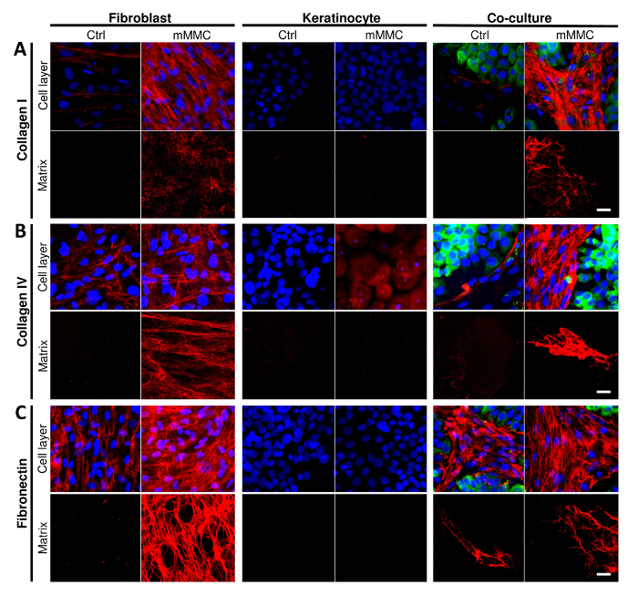
Figure 1: Mixed macromolecular crowding (mMMC) enhances the deposition of dermal-epidermal junction components in vitro. (A) Collagen I deposition is enhanced by mMMC (cell layer and matrix) in fibroblasts only. Crowding of co-cultures produce the most collagen I and show that keratinocytes stimulated collagen I production by fibroblasts. (B) Collagen IV deposition by fibroblasts is enhanced by crowding. This is seen even more clearly in crowded co-cultures. Of note, keratinocytes stained for collagen IV show mostly cell-associated or intracellular collagen IV, but not a pericellular matrix. In co-cultures, both cell types segregate with collagen IV being predominantly associated with fibroblasts sparing keratinocyte islands. (C) Fibronectin deposition was only seen with fibroblasts, and therein strongly enhanced by crowding (cell layer and matrix). In co-cultures, a reticular mesh of fibronectin was associated with fibroblasts only, sparing islands of keratinocytes. Scale bars = 20 µm. This figure has been modified from Benny et al., 2015. Please click here to view a larger version of this figure.
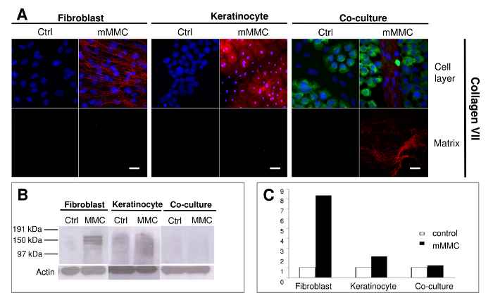
Figure 2: Mixed macromolecular crowding (mMMC) facilitates the deposition of anchoring fibril building collagen VII. (A) A reticular deposition pattern of collagen VII deposition is evident with fibroblasts only under mMMC. In co-cultures, extracellular collagen VII is strongly associated with fibroblast colonies in between keratinocyte islands. Keratinocytes show pericellular and intracellular collagen VII more strongly expressed in the presence of mMMC. After cell lysis, collagen VII footprints are seen in a fine granular layer in mMMC-treated fibroblast cultures, but a discernible fibrillar deposition is retrieved from co-cultures. (B) Immunoblot analysis of lysed cell layers shows that both crowded fibroblasts and keratinocyte cultures contain significantly more collagen VII compared with uncrowded controls. The retrieved collagen VII is mainly pericellular derived. (C) Densitometric analysis of B shows that mMMC increases the amount of cell-associated collagen VII by a factor of 8 in fibroblasts and a factor of 2 in keratinocytes. Scale bars = 20 µm. This figure has been modified from Benny et al., 2015. Please click here to view a larger version of this figure.
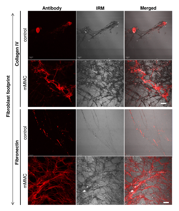
Figure 3: Fibroblast footprints contain more total ECM as visualized by interference reflection microscopy (IRM). On analysis of individual ECM components (collagen IV and fibronectin), culture under mMMC enhanced the extracellular deposition. To visualize the total ECM deposited, IRM was used to quantify all ECM as antibody staining had its limitations. IRM clearly showed the total matrix quantity and pattern under mMMC as compared with control conditions. Scale bar = 20 µm. This figure has been modified from Benny et al., 2015. Please click here to view a larger version of this figure.
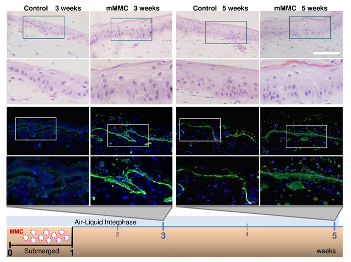
Figure 4: Mixed macromolecular crowding (mMMC) during the submerged phase enhances maturation of the dermal-epidermal junction (DEJ) in skin equivalents. Fibroblast-containing collagen gels were seeded with keratinocytes on top and kept submerged for 1 week, then lifted to an air-liquid interface. In the classical protocol, collagen VII (green) was absent after a total of 3 weeks in culture, but appeared in skin equivalents after 5 weeks. In contrast, under mMMC, collagen VII was already strongly evident after 3 weeks and even more strongly stained after 5 weeks compared with standard cultures. Hematoxylin and eosin (20X magnification) staining confirmed that with this rapid protocol, stratification and maturity of the skin equivalent were maintained and accelerated. Scale bar = 100 µm. This figure has been modified from Benny et al., 2015. Please click here to view a larger version of this figure.
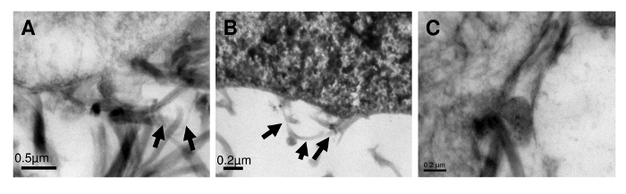
Figure 5: Evidence of de novo formation of anchoring fibrils in skin equivalents generated under mMMC. Ultrastructural studies of the nascent dermal-epidermal junction of organotypic co-cultures after a 3 week culture protocol with mMMC (A, B) suggests structures akin to anchoring fibrils (arrows) that are absent in non-crowded skin equivalents (C) after 3 weeks of culture. This figure has been modified from Benny et al., 2015. Please click here to view a larger version of this figure.
Enhanced extracellular matrix is obtained upon introduction of macromolecular crowders to cell culture, owing to the excluded volume effect which increases the propeptide cleavage by proteinases. This results in extracellular matrix, in particular collagen, to be processed faster and deposited on the culture surface. While other groups have obtained thick fibroblast cell layers, it involved a culture time of several weeks20. In contrast, MMC described in this Protocol, dramatically shortens the culture time while increasing the amount of ECM. The increase in reaction rates in crowded cell cultures has also been reported in chondrocyte matrix deposition21, extracellular matrix organization22,23 and adipogenesis24.
After decellularization of crowded cell layers, cell-derived matrices are harvested and used for various tissue engineering applications, one of which is the seeding of secondary cell cultures on top of the cell-derived matrix25,26. As the matrices are cell-derived and natural products, degradation might occur if kept at non-ideal conditions for extended periods of time. A significant benefit of the cell-derived matrix, obtained under MMC, is that it is cell-derived and cell-specific. This could be a potential alternative to commercially available coatings which often are expensive and from animal sources. The skin equivalents must be handled with care, especially prior to fixation.
The use of interference reflection microscopy (IRM) in this Protocol shows, in much greater detail, the cell-derived matrix obtained with MMC. As compared with conventional fluorescent immunocytochemistry, only two or three proteins can be viewed at a time. With IRM, the total matrix can be visualized, with minimal manipulation to the sample. When immunocytochemistry was merged with the IRM field, it provided important information as to the ECM protein deposited in relation to the total amount of ECM. For example, it was observed that fibroblasts produced most ECM components, but a fibroblast-derived matrix was largely composed of fibronectin.
Fibroblasts were also found to be the main producers of collagen VII, a feat which could be enhanced by MMC in vitro. This Protocol could have implications in therapies pertaining to the skin disorder, recessive dystrophic epidermolysis bullosa, where there is defective collagen VII.
Finally, the application of MMC in a 3D model to generate organotypic skin co-cultures, provides a useful tool in the advancement of skin research. Since MMC allows for the generation of skin equivalents with complete stratification, a mature dermal-epidermal junction delivered in a condensed time frame, this would be largely beneficial in the modeling of skin diseases, the study of skin biology in vitro, as well as the production of skin grafts for burn and chronic wounds.
The authors have nothing to disclose.
This work was supported by the Biomedical Research Council, Singapore through core support to the Institute of Medical Biology and grant SPF2013/005. M.R. was supported by a NUS Faculty Research Committee Grant (Engineering in Medicine) (M.R.) R-397-000-081-112, and the NUS Tissue Engineering Program (NUSTEP). The authors would like to thank Professor Irene Leigh for providing the collagen VII antibody. Electron microscopy work was carried out at the National University of Singapore Electron Microscopy Unit.
Ascorbic acid | Wako | 013-12061 | Cell culture media additive |
Cell culture inserts (6-well format) | Greiner Bio-One | 657610 | Organotypic cultures |
Citrate solution | Dako | S2369 | DakoCytomation Target Retrieval Solution Citrate, pH 6 (x10) |
Collagen (rat tail) | Corning | 354236 | Organotypic cultures |
CnT-57 | CELLnTEC | CnT-57 | Cell culture media |
DAB Substrate + chromogen kit | Dako | K3468 | Histology |
Deep-well plate (6-well format) | Corning | 355467 | Organotypic cultures |
ECL detection reagent | GE Healthcare Life Sciences | RPN2106 | Amersham ECL Western Blotting Detection Reagent |
Electron microscope (TEM) | JEOL | JEM-1010 (100kV) | Transmission electron microscopy |
Fibroblast media (FM) | High Glucose-DMEM + 10% Fetal Bovine Serum + 1% Penicillin Streptomycin | ||
Ficoll PM70 | GE Healthcare Life Sciences | 17-0310-05 | Macromolecular crowder |
Ficoll PM400 | GE Healthcare Life Sciences | 17-0300-05 | Macromolecular crowder |
Keratinocyte serum-free media (KSFM) | Life Technologies | 17005-042 | Cell culture media |
Lysis buffer | ThermoFisher Scientific | 89900 | Lysis buffer for protein extraction. A protease inhibitor (Roche, #11836170001) was added to this lysis buffer. |
Microscope | Zeiss | LSM510 | Red, green and blue channels to visualize AF594, AF488 and DAPI fluorescent immunostainings (40X mag). |
Mounting media (Hydromount) | National Diagnostics | HS-106 | Fluorescent staining |
Mounting media (Cytoseal) | ThermoFisher Scientific | 8310-16 | Histology (HRP) |
OCT compound (Tissue Tek) | Sakura | 4583 | Embedding for cryotomy |
Penicillin-streptomycin antibiotics | Sigma Aldrich | A5955 | Cell culture media additive |
Primary antibodies | |||
Anti-Collagen I antibody | Abcam | #ab6308 | |
Anti-Collagen Type IV | Novocastra | #NCL-COLL-IV | |
Anti-collagen VII | LH7.2 (in house) | ||
Anti-Fibronectin antibody | Abcam | #ab2413 | |
Reducing agent | ThermoFisher Scientific | NP0009 | Western blot |
Sample buffer | ThermoFisher Scientific | NP0008 | Western blot |
Secondary antibodies (Immunostaining) | |||
4′,6-Diamidino-2-phenylindole dihydrochloride (DAPI) | Sigma Aldrich | #D9542 | |
AlexaFluor 594 goat-anti-rabbit | ThermoFisher Scientific | #A-11037 | |
AlexaFluor 594 goat-anti-mouse | ThermoFisher Scientific | #A-11005 | |
AlexaFluor 488 chicken-anti-rabbit | ThermoFisher Scientific | #A-21441 | |
AlexaFluor 488 goat-anti-mouse | ThermoFisher Scientific | #A-11001 | |
Secondary antibodies (HRP) | Dako | Envision + System – HRP Labelled Polymer Anti-Rabbit and Anti-Mouse | undiluted |
Sodium deoxycholate | Prodotti Chimicie Alimentari | Decellularization | |
Stratification media | |||
Dulbecco's Modified Eagle's Medium | Used during the air-liquid interface of organotypic cultures. This media is added to the outside of the cell-culture insert. | ||
Ham’s F12 | Used during the air-liquid interface of organotypic cultures. This media is added to the outside of the cell-culture insert. | ||
10% fetal bovine serum | Used during the air-liquid interface of organotypic cultures. This media is added to the outside of the cell-culture insert. | ||
100U/mL penicillin and 100U/mL streptomycin | Used during the air-liquid interface of organotypic cultures. This media is added to the outside of the cell-culture insert. | ||
0.4mg/mL hydrocortisone | Used during the air-liquid interface of organotypic cultures. This media is added to the outside of the cell-culture insert. | ||
5mg/mL insulin | Used during the air-liquid interface of organotypic cultures. This media is added to the outside of the cell-culture insert. | ||
1.8·10-4 M adenine | Used during the air-liquid interface of organotypic cultures. This media is added to the outside of the cell-culture insert. | ||
5mg/mL transferrin | Used during the air-liquid interface of organotypic cultures. This media is added to the outside of the cell-culture insert. | ||
2·10- 11 M triiodothyronine | Used during the air-liquid interface of organotypic cultures. This media is added to the outside of the cell-culture insert. | ||
Trypsin | Biopolis Shared Facilities | 0.125% Trypsin/Versene pH 7.0 + 0.3 | Used to trypsinize cells |