We describe a method to collect quantifiable hemolymph efficiently from small arthropods for subsequent analysis.
Arthropods are known to transmit a variety of viruses of medical and agricultural importance through their hemolymph, which is essential for virus transmission. Hemolymph collection is the basic technology for studying virus-vector interactions. Here, we describe a novel and simple method for the quantitative collection of hemolymph from small arthropods using Laodelphax striatellus (the small brown planthopper, SBPH) as a research model, as this arthropod is the main vector of rice stripe virus (RSV). In this protocol, the process begins by gently pinching off one leg of the frozen arthropod with fine-tipped tweezers and pressing the hemolymph out of the wound. Then, a simple micropipette consisting of a capillary and a pipette bulb is used to collect the transudative hemolymph from the wound according to the principle of capillary forces. Finally, the collected hemolymph can be dissolved into a specific buffer for further study. This new method for collecting hemolymph from small arthropods is a useful and efficient tool for further research on arboviruses and vector-virus interactions.
Both animal and plant viruses can be transmitted by arthropods, and these viruses pose a severe threat to human health and cause tremendous economic losses in agriculture1,2,3. Importantly, the arthropod hemolymph, which serves as the circulatory system and a vital element of the immune system in arthropods, plays an important role in regulating arboviral transmission. Viruses acquired through the arthropod guts are transported to other tissues only after successfully escaping the adverse hemolymph environment4,5,6,7. The lifecycle of viruses in the arthropod hemolymph involves virus survival in the fluid plasma, entry into the hemocyte, and transport to other tissues, and various virus-vector interaction mechanisms occur in the hemolymph8,9,10,11,12. For example, the vertical transmission of RSV by the SBPH is dependent on a molecular interaction between the SBPH vitellogenin protein and the RSV (rice stripe virus) capsid protein13,14. Some viruses may escape the immune response of the hemolymph by binding specific vector factors15,16,17,18. Therefore, investigating vector-virus interactions in the hemolymph of arthropods is important for developing a better understanding of arbovirus transmission.
The hemolymph of some small insects, such as planthoppers, leafhoppers, and some mosquitoes, is difficult to collect due to their size. To address this issue, several methods have been developed to collect hemolymph, including inserting a syringe needle directly into the insect body to extract a microvolume of the hemolymph, collecting exudate from the wound site with fine-tipped tweezers, and direct centrifugation. These methods have enabled the measurement of relative gene expression levels and viral titers within the hemolymph19,20,21. However, an effective method for quantifying the hemolymph volume, which is necessary for hemocyte counting, protein quantification, and enzyme activity analysis, is currently not available for these small insects.
The SBPH (small brown planthopper) is a type of small insect vector with a body length of about 2-4 mm. The SBPH is capable of transmitting a variety of plant viruses, including RSV, maize rough dwarf virus, and rice black streaked dwarf virus22,23,24. The interaction between the SBPH and RSV has been studied in depth over the past decade. To facilitate working with SBPHs, we developed a novel and simple method of collecting hemolymph. This method, which is based on the principle of capillary forces, uses a capillary with a scale mark to acquire the insect's hemolymph in a precise and quantifiable manner. This allows us to collect a specific volume of hemolymph from small insects efficiently and to study the hemolymph environment of small vectors in more detail.
1. Insect rearing
2. Dissection of the SBPHs for hemolymph collection
3. Hemolymph collection using micropipettes
4. Coomassie Blue staining
5. Protein concentration determination
6. Microscopic detections
7. Cell quantification
8. Statistical analyses
Micropipette model and hemolymph collection
We have developed a simple micropipette whose action is based on the capillary forces of the capillary tube. The micropipette is composed of a capillary tube and a pipette bulb (Figure 1A). Capillary tubes are available in different volume sizes ranging from 1 µL to 20 µL, and the capillary tube volumes are selected according to the requirements. Capillary tubes with smaller volumes are not suggested because the extra fine apertures of smaller-volume tubes can make it difficult to absorb liquid such as hemolymph. The pipette bulb contains a hole on the top that cannot be plugged during the hemolymph collection. This pipette bulb is convenient for holding the micropipette during the liquid collection (Figure 1B) and also assists in transferring the collected liquid from the capillary tube into the collection buffer.
In order to collect hemolymph easily, in this work, the SBPHs were firstly frozen in ice or in the refrigerator. These frozen SBPHs were then localized on a slide under a stereomicroscope, and one of the legs of each SBPH was pulled off with fine-tipped tweezers (Figure 1C). To ensure a large wound and optimal hemolymph collection, it is best to pull off the leg at the root (Figure 1Ca, b). In order to minimize the risk of contamination by the fat body, only clear liquid drops without any white floccule were collected (Figure 1Cc). The micropipette was used to absorb the desired volumes of hemolymph (Figure 1Cd). To collect 1 µL of hemolymph, approximately 30-40 larval SBPHs or 8-15 adult SPBHs had to be dissected.
Analysis of the collected hemolymph
To assess the accuracy of the micropipette as a method for evaluating the volume of collected hemolymph, we tested the protein concentrations of different samples. Hemolymph from larvae was collected using a micropipette with a capillary volume of 1 µL, and three protein samples were collected separately and tested by running an SDS-PAGE gel. The results showed that the amount of protein in the three lanes was nearly equal (Figure 2A). For larvae, the total protein content was 3.707 mg/mL ± 1.382 mg/mL. We also collected the same volume of hemolymph from adult female and male SBPHs, and these showed protein concentrations of 3.515 mg/mL ± 1.400 mg/mL and 3.621 mg/mL ± 0.860 mg/mL, respectively (Table 1). There were no significant differences in the protein among the three samples (Figure 2B).
In addition, we also observed the hemocytes inside the collected larval hemolymph to assess the quality and purity of the hemolymph samples. The hemocytes varied in size between 2-20 nm, and no fat body was detected (Figure 2C). The majority of the cells identified were plasmatocytes, ranging from 5-15 nm in diameter and often appeared in aggregates25. We then counted the cell concentrations of the hemolymph from larvae, adult females, and adult males, and the cell concentrations identified were 1.794 x 10⁵/µL ± 0.614 x 10⁵/µL, 1.256 x 10⁵/µL ± 0.603 x 10⁵/µL, and 1.553 x 10⁵/µL ± 0.474 x 10⁵/µL, respectively (Table 2). The hemocyte cell count in the larvae, adult females, and adult males showed no significant differences (Figure 2D). These results indicate that the micropipette collection method is a reliable and accurate way to collect hemolymph from SBPHs.
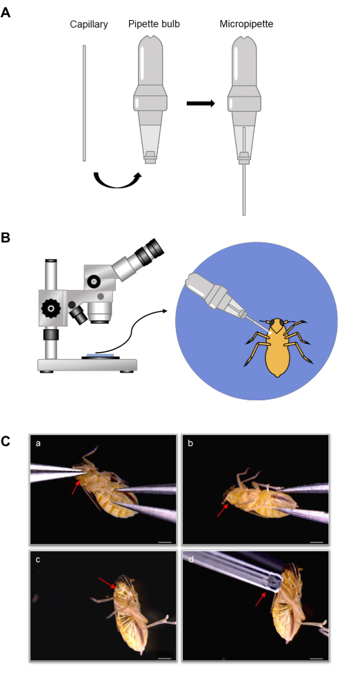
Figure 1: Schematic of the micropipette model and hemolymph collection. (A) Micropipette composition. The micropipette consists of a capillary and a pipette bulb. The capillary volumes range from 1-20 µL. The pipette bulb has a small hole on the top and the bottom. The capillary is inserted into the bottom hole, and the scale is visible. (B) Overview diagram of hemolymph collection with a micropipette. The insect abdomen is kept facing up while a leg is detached, hemolymph outflow is induced, and the hemolymph is collected into the pipette under a stereomicroscope. (C) Process of hemolymph collection with a micropipette. One of the legs is pulled off at the root with fine-tipped tweezers (a), and the insect's body is pressed to make hemolymph flow out (b,c). The hemolymph from the wound is collected into the capillary (d). Scale bar = 500 µm. Please click here to view a larger version of this figure.
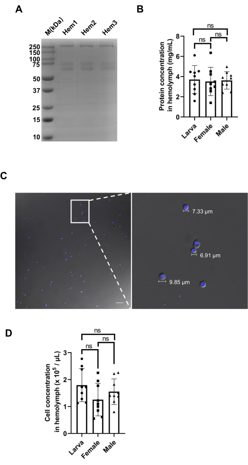
Figure 2: Analysis of the SBPH hemolymph. (A) Coomassie Blue staining showing three replicates of collected larval hemolymph. Hem indicates hemolymph. (B) Total protein concentrations of the hemolymph of larval, female, and male SBPHs. (C) Microscopic images showing the cells present in the hemolymph in SBPHs at 20x and 60x magnification, respectively. The nucleus was stained with DAPI (blue). Scale bar = 50 µm. (D) Hemocyte density of larval, female, and male SBPHs. The mean and SD were calculated from three independent experiments. Please click here to view a larger version of this figure.
Hemolymph | Protein concentration (mg/mL) |
Larva | 3.707±1.382 |
Female | 3.515±1.400 |
Male | 3.621±0.860 |
Table 1: Protein concentrations of hemolymph from different SBPHs. The data were obtained from three biological replicates.
Hemolymph | Cell concentration (105 / μL) |
Larva | 1.794±0.614 |
Female | 1.256±0.603 |
Male | 1.553±0.474 |
Table 2: The total concentrations of hemocytes in the hemolymph from different SBPHs. The data were obtained from three biological replicates.
Supplementary Figure 1: Determining the number of cells. The four corner squares (1, 2, 3, and 4) and the central square (5) are counted on the cell counting chamber. For border cells, only the two boundaries (top and left) are counted. Please click here to download this File.
Hemolymph is the medium of the circulatory system in arthropods, and arboviruses can only invade other arthropod tissues if they are able to survive the hostile hemolymph environment. Collecting a high-quality sample of hemolymph is the first step in studying the vector-virus interactions that occur in the hemolymph. It has been reported that insect hemolymph can be obtained from several sites on the insect's body, including a wound on the front leg, a minor incision in the head area, or a tear wound at the abdomen26,27,28,29. Different collection methods may work for certain acquisitions. The method involving creating a small incision at the head-neck joint is more suitable for hemolymph collection from large insects such as honeybees26. Creating a wound in the head area of the SBPH is challenging as it may damage other organs and introduce contamination from other tissues. After making a tear wound at the abdomen, the insects are immersed in the buffer, and then the buffer is centrifuged to extract the hemolymph. This is a convenient method to collect lipids from small insects21. However, as there is a high amount of fat body within the hemocoel of SBPHs, a wound on the body part may cause fat body leakage from the wound and lead to the contamination of the hemolymph. This method is more suitable for collecting hemolymph from adult SBPHs, which have less fat body than larvae. To effectively prevent fat body contamination, it is recommended to make a wound at the leg site rather than directly on the body. In previous studies of SBPH hemolymph, fine-tipped tweezers were used to facilitate the collection of small drops of liquid from the wound13. Although the collected hemolymph was clear from impurities, the volume of hemolymph was not measured. In this study, we developed a simple micropipette that can be used to accurately and quantitatively collect hemolymph from very small insect vectors.
For successful hemolymph collection, several critical steps must be taken into account. Firstly, it is essential to anesthetize the insects at 4 °C for 15 min in order to produce hemolymph that is free of fat body. Secondly, it is simpler to obtain pure hemolymph by pulling off the first leg. Thirdly, the collection of the hemolymph should be done in a continuous and swift manner, as otherwise the hemolymph may coagulate at the bottom and block the capillary.
The capillary collection technique has been widely used for animal blood collection30,31. We modified the technique to suit the distinct characteristics of insect hemolymph. As SBPHs possess a small body size, a capillary with a minimal volume of 1 µL was chosen. Using a capillary with a volume lower than 1 µL is not recommended. It is noteworthy that hemolymph contains a high density of proteins and a considerable number of cells (Figure 2), which elevate its liquid density and make it difficult to absorb hemolymph via capillaries with smaller volumes.
The method of collecting hemolymph by capillary is a simple, cost-effective, and viable technique that enables the reliable and precise quantification of the hemolymph. This newly developed method could also be applied for collection of other trace fluids from small vectors, such as honeydew. It is vital to highlight that the insects remain alive after the extraction of hemolymph using this method. This is beneficial for studies involving other insect organs or for repeated hemolymph collection. This work presents a valuable technology for the study of entomology and virus-vector interactions.
The authors have nothing to disclose.
This work was supported by the National Key R&D Program of China (No. 2022YFD1401700) and by the National Science Foundation of China (No. 32090013 and No. 32072385).
10% SDS-PAGE protein gel | Bio-rad | 4561035 | Protein separation and detection |
4% paraformaldehyde | Solarbio | P1110 | For fixation of the cells or tissues |
Bradford dye reagent | Bio-rad | 5000205 | Protein concentration detection |
Capillary | Hirschmann | 9000101 | For collecting hemolymph |
Cell counting chamber | ACMEC | AYA0810 | Hemocytes counting |
Glass slide | Gitoglas | 10127105A | For holding insects |
Glass slide coated with silane | Sigma | S4651-72EA | For holding microscope samples |
Gold antifade reagent with DAPI | Invitrogen | P36935 | Nucleus staining |
Microscope cover glass | Gitoglas | 10212424C | For microscopic observation |
Pipette bulb | Hirschmann | 9000101 | For collecting hemolymph |
Prism 8.0 software | GraphPad Software | / | Statistical analyses |
Stereomicroscope | Motic | SMZ-168 | For insect dissection |
Tweezers | Tianld | P5622 | For insect dissection |
Zeiss inverted microscope | Zeiss | Observer Z1 | Hemocytes observation |