Dual camera emission splitting systems for two-color fluorescence microscopy generate real-time image sequences with exceptional optical and temporal resolution, a requirement of certain live cell assays including parallel plate flow chamber adhesion assays. When software is employed to merge images from simultaneously acquired emission channels, pseudocolored image sequences are produced.
Multi-color immunofluorescence microscopy to detect specific molecules in the cell membrane can be coupled with parallel plate flow chamber assays to investigate mechanisms governing cell adhesion under dynamic flow conditions. For instance, cancer cells labeled with multiple fluorophores can be perfused over a potentially reactive substrate to model mechanisms of cancer metastasis. However, multi-channel single camera systems and color cameras exhibit shortcomings in image acquisition for real-time live cell analysis. To overcome these limitations, we used a dual camera emission splitting system to simultaneously capture real-time image sequences of fluorescently labeled cells in the flow chamber. Dual camera emission splitting systems filter defined wavelength ranges into two monochrome CCD cameras, thereby simultaneously capturing two spatially identical but fluorophore-specific images. Subsequently, psuedocolored one-channel images are combined into a single real-time merged sequence that can reveal multiple target molecules on cells moving rapidly across a region of interest.
Methods for analysis of molecules on the cell surface, such as immunostaining, employ probes that are chemically conjugated to fluorophores, allowing detection of target molecules. Live cell imaging and hydrodynamic flow-based cell adhesion assays are typically recorded with monochrome CCD cameras designed to capture physiological processes at the cellular and/or molecular level 1, 2. These cameras are highly sensitive, deliver fast frame rates (greater than 30 frames per second), and provide exceptional temporal resolution (due to fast frame rates and short exposure times). However, monochrome cameras can only capture a single emission channel (detecting a single fluorophore) to collect images. Single camera emission splitting systems can be incorporated to capture multiple emission channels but often reduce the field of view and require the same exposure time for imaging all channels. To capture the full color spectrum from cells labeled with multiple fluorophores, a color camera can be used as an alternative. However, color cameras are not generally capable of providing the temporal resolution desired for live cell imaging in certain applications. Another imaging device is needed for applications in which it is advantageous to image live cells at multiple wavelengths while retaining a high temporal resolution. A prime experimental application is the parallel plate flow chamber adhesion assay, in which cells are perfused at physiologically relevant conditions over a potentially reactive substrate 1, 3. Cells in flow that express specific cell surface molecules may adhere and roll on the substrate, such as a cell monolayer expressing adhesion molecules or surface-adsorbed extracellular matrix proteins 4, 5. Rolling cells may undergo rotational and translational movement in fractions of a second. Molecular features on rolling and adherent cells, such as clusters of cell surface molecules, also have the potential to undergo active reorganization on the cell surface. Thus, imaging systems must provide an exceptional temporal resolution (30 frames per second or greater and “near zero” exposure times) to generate an image sequence that illustrates the step-by-step progression of cell rolling 6, 7. Dual camera emission splitting systems are capable of meeting these demands for imaging cells labeled with multiple fluorophores.
Dual camera emission splitting systems split and filter fluorescence channels into two similar cameras to simultaneously capture two spatially identical but fluorophore-specific images while retaining the full field of view. This technology enables direct comparison of the image captured in real-time in each channel and allows the user to quickly switch between camera models with different imaging capabilities. This feature is useful for making adjustments to image capture settings in one camera that better allow the system to capture fluorophores with different intensities, lifetimes, and extinction coefficients 8. Coupled with imaging software, dual camera emission splitting systems allow the real-time recording of live cell imaging assays in multiple wavelengths and may enhance in vitro assays that use fluorescence to study cell behavior.
1. Software Installation
2. Installation of Dual Camera System Capable of Two Color Simultaneous Real-time Imaging
3. Preparation of Cancer Cells for Parallel Plate Flow Chamber Adhesion Assay
4. Preparation of Reactive Substrate for Parallel Plate Flow Chamber Adhesion Assay
5. Two-color Image Acquisition
A parallel plate flow chamber adhesion assay was used to demonstrate a dual camera emission splitting system that simultaneously captured real-time image sequences in two emission channels (Figure 1). The dual camera emission splitting system detected BT-20 cells that were fluorescently labeled with anti-human CD24 and HECA-452 (detecting sialofucosylated antigens) monoclonal antibodies and appropriate secondary antibodies (Figure 2). Some rolling cells displayed red signals (CD24) yet undetectable green signals (HECA-452), others displayed green signals and undetectable red signals, and yet other cells displayed both red and green signals (Figure 2). Merged emission channels revealed the distribution and colocalization of CD24 and sialofucosylated molecules on the surface of BT-20 cells rolling on and adhering to CHO-E monolayers (Figures 3 and 4). The dual camera emission splitting system originally had a slight image alignment error due to the alignment of the cameras, but this error was corrected with digital adjustments (Figure 5) using the SimulPix module of StreamPix 5 . Altogether, the dual camera emission splitting system has the spatial, temporal, and optical resolution needed to reveal cellular and molecular features in applications such as flow chamber adhesion assays, in which cells move rapidly through the field of view.
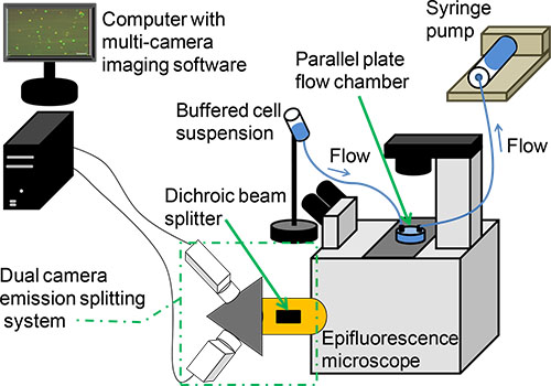
Figure 1. Illustration of a dual camera emission splitting system for acquiring images in a parallel plate flow chamber adhesion assay. A syringe pump is used to perfuse cells over substrate in the parallel plate flow chamber. The dual camera emission splitting system connected to an inverted Epifluorescence microscope uses a dichroic mirror and filter cubes to split and filter emission channels into two cameras. These cameras simultaneously capture two spatially identical but fluorophore-specific image sequences. A computer equipped with multi-camera imaging software is used to merge and record image sequences from each emission channel.
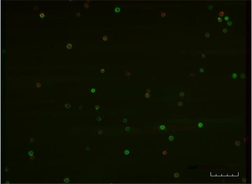
Figure 2. A dual camera emission splitting system was used for real-time simultaneous imaging of a parallel plate flow chamber adhesion assay. BT-20 cells labeled with (1) anti-human CD24 and anti-mouse IgG AlexaFluor 568 (pseudocolored red, emission max, 603 nm; camera 1, 620±60 nm) and (2) HECA-452 and anti-rat IgM AlexaFluor 488 (pseudocolored green, emission max 519 nm; camera 2, 535±40 nm) were perfused over a CHO-E monolayer at wall shear stress = 1 dyne/cm2. Dual camera merged images reveal heterogeneity within a rolling cell population and can be used to characterize rolling behaviors based on expression of cell surface molecules. Scale bar = 100 μm. Image acquired with a 10X objective.
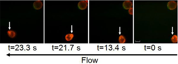
Figure 3. A time sequence of merged images captured by a dual camera emission splitting system showing BT-20 cells expressing CD24 (pseudocolored red) and sialofucosylated molecules (pseudocolored green) rolling on a CHO-E monolayer at wall shear stress = 1 dyne/cm2. Cameras maintained the optical and temporal resolution to track cell features on a cell tumbling "end over end" as it rolled on the reactive substrate in the direction of flow. White arrows indicate a tracked cluster of cell surface molecules. Emission signals in each channel were pseudocolored and merged in the imaging software. Note that colocalized signals appear yellow/orange. Still images were exported from real-time image sequences at time points shown. Scale bar = 10 μm. Images acquired with a 40X objective.
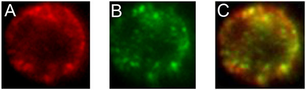
Figure 4. Dual camera emission splitting system reveals colocalization of CD24 (pseudocolored red) and sialofucosylated molecules (pseudocolored green) expressed by a representative BT-20 breast cancer cell rolling on a CHO-E monolayer. Heterogeneous expression of (A) CD24 and (B) sialofucosylated molecules on the cell surface is emphasized in each image. (C) Colocalization of molecules appears as yellow/orange pseudocolored spots when red and green pseudocolored emission channels are merged. Cell diameter is approximately 10 μm. Images acquired with a 40X objective.
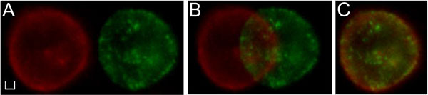
Figure 5.Adjustments to the horizontal, vertical, and rotational spatial settings of images captured in camera 1 and camera 2 can improve the spatial alignment in the merged image. Proper alignment is critical to accurately portray how cells are adhering and rolling in the adhesion assay. (A) A merged image of a single BT-20 cell is misaligned due to camera positioning. (B) Spatial alignment is improved with software adjustments in the SimulPix module of StreamPix 5. (C) Further software adjustments achieve near-perfect alignment. Scale bar = 2 μm. Images acquired with a 40X objective.
The dual camera emission splitting system has the spatial, temporal, and optical resolution needed to capture high quality images in applications where cell or molecular movement is rapid. In generating the representative results, parameters in the dual camera emission splitting system, including software settings and camera settings, were optimized to obtain a merged image sequence in which rolling and adherent cells were spatially and temporally aligned. This optimization step is critical, as misalignment can result in images that do not properly convey the physical phenomenon under investigation, e.g. a single cell observed to roll in both the green and red channels may appear as two rolling cells in the merged workspace. Spatial alignment should be achieved in the setup of the dual camera emission splitting system when cameras are physically aligned as described in step 2.4 of the protocol. Digital corrections to the spatial alignment of images captured by the cameras can be applied for horizontal, vertical, or rotational offset(s) through digital alignment settings in the imaging software (step 5.7 and Figure 5).
Temporal alignment between images captured by camera 1 and camera 2 can be achieved by adjusting the “live settings” of the cameras: exposure time, offset, and gain. Gain and offset can be adjusted without affecting the temporal alignment of the merged images, yet deviations in the user-defined exposure time between cameras can result in misalignment. Thus gain can be adjusted to compensate for differences between exposure times used to collect images in each camera. Ideally, differences in the exposure time between cameras should only be allowed when gain adjustments appear to significantly compromise image quality. Alternatively, antibody titration experiments can be performed to optimize cell labeling to compensate for differences in exposure time required by each camera (a) due to inherent properties of each fluorophore or (b) due to differences in the molecular expression levels detected by each antibody/fluorophore. However, it should also be noted that antibody titration experiments are best-practice to prevent bleed-through artifacts (step 5.4.5)11.
Although the dual camera emission splitting system was used to simultaneously image two target molecules in separate emission channels, alternative technologies are able to simultaneously capture images in multiple emission channels. These imaging systems range from single color cameras to confocal microscopes, and each has advantages and disadvantages relative to image quality, speed, and cost 12-17. In particular, state-of-the-art laser scanning confocal microscopes that incorporate resonant scanners are impressive multi-spectral imaging systems capable of acquiring images at hundreds of frames per second. These confocal systems are extremely powerful, but their cost may limit the number of researchers that have access to these systems through core facilities. Ultimately, the investigational needs and resources of each lab dictate which multi-channel imaging system is most appropriate.
The methods described herein explain how to obtain real-time image sequences in two emission channels using a dual camera emission splitting system and imaging software. Spatial and temporal alignment of images captured in two emission channels by similar monochrome CCD cameras is essential for production of high quality real-time merged image sequences that simultaneously reveal multiple target molecules on cells. Applications for the dual camera emission splitting system include any two-color live cell analysis that demands exceptional optical and temporal resolution of a full field of view, such as flow assays, translocation of cell surface molecules, cytoskeletal remodeling, vesicular transport, and 3D particle tracking .
The authors have nothing to disclose.
The authors wish to thank Dr. Douglas Goetz (Department of Chemical and Biomolecular Engineering, Ohio University) and Dr. Fabian Benencia (Department of Biomedical Sciences, Ohio University) for insightful discussions and manuscript review. We also thank Dr. Christopher Huppenbauer for helpful technical discussions (W. Nuhsbaum Inc.). This work was supported by grants from the National Science Foundation (CBET-1106118) and the National Institutes of Health (1R15CA161830-01).
Reagent/Material | |||
BT-20 cells | ATCC | HTB-19 | |
CHO-E cells | Gift from Dr. R. Sackstein (Brigham and Women’s Hospital, Harvard Medical School) | ||
MEM | Thermo Scientific | SH30024.01 | |
FBS | Thermo Scientific | SH30396.03 | |
Penicillin-streptomycin | Thermo Scientific | SV30010 | |
0.25% Trypsin / 0.1% EDTA | Thermo Scientific | SV30031.01 | |
DPBS | Thermo Scientific | SH30028.02 | |
DPBS+ | Life Technologies | 14080-055 | |
BSA | Sigma | A9647 | |
HECA-452 monoclonal antibody | BD Biosciences | 555946 | |
Anti-human CD24 monoclonal antibody | BD Biosciences | 555426 | |
Anti-rat IgM AlexaFluor 488 | Invitrogen | A21212 | |
Anti-mouse IgG AlexaFluor 568 | Invitrogen | A11004 | |
[header] | |||
Equipment | |||
EXi Blue Fluorescence Microscopy Digital CCD Camera | Q Imaging | EXI-BLU-R-F-M-14-C | |
Retiga EXi FAST 1394 Digital CCD Camera | Q Imaging | RET-EXi-F-M-12-C | |
DC2 Emission Splitter | Photometrics | DC2 | |
Inverted Fluorescence Microscope | Leica | DMI6000 B | |
Streampix 5 software | Norpix |