We present a combination of Cryo-electron microscopy, lipid nanotechnology, and structure analysis applied to resolve the membrane-bound structure of two highly homologous FVIII forms: human and porcine. The methodology developed in our laboratory to helically organize the two functional recombinant FVIII forms on negatively charged lipid nanotubes (LNT) is described.
Cryo-electron microscopy (Cryo-EM)1 is a powerful approach to investigate the functional structure of proteins and complexes in a hydrated state and membrane environment2.
Coagulation Factor VIII (FVIII)3 is a multi-domain blood plasma glycoprotein. Defect or deficiency of FVIII is the cause for Hemophilia type A - a severe bleeding disorder. Upon proteolytic activation, FVIII binds to the serine protease Factor IXa on the negatively charged platelet membrane, which is critical for normal blood clotting4. Despite the pivotal role FVIII plays in coagulation, structural information for its membrane-bound state is incomplete5. Recombinant FVIII concentrate is the most effective drug against Hemophilia type A and commercially available FVIII can be expressed as human or porcine, both forming functional complexes with human Factor IXa6,7.
In this study we present a combination of Cryo-electron microscopy (Cryo-EM), lipid nanotechnology and structure analysis applied to resolve the membrane-bound structure of two highly homologous FVIII forms: human and porcine. The methodology developed in our laboratory to helically organize the two functional recombinant FVIII forms on negatively charged lipid nanotubes (LNT) is described. The representative results demonstrate that our approach is sufficiently sensitive to define the differences in the helical organization between the two highly homologous in sequence (86% sequence identity) proteins. Detailed protocols for the helical organization, Cryo-EM and electron tomography (ET) data acquisition are given. The two-dimensional (2D) and three-dimensional (3D) structure analysis applied to obtain the 3D reconstructions of human and porcine FVIII-LNT is discussed. The presented human and porcine FVIII-LNT structures show the potential of the proposed methodology to calculate the functional, membrane-bound organization of blood coagulation Factor VIII at high resolution.
Blood coagulation Factor VIII (FVIII) is a large glycoprotein of 2,332 amino acids organized in six domains: A1-A2-B-A3-C1-C2 3. Upon Thrombin activation FVIII acts as the cofactor to Factor IXa within the membrane-bound Tenase complex. Binding of activated FVIII (FVIIIa) to FIXa in a membrane-depending manner enhances FIXa proteolytic efficiency more than 105 times, which is critical for efficient blood coagulation4. Despite the important role FVIII plays in coagulation and the Tenase complex formation, the functional membrane-bound FVIII structure is yet to be resolved.
To address this, single lipid bilayer nanotubes (LNT) rich in phosphatidylserine (PS), capable of binding FVIII with high affinity8,9 and resembling the activated platelet surface have been developed10. Consecutive helical organization of FVIII bound to LNT has been proven to be effective for structure determination of FVIII membrane-bound state by Cryo-EM5. Functionalized LNT are an ideal system to study protein-protein and protein-membrane interactions of helically organized membrane-associated proteins by Cryo-EM11,12. Cryo-EM has the advantage over traditional structural methods such as X-ray crystallography and NMR, as the specimen is preserved at closest to the physiological environment (buffer, membrane, pH), without additives and isotopes. In the case of FVIII, studying the membrane-bound structure with this technique is even more physiologically relevant, as the LNT resemble closely by size, shape and composition the pseudopodia of the activated platelets where the Tenase complexes assemble in vivo.
Defects and deficiency of FVIII cause Hemophilia A, a severe bleeding disorder affecting 1 in 5,000 males of the human population4,6. The most effective therapy for Hemophilia A is life-long administration of recombinant human FVIII (hFVIII). A significant complication of the recombinant FVIII Hemophilia A therapy is the development of inhibitory antibodies to the human form affecting approximately 30% of Hemophilia A patients13. In this case, porcine FVIII (pFVIII) concentrate is used, as porcine FVIII displays low cross-reactivity with inhibitory antibodies against human FVIII and forms functional complexes with human FIXa7. Establishing the membrane-bound organization of both porcine and human FVIII forms is important to understand the structural basis of FVIII cofactor function and implications for blood hemostasis.
In this study, we describe a combination of lipid nanotechnology, Cryo-EM, and structure analysis designed to resolve the membrane-bound organization of two highly homologous FVIII forms. The presented Cryo-EM data and 3D structures for helically organized porcine and human FVIII on negatively charged LNT show the potential of the proposed nanotechnology as basis for structure determination of FVIII and membrane-bound coagulation factors and complexes in a physiological membrane environment.
1. Sample Preparation
2. Cryo-electron Microscopy of FVIII-LNT
3. 3D Reconstruction
NOTE: The image analysis software used for the 2D and 3D analysis: EMAN2 and IHRSR are freely available. EMAN2 can be downloaded from http://blake.bcm.edu/emanwiki/EMAN2/Install. The IHRSR software can be obtained from Professor Egelman: egelman@virginia.edu. The final IHRSR refinements are run on the Texas advanced computing center cluster: http://www.tacc.utexas.edu/ at the University of Texas, Austin. The 3D reconstruction algorithm shown on Figure 1 consists of two main steps: First selecting a homogenous set of helical segments (particles) with the 2D reference free alignment (RFA) algorithms implemented in EMAN 2, second achieving a 3D reconstruction based on the helical parameters and back projection algorithms incorporated in IHRSR. The first step utilizes the programs developed for selecting homogenous particle sets for 3D reconstruction with Single Particle SPA (algorithms) for which EMAN2 has been specifically developed and distributed: http://blake.bcm.edu/emanwiki/EMAN2. This step has been adapted to the Cryo-EM data. The second step is achieved with the IHRSR algorithm, which is specifically designed for the type of helical assemblies obtained with the recombinant Factor VIII forms. This algorithm has been documented extensively through the scientific literature12.
4. Electron Tomography
Recombinant human and porcine FVIII were successfully organized helically on negatively charged single bilayer LNT, resembling the activated platelet surface. The helical organization of the human and porcine FVIII-LNT was consistent through the collected digital micrographs (Figure 2). The control LNT and the human and porcine FVIII-LNT helical tubes were selected and segmented with the e2helixboxer.py GUI and initial data sets created with the e2workflow.py GUI, Single particle option (Table 1).
The helical order of the membrane-bound human and porcine FVIII-LNT was evaluated from the Fourier transform of the class averages with the e2display.py GUI (EMAN2) (Figure 3). The lipid bilayer in the best control LNT 2D class averages is well defined. The inner and outer leaflet and lower density of the membrane hydrophobic core are clearly visible (Figure 3A). The projected density of the membrane-bound human and porcine FVIII molecules oriented towards and perpendicular to the membrane surface is well defined and clearly shows the variations in the helical organization between the two proteins (Figures 3B and 3C). The more pronounced twist for the human FVIII-LNT helical tubes indicates that the protein-protein interactions between adjacent membrane-bound FVIII molecules are consistently different for the two FVIII forms (Figures 3B and 3C). Particles from class averages showing good helical organization (helical diffraction pattern) were merged in the e2display.py GUI to form an intermediate particle set (Table 1). The particles from the intermediate particle sets were again classified in 50 classes with the same constraints. The particles from class averages with the same diameter were merged in the final data sets (Table 1).
Initial 3D reconstructions for the human and porcine FVIII-LNT were carried out with 1,000 representative particles from the final human and porcine FVIII-LNT data sets. One hundred consecutive IHRSR iterations were run for each 3D reconstruction with a featureless cylinder (160 Å inner and 500 Å outer diameter), as initial volume. The axial rise ( Δz) calculated from the combined Fourier transform of the helical segments (particles set) is equal to 41 Å for human FVIII-LNT and 36 Å for porcine FVIII-LNT (Figures 4A and 4B). The initial azimuthal angle (ΔΦ) defined from the iterative search is estimated at 40.0° for the human FVIII-LNT and at 35.0° for the porcine FVIII-LNT. The final volumes are inspected for convergence of the helical parameters and correspondence between class averages and projections from the final reconstruction, also following the criteria described in5. The selected 3D reconstructions and corresponding helical parameters are imposed as initial volumes and initial helical parameters for a second IHRSR refinement of 100 cycles which converged to a four-start helical organization for the human FVIII-LNT with Δz = 41.1 Å and ΔΦ = 42.0° and a five-start helical organization for the porcine FVIII-LNT with Δz = 35.5 Å and ΔΦ = 34.8°. A final 100 IHRSR iterations imposing a 4-fold and a 5-fold helical symmetry for the human and porcine FVIII-LNT reconstructions respectively are carried out with initial volumes and corresponding helical parameters from the last asymmetric IHRSR refinements (Figures 4C and 4D). The final volumes show 8 human FVIII and 10 porcine FVIII membrane-bound molecules organized around the helical axis (Figure 5A). Each human FVIII molecule is translated 41.2 Å and rotated 42.0° from the previous one and each porcine FVIII molecule is translated 35.9 Å and rotated 35.2º from the previous one, corresponding to the helical parameters of the final 3D reconstructions (Figure 5B).
The reconstructed electron tomograms confirm the difference in the helical organization between the human and porcine FVIII-LNT obtained at the same experimental conditions. Comparison of the top views from the reconstructed tomograms and the 3D volumes from the helical reconstruction viewed in the direction perpendicular to the helical axis, further validates the correctness of the 3D reconstructions refined with the IHRSR helical parameters (Figure 6). The asymmetric 2D unit cell dimensions for the human FVIII-LNT 3D reconstruction are: a = 17.8 nm, b = 8.2, γ = 84° and for the porcine FVIII-LNT 3D reconstruction: a = 18.4, b = 7.2 and γ = 70° (Figure 6). The unit cell dimensions of human FVIII organized in membrane-bound 2D crystals are: a = 8.1, b = 7.0 and γ = 67º, which corresponds to the surface covered by one FVIII molecule viewed toward the membrane-surface20. Comparing the unit cell dimensions between FVIII organized in 2D and helical crystals indicates that both human and porcine FVIII molecules form dimers when helically organized on the LNT surface.
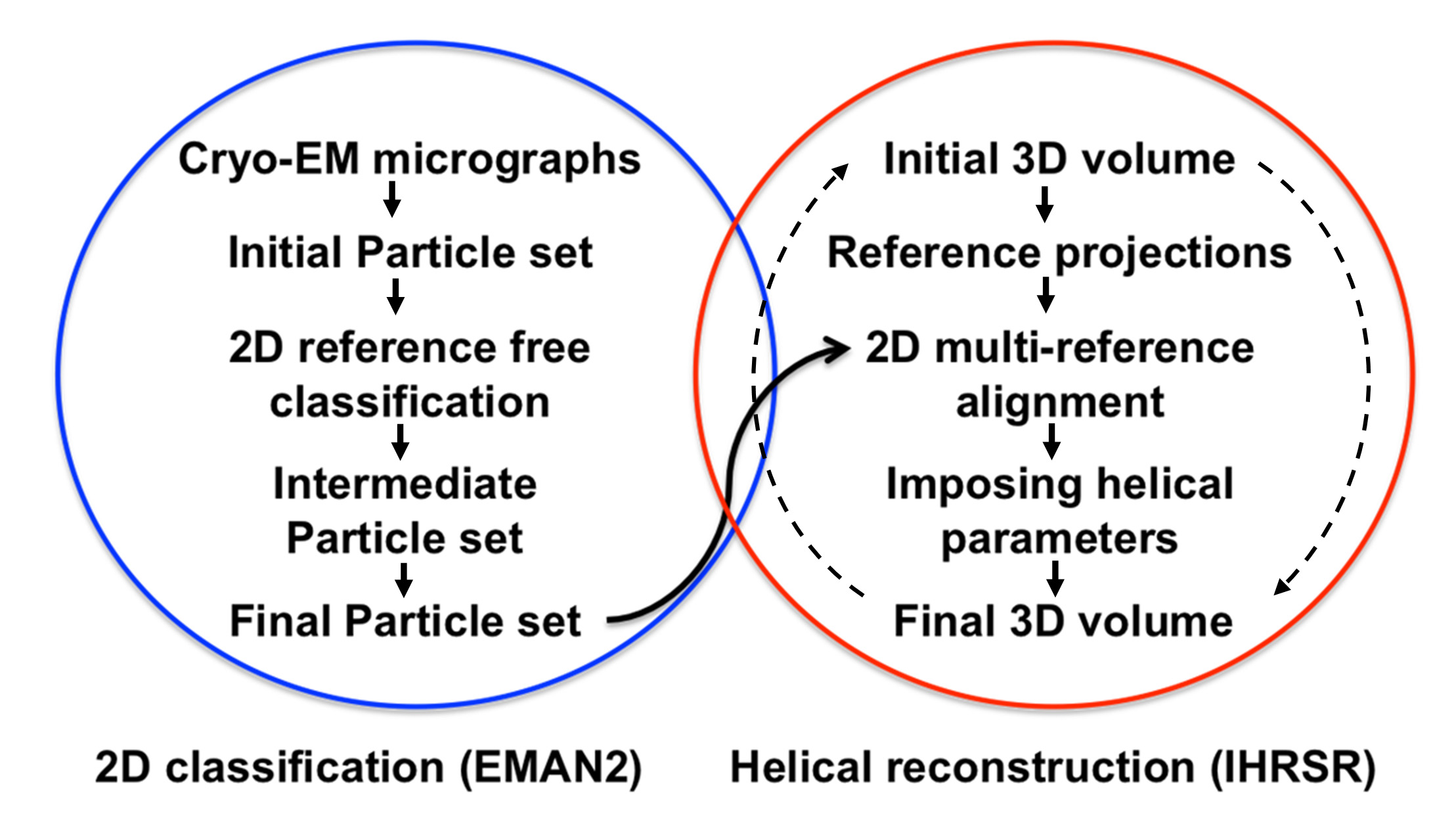
Figure 1. Structure analysis flow chart. The steps followed for the 2D classification analysis based on reference free alignment algorithms implemented in EMAN216 are circled in blue. The steps followed for the 3D analysis carried out with the iterative helical real space reconstruction algorithms (IHRSR) are circled in red. The iterative IHRSR cycles are denoted with dashed arrows.
Figure 2. Cryo-EM digital micrographs. (4,096 x 4,096 pixels, 2.9 Å/pix) of lipid nanotubes (LNT) with and without bound FVIII. A. Control LNT. B. Human FVIII-LNT. C. Porcine FVIII-LNT. The edge of the hole in the carbon film in which the FVIII-LNT are suspended in amorphous ice is indicated with a white star. The protein and lipid densities are in black. The magnified views (insets) of 512 x 512 cropped areas (white dashed square) illustrate the difference in the helical organization of the human and porcine FVIII, respectively. The scale bar is 100 nm. Please click here to view a larger version of this figure.
Figure 3. Representative 2D class averages (top row) and corresponding Fourier transforms (bottom row) from the intermediate particle sets (Table 1) classified in 50 classes. A. Control LNT B. Human FVIII-LNT C. Porcine FVIII-LNT. The class number and number of particles included in each class are indicated. The difference in helical order between the human and porcine FVIII is clearly seen on the images and confirmed by the diffraction patterns obtained from the Fourier transforms of these images. Please click here to view a larger version of this figure.
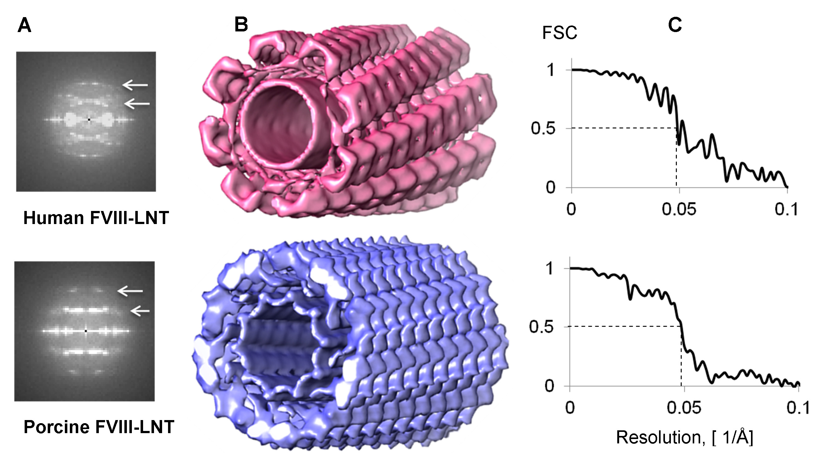
Figure 4. 3D helical reconstructions of human and porcine FVIII-LNT. A. Combined Fourier transform from 1,000 helical segments. The first and second layer line are centered at 1/82 Å-1 and 1/41 Å-1 for human FVIII-LNT, and at 1/72 Å-1 and 1/36 Å-1 for porcine FVIII-LNT (white arrows). B. Surface representation of human in pink (Δz = 41.1 Å, ΔΦ = 42.0º) and porcine in blue (Δz = 35.9 Å, ΔΦ = 35.2º) FVIII-LNT 3D helical reconstructions. Both volumes are presented at 0.005 contour level (minimum density is 0 and maximum density 0.02, as calculated in UCSF Chimera, Volume viewer option21). The length of the FVIII-LNT tube is 256 pixels at 2.9 Å/pix. C. Fourier Shell Correlation (FSC) plots for human and porcine FVIII-LNT showing a resolution of 20.5 Å at FSC = 0.5.
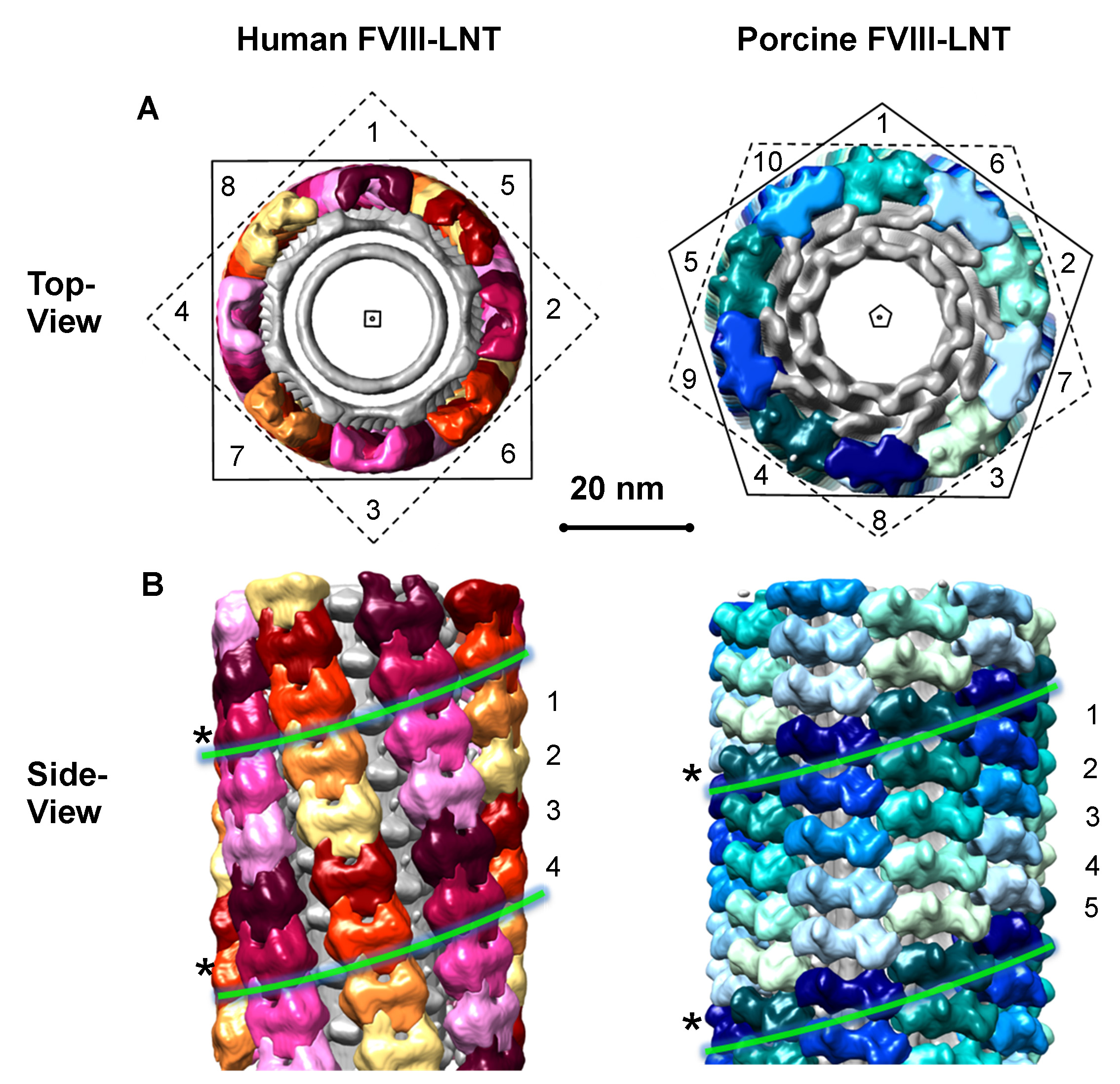
Figure 5. Helical organization of human and porcine FVIII-LNT. Segmented surface representation of human and porcine FVIII-LNT helical reconstructions, shown in Figure 4B. The volumes are segmented after imposing 4-fold symmetry to the human FVIII-LNT and 5-fold symmetry to the porcine FVIII-LNT. The asymmetric units are color coded yellow-red for the human FVIII-LNT and blue-green for the porcine FVIII-LNT. A. Views along the helical axis indicated with a square for the human and as a pentagon for the porcine FVIII-LNT. The human FVIII-LNT structure shows 8 molecules organized around the outer LNT membrane and the porcine FVIII structure shows 10 molecules organized around the outer LNT membrane, indicated with numbers. B. Views perpendicular to the helical axis. The human FVIII-LNT is a 4-start helical structure and the porcine FVIII-LNT is a 5-start helical structure. The individual one start helices are indicated with numbers and color-coded. We have emphasized one of the helices from each structure with a (*) and green lines. The scale bar is 20 nm.
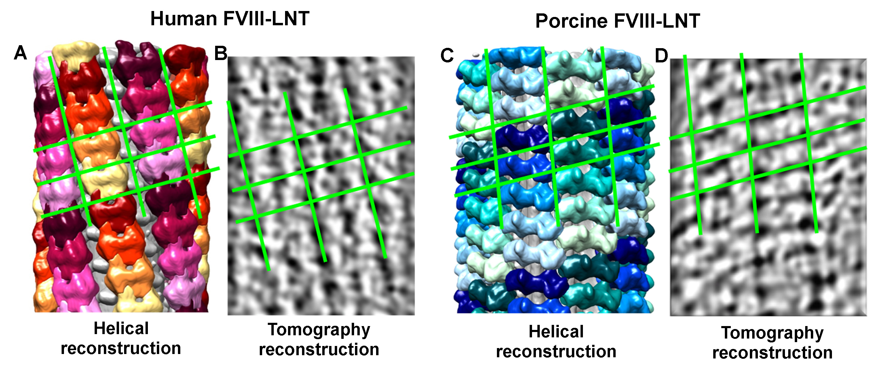
Figure 6. Comparison between the helical and tomography 3D reconstructions. The human FVIII-LNT (A) and porcine FVIII-LNT (C) helical 3D reconstructions are shown perpendicular to the helical axis. Each unit cell and individual helices are color-coded as in Figure 5. B. and D. are density representations of the 3D tomography reconstructions, viewed perpendicular to the helical axis. The 2D lattice reflecting the helical arrangement of the FVIII molecules is shown with green lines.
SAMPLES | cLNT | hFVIII-LNT | pFVIII-LNT |
Initial Micrographs | 61 | 474 | 542 |
Initial Particle sets | 29113 | 60395 | 64665 |
Defocus (nm) | -4,051 ± 502 | -3,643 ± 737 | -3,443 ± 1,086 |
Intermediate Particle sets | 25,907 | 27,305 | 22,773 |
Final Particle sets | 25,907 | 10,455 | 10,430 |
Table 1. 2D analysis statistics following the algorithm presented in the flowchart on Figure 3.
In this work a methodology is presented to differentiate between two membrane-bound organizations of highly homologous proteins: human and porcine FVIII self-assembled on lipid nanotubes in the conditions encountered in the human body.
In the described procedure, human and porcine FVIII are successfully organized helically on lipid nanotubes, which is the most critical step. The next critical step is to preserve the sample in thin amorphous ice by flash freezing at near liquid N2 temperature. Preserving the sample in amorphous ice and LN2 temperature keeps the helical tubes hydrated and the protein-lipid macromolecular assemblies physiologically active. The final critical step is acquiring Cryo-EM data of sufficient quantity and quality for a high-resolution 3D structure at near LN2 temperature. Collecting data at near LN2 temperature further prevents dehydration of the sample in the high vacuum of the microscope and radiation damage from the electron beam.
To calculate the membrane-bound structure of the FVIII the first critical step is to obtain homogenous particles (helical segments) sets by applying 2D reference free classification and combine particles from classes with the same diameter and degree of order. The second critical step is to impose the right initial volume and helical parameters (rise and azimuthal angle) for the helical reconstruction. The third and final critical step is to validate the helical structure by comparing the 3D maps obtained by the helical and electron tomography (without imposed symmetry) reconstructions of the same specimen.
The presented methodology is unique in its capacity to resolve the functional structure of membrane-associated proteins at near physiological conditions. The LNT developed in our laboratory can be successfully used as a platform for helical organization of functional membrane-bound blood coagulation factors and achieve better resolution than for FVIII organized in membrane-bound 2D crystals and as single particles. Our goal is to further increase the resolution of our helical reconstructions by improving the homogeneity and quality of the final particle sets. Collecting more Cryo-EM micrographs at better Cryo-EM conditions (Field emission gun, energy filter, DE camera detectors) of FVIII-LNT helical filaments and therefore including larger initial particle sets for the 2D reconstruction will achieve this. Improving the FVIII-LNT helical assembly and 3D reconstruction algorithms will allow us to obtain sub-nanometer and near atomic resolution, which will unambiguously define the membrane-bound organization of this critical for blood coagulation protein.
Organizing helically homologous FVIII forms gives us also the opportunity to characterize how difference in sequence can correlate to differences in structure and function. Resolving the human and porcine FVIII membrane-bound structures by the methods described in this article can help identify the sequences, which when modified will improve the recombinant FVIII function. This knowledge will have significant clinical implications for drug discovery in both Thrombosis and Hemostasis fields.
The authors have nothing to disclose.
This work is supported by a National Scientist Development grant from the American Heart Association: 10SDG3500034 and UTMB-NCB start up funds to SSM. The authors acknowledge the Cryo-EM and Scientific Computing facilities at the Sealy Center for Structural Biology at UTMB (www.scsb.utmb.edu), as well as Drs. Steve Ludtke and Ed Egelman for help with the 2D and 3D helical reconstruction algorithms.
JEM2100 with LaB6 | JEOL Ltd. | JEM-2100 | operated at 200 kV |
with TEMCON software | JEOL Ltd. | ||
Gatan626 Cryo-holder | Gatan, Inc. | 626.DH | cooled to -175 °C |
with temperature controler unit | Gatan, Inc. | ||
Gatan 4K x 4K CCD camera | Gatan, Inc. | US4000 | 4096 x 4096 pixel at 15 microns/pixel physical resolution |
Solarus Model 950 plasma cleaner | Gatan, Inc. | ||
Vitrobot Mark IV | FEI | ||
Materials | |||
Carbon coated 300 mesh 3mm copper grid | Ted Pella | 01821 | plasma cleaned for 10 s on high power |
Quantifoil R2/2 300 mesh | Electron Microscopy Sciences | Q225-CR2 | Carbon coated 300 mesh Cu grids with 2 mm in diameters holes |
Uranyl acetate dihydrate | Ted Pella | 19481 | 1% solution, filtered |
Galactosyl ceramide | Avanti Polar Lipids Inc. | 860546 | |
Dioleoyl-sn-glycero-phospho-L-serine | Avanti Polar Lipids Inc. | 840035 | |
Software | |||
EM software Digital Micrograph | Gatan, Inc. | http://www.gatan.com/DM/ | |
EM software EMAN | free download | http://blake.bcm.edu/emanwiki/EMAN/ | |
EM software Spider | free download | http://spider.wadsworth.org/spider_doc/spider/docs/spider.html | |
EM software IHRSR | free download | Programs available from Edward H. Egelman http://people.virginia.edu/~ehe2n/ | |
EM software (IMOD) | free download | http://bio3d.colorado.edu/imod/ | |
EM software (SerialEM) | free download | ftp://bio3d.colorado.edu/pub/SerialEM/ | |
UCSF-Chimera | free download | http://www.cgl.ucsf.edu/chimera/download.html |