In this paper, we demonstrate a standard method for producing an embolic middle cerebral artery occlusion with homologous blood clots (fibrin-rich) in adult rat. This model closely mimics human ischemic stroke and is suitable for preclinical study of thrombolytic therapy for ischemic stroke.
Clinically, thrombolytic therapy with use of recombinant tissue plasminogen activator (tPA) remains the most effective treatment for acute ischemic stroke. However, the use of tPA is limited by its narrow therapeutic window and by increased risk of hemorrhagic transformation. There is an urgent need to develop suitable stroke models to study new thrombolytic agents and strategies for treatment of ischemic stroke. At present, two major types of ischemic stroke models have been developed in rats and mice: intraluminal suture MCAO and embolic MCAO. Although MCAO models via the intraluminal suture technique have been widely used in mechanism-driven stroke research, these suture models do not mimic the clinical situation and are not suitable for thrombolytic studies. Among these models, the embolic MCAO model closely mimics human ischemic stroke and is suitable for preclinical investigation of thrombolytic therapy. This embolic model was first developed in rats by Overgaard et al.1 in 1992 and further characterized by Zhang et al. in 19972. Although embolic MCAO has gained increasing attention, there are technical problems faced by many laboratories. To meet increasing needs for thrombolytic research, we present a highly reproducible model of embolic MCAO in the rat, which can develop a predictable infarct volume within the MCA territory. In brief, a modified PE-50 tube is gently advanced from the external carotid artery (ECA) into the lumen of the internal carotid artery (ICA) until the tip of the catheter reaches the origin of the MCA. Through the catheter, a single homologous blood clot is placed at the origin of the MCA. To identify the success of MCA occlusion, regional cerebral blood flow was monitored, neurological deficits and infarct volumes were measured. The techniques presented in this paper should help investigators to overcome technical problems for establishing this model for stroke research.
Stroke is the third leading cause of death in the United States, but treatment options for acute stroke remain limited. At present, intravenous infusion of recombinant tissue plasminogen activator (tPA) to dissolve the blood clots is the most efficient therapy for acute ischemic stroke. However, the use of tPA is limited by its narrow therapeutic window and by increased risk of intracerebral hemorrhage. Therefore, a model of stroke suitable for thrombolytic research is urgently needed.
The middle cerebral artery (MCA) is the artery most often occluded in stroke in humans. Focused on this artery, many animal models of ischemic stroke have been established. At present, two major types of rodent focal ischemia models by occluding MCA have been developed: suture MCAO model and embolic MCAO model. Although MCAO models via the intraluminal suture technique have been widely used in mechanism-driven stroke research, these suture models do not mimic human stroke, as up to 80% of human strokes are caused by thrombosis or embolism. However, embolic stroke model using blood clots closely mimics human stroke and is considered suitable for thrombolytic study. This embolic model was first developed in rats by Overgaard et al.1 in 1992 and further characterized by Zhang et al. in 19972 and by Dinapoli et al. in 20063. Although embolic MCAO has gained increasing attention, there are technical problems faced by many laboratories.
In this article, we demonstrate a standard method for producing embolic MCAO with homologous blood clots in the adult rat, which can develop a predictable infarction within the MCA territory. The techniques presented in this article should help investigators to overcome technical problems for establishing this model for stroke research.
Ethics Statement: Male adult Sprague-Dawley rats (weighing 330-380 g) were used in this protocol. This protocol was approved by the Institutional Animal Care and Use Committee (IACUC) at the LSU Health Science Center-Shreveport and is in compliance with the 'Guide for the Care and Use of Laboratory Animals' (eighth edition, National Academy of Sciences, 2011).
1. Preparation of Modified PE-50 Tube
2. Preparation of Homologous Blood Clots
3. Embolic Middle Cerebral Artery Occlusion (Figure 3A,B)
4. Monitoring Regional Cerebral Blood Flow (rCBF)
5. Post-operative Care
6. Neurological Deficit Score
Laser Doppler flowmetry (LDF) was used to monitor rCBF during induction of cerebral ischemia6,7. Many laboratories including our laboratory have been using rCBF to identify animals with successful MCA occlusion, but the thresholds of baseline varied between laboratories which are related to the site of measurement. The probe of the LDF is positioned at 2 mm posterior and 5 mm lateral to the bregma as described previously6. rCBF was monitored at 0, 5, 15, 30, 60, 90, and 120 min after injection of the clot. On the basis of this data, only animals that exhibit a reduction in rCBF >70% of baseline are considered successful embolic occlusion of the MCA (Figure 5). At 2 hr after injection of clot, a standard rat dose of tPA (10 mg/kg)7,8 was administered intravenously with a 10% bolus and 90% continuous infusion over 30 min using a syringe infusion pump7,8. We observed that rCBF levels gradually increased to >70% of the baseline within 30 min of tPA therapy (Figure 5).
At 24 hr after embolization, the animals were euthanized and transcardially perfused with 200 ml of PBS to remove intravascular blood. Previous studies2,3,7,8 and our data (Figure 6) have shown substantially larger tissue infarctions produced at 24 hr after embolization. The brains were collected and taken pictures. In saline-treated group, the blood clot was readily visualized at the origin of the MCA and ACA, but the clot was almost completely dissolved in tPA-treated group (Figure 6A). After that, the brain was sliced into seven 2 mm coronal sections with a rat brain matrix on ice. Incubate the brain slices in 2% 2,3,5-triphenyltetrazolium chloride (TTC) at 37 °C for 30 min9,10. After TTC staining, the coronal sections were placed on a plate and photographed (Figure 6B). The area of infarction in each slice was determined by the computerized image analysis system (National Institutes of Health Image), and the average infarct volume was calculated by multiplying the distance between sections. This embolic stroke model produced tissue infarction within the MCA territory as seen in the neocortex and striatum regions (Figure 6B). Early reperfusion was established by intravenous administration of tPA at 2 hr after embolization, the infarct volumes were significantly reduced in the tPA-treated group (229.1 ± 45.7 mm3, n = 12) compared with the saline-treated group (394.2 ± 68.2 mm3, n = 9) (P < 0.01). The 24 hr mortality rate is 16% (4/25).
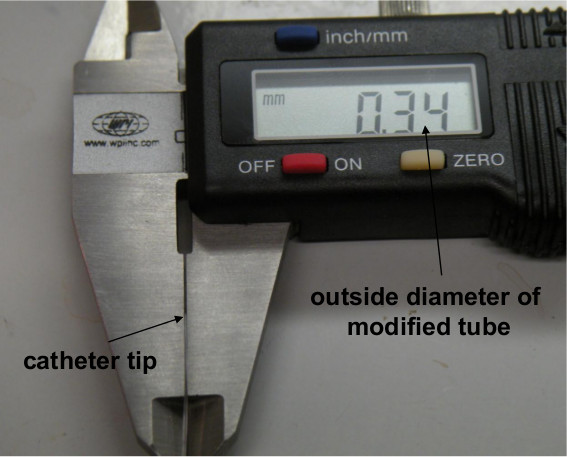
Figure 1. Measurement of the outside diameter of the modified PE-50 tube using a digital caliper.
Figure 2. Washing blood clot in a PE-10 tube. The blood clot was washed by alternative pushing the two syringes connected to the end of PE-50 tube.
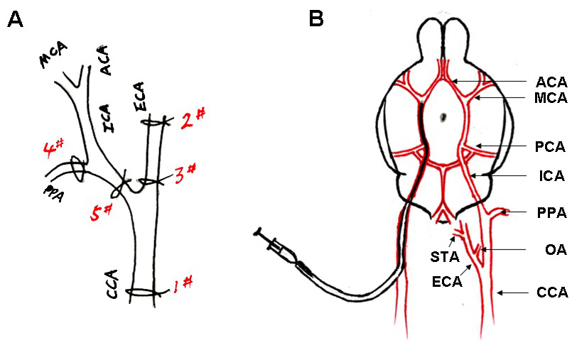
Figure 3. (A) Simplified scheme of rat right hemisphere isolated arteries showing successive sutures to prepare the introduction of modified PE-50 tube. (B) Scheme of the arterial architecture in the rat brain, and the catheter advanced from the ECA to the ICA of the rat. A single blood clot (black) was contained in the tube.
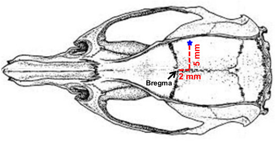
Figure 4. The burr hole (1.5 mm diameter) located at 2 mm posterior and 5 mm lateral to the bregma.
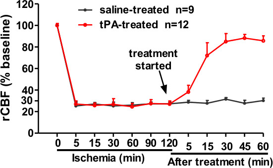
Figure 5. Regional cerebral blood flow (rCBF) was measured using a laser Doppler flowmeter (LDF). The clot injection led to >70% rCBF reduction of the baseline value. tPA (10 mg/kg) treated at 2 hr after the clot injection restored rCBF close to baseline after 30 min of tPA treatment. Data was expressed as mean ± SD.
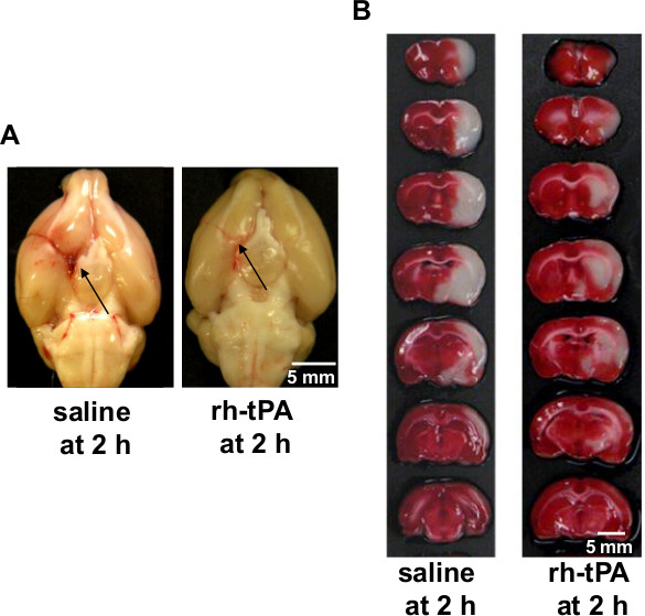
Figure 6. (A) Representative pictures showing the blood clots (black arrows) in the origin of the middle cerebral artery (MCA) and anterior cerebral artery (ACA) at 24 hr after stroke. Left, saline-treated rat; Right, tPA-treated rat, bar = 5 mm. (B) Representative images showing TTC-stained brain slices at 24 hr after stroke. Left, saline-treated rat; Right, tPA-treated rat, bar = 5 mm.
In this study, we demonstrated a standard method for performing an embolic MCAO stroke model in the rat, in which the origin of the MCA is occluded by a fibrin-rich clot. The major advantage of this model is: the occlusion of the stem of MCA with a fibrin-rich blood clot is similar to thromboembolic stroke in humans, the embolic stroke model is suitable for performing preclinical investigation of fibrinolytic therapy, and that this model can develop a reproducible and predictable infarct volume within the territory supplied by the MCA.
For performing the embolic MCAO model, the clot introduction, stability and lodgment are difficult to manage, which leads to variations in infarct size and affected brain regions2,3. Previous studies demonstrated that reproducible ischemic lesions in the MCA territory could only be achieved when the obstructing clots are lodged in the stem of MCA2,3. To precisely lodge the clot and produce a reproducible embolic stroke model, in this study, we demonstrate how to prepare a modified PE-50 tube and how to introduce the tip of the modified tube to the origin of MCA and how to inject a fibrin-rich clot into the MCA. Consistent with the previous report2, the injected blood clot was readily visualized at the origin of the MCA in all saline-treated rats (n = 9) but was largely dissolved in all tPA-treated rats (n = 12) at 24 hr after embolization.
To evaluate the success of MCAO, the rCBF, neurological deficits, and the pattern and distribution of cerebral lesions were assessed. After the clot injection, rCBF was decreased to 30% of baseline and this decrease of rCBF persisted for at least 2 hr after embolization, consistent with previous reports6,7. After treatment with tPA at 2 hr after embolization, the levels of rCBF were restored close to baseline after 30 min of tPA treatment. Neurological score was measured 10 min before drug treatment using the modified Bederson score to help evaluating the success of MCAO. This neurological scoring is a simple and quick way to detect global neurological function in acute phase of stroke. Animals showing normal behaviors (score = 0) were excluded from drug treatment and further analysis. Moreover, we demonstrated that the tissue lesion was produced mostly within the MCA territory as seen in the neocortex and striatum regions, and tPA treatment (at 2 hr) significantly reduced the infarction size at 24 hr after stroke. Together, our data demonstrated that the embolic stroke model presented in this work can develop a predictable infarct volume within the MCA territory.
Finally, we note that there are several technical concerns that may affect the success of the embolic MCAO model. A common problem encountered in performing embolic MCAO model is early spontaneous reperfusion after embolization. The occurance of spontaneous reperfusion is likely to be associated with the fragile of extravascular coagulated clot and the length of the clot used to occlude the MCA2,3,10. We believe that the method we described (step 2) to prepare blood clot would minimize the fragile of extravascular coagulated clot. The length of single blood clot used to occlude MCA varied from lab to lab between 25-mm to 50-mm length2,3,6-8,10. We found that the use of a 35-40 mm long clot ideally occluded the MCA and produced highly reproducible infarct volume. Modifying the PE-50 tube with the tip diameter between 0.30-0.34 mm. If the tip diameter is >0.34 mm, it cannot reach the origin of the MCA. If the tip diameter is <0.30 mm, the clot inside the modified PE-50 tube is difficult to go through the tip. The mortality rate is closely related to severe brain swelling and intracerebral hemorrhage. In this work, the 24 hr mortality rate is 16% (4/25; see representative results), and all dead rats showed severe brain swelling although we didn’t see intracerebral hemorrhage (possibly due to limited sample size). A controllable mortality factor is body temperature during the surgery. In controlling the body temperature between 36.5-37.5 °C from the start of surgery until full recovery from anesthesia. Body temperature significantly affects on the extent of brain tissue damage. Hypothermia decreases and hyperthermia increases the infarct volume11,12. Performing the surgery in a short time period (about 20-25 min) according the protocol in order to produce highly reproducible infarct size. Infectious complications are a leading cause of death in patients with acute stroke13. To reduce the incidence of infectious complications and enhance the survival rate after stroke, aseptic technique should be used during surgery.
In conclusion: the techniques presented in this article should help investigators to overcome technical problems for establishing this model for stroke research.
The authors have nothing to disclose.
This work was supported by National Institutes of Health grants HL087990 (G.L.).
rh-tPA | Chemical | Genentech | |
2,3,5-triphenyltetrazolium chloride | Chemical | Sigma | T8877 |
Anesthesia vaporizer | Equipment | Soma Technology | Drager Vapor 19.1 |
Rechargeable high speed micro drill | Equipment | Fine Science Tools | 18000-17 |
Curved scissors | Equipment | Fine Science Tools | 14117-14 |
Dumont forceps(Medical #7) | Equipment | Fine Science Tools | 11270-20 |
Dumont forceps(Medical #5) | Equipment | Fine Science Tools | 11251-35 |
Vannas-style spring scissors | Equipment | Fine Science Tools | 15000-03 |
Veterinary recovery chamber | Equipment | Peco Services | V1200 |
Genie plus syringe pump | Equipment | Kent Scientific Corporation | |
Rat brain matrix | Equipment | Kent Scientific Corporation | RBMA-310C |
Digital caliper | Equipment | World Precision Instruments | 501601 |
Dissecting microscope | Equipment | World Precision Instruments | PZMTIII-BS-LWD |
Hamilton syringe | Equipment | Hamilton | model 710 |
Homeothermic blanket control unit | Equipment | Harvard Apparatus | |
Electric clipper | Equipment | Braintree Scientific | CLP-9931 |
Veterinary electrosurgical unit | Equipment | MACAN Manufacturing Company | MV-9 |
Blood flowmeter | Equipment | Adinstruments | |
PowerLab 4/30 | Equipment | Adinstruments | |
LabChart 7.2 | software | Adinstruments | |
1ml syringe | Consumable | Becton, Dickinson and Company | 309659 |
23G needle | Consumable | Becton, Dickinson and Company | 305143 |
30G needle | Consumable | Becton, Dickinson and Company | 305106 |
PE-50 tubing | Consumable | Becton, Dickinson and Company | 427517 |
PE-10 tubing | Consumable | Becton, Dickinson and Company | 427400 |
6-0 Silk suture | Consumable | Harvard apparatus | 723287 |
5-0 Silk suture | Consumable | Harvard Apparatus | 517607 |