Here, we present a protocol demonstrating a hemorrhagic shock model in swine that uses aortic occlusion as a bridge to definitive care in trauma. This model has application in testing a wide range of surgical and pharmacological therapeutic strategies.
Hemorrhage remains the leading cause of preventable deaths in trauma. Endovascular management of non-compressible torso hemorrhage has been at the forefront of trauma care in recent years. Since complete aortic occlusion presents serious concerns, the concept of partial aortic occlusion has gained a growing attention. Here, we present a large animal model of hemorrhagic shock to investigate the effects of a novel partial aortic balloon occlusion catheter and compare it with a catheter that works on the principles of complete aortic occlusion. Swine are anesthetized and instrumented in order to conduct controlled fixed-volume hemorrhage, and hemodynamic and physiological parameters are monitored. Following hemorrhage, aortic balloon occlusion catheters are inserted and inflated in the supraceliac aorta for 60 min, during which the animals receive whole-blood resuscitation as 20% of the total blood volume (TBV). Following balloon deflation, the animals are monitored in a critical care setting for 4 h, during which they receive fluid resuscitation and vasopressors as needed. The partial aortic balloon occlusion demonstrated improved distal mean arterial pressures (MAPs) during the balloon inflation, decreased markers of ischemia, and decreased fluid resuscitation and vasopressor use. As swine physiology and homeostatic responses following hemorrhage have been well-documented and are like those in humans, a swine hemorrhagic shock model can be used to test various treatment strategies. In addition to treating hemorrhage, aortic balloon occlusion catheters have become popular for their role in cardiac arrest, cardiac and vascular surgery, and other high-risk elective surgical procedures.
Hemorrhage continues to be the dominant cause of preventable deaths in patients undergoing traumatic events, accounting for 90% of trauma-related deaths in the military setting and 40% of post-traumatic deaths in the civilian population1,2. Although direct pressure can treat compressible hemorrhage, non-compressible torso hemorrhage remains difficult to treat and can be lethal without prompt hemostatic control. The historical approach of resuscitative thoracotomy or laparotomy with aortic cross-clamping has proved to be extremely invasive3,4. This intervention also requires a complex selection algorithm to determine the candidacy of patients that have undergone traumatic insults5.
In recent years, there has been a resurgence of interest in a previously described approach—resuscitative endovascular balloon occlusion of the aorta (REBOA)6,7,8. Although REBOA has conferred a short-term survival advantages in hemorrhage, a prolonged complete occlusion of the aorta during balloon inflation poses serious concerns that include irreversible end-organ ischemia9,10. In an attempt to overcome this potential morbidity, alternative endovascular strategies to manage hemorrhage are being devised. One such strategy that has seen a growing attention is a partial occlusion of the aorta11,12. The idea of partial aortic balloon occlusion affords the perfusion of vascular beds distal to the site of occlusion, improved physiologic proximal aortic MAPs, and a gradual afterload reduction following the balloon deflation. These changes in parameters are desired modifications to the physiological characteristics of a bleeding animal. Prior to this method's translation to humans, complete and partial aortic balloon occlusion catheters have been heavily tested in swine models of hemorrhagic shock11,12,13.
Swine have been used in studies entailing hemorrhagic shock for many years. Most of the current understanding of the pathophysiology of hemorrhagic shock is derived from studies that have utilized animal models, including swine. Their physiology and homeostatic responses in the setting of pathologic volume depletion following hemorrhage, especially those pertaining to blood clotting and cardiovascular responses, have been well-documented and are like those in humans14. Swine models of hemorrhagic shock also provide opportunities to investigate treatment strategies for hemorrhagic shock and other traumatic injuries.
In this study, we demonstrate a clinically realistic model of hemorrhagic shock in swine to evaluate endovascular treatment strategies, including complete and partial aortic balloon occlusion. We hypothesize that a partial occlusion of the aorta results in a better physiologic and laboratory profile compared to a complete occlusion of the aorta in swine undergoing a controlled fixed-volume hemorrhage.
We aimed to compare the physiologic effects of partial and complete aortic occlusion as a treatment for hemorrhagic shock in a swine model. Partial aortic occlusion was achieved using a selective aortic balloon occlusion in trauma (SABOT) catheter (Figure 1). The SABOT catheter is a two-balloon system that allows intra-luminal blood flow, thereby providing a partial aortic flow to the vascular beds distal to the occlusion. Complete aortic occlusion was achieved using a single-balloon aortic occlusion catheter (e.g.,CODA) (Figure 1). Treatment groups were randomized to undergo resuscitative aortic occlusion either with the complete or with the partial aortic balloon occlusion catheters (n = 2/group).
The major steps of the model include the induction of anesthesia and intubation, the maintenance of anesthesia, instrumentation, 35% TBV hemorrhage (20 min total; half over the first 7 min, and half over the remaining 13 min), aortic balloon occlusion and whole-blood resuscitation (60 min of occlusion; 20% whole-blood resuscitation during the last 20 min of the occlusion), critical care monitoring (240 min) with hemodynamic observation, and euthanasia with tissue harvesting. Figure 2 demonstrates the model utilized in this experiment.
In conducting research using animals, the investigators adhered to the Animal Welfare Act Regulations and other Federal statutes relating to animals and experiments involving animals and the principles set forth in the current version of the Guide for Care and Use of Laboratory Animals of the National Research Council. This study protocol was approved by the University of Michigan Institutional Animal Care and Use Committee (IACUC). The experiments were conducted in compliance with all regulations and guidelines regarding animal welfare in research.
1. Animal Selection and Acclimation
2. Anesthesia and Intubation
3. Surgical Site Sterilization (Preparation and Draping)
4. Cannulation
5. PA Catheter Insertion
6. Cystostomy Tube Placement
7. Complete and Partial Aortic Balloon Catheter Insertion
8. Intraoperative Hemodynamic and Laboratory Monitoring
9. Hemorrhage
10. Aortic Balloon Occlusion Catheter Inflation and Whole-Blood Resuscitation
11. Critical Care, Observation, and Recovery
12. Necropsy
Hemodynamic and Physiological Parameters:
The MAP decreased immediately after the hemorrhage (Figures 3A – 3D). During the balloon inflation phase, animals in the complete occlusion group experienced a higher proximal MAP compared to the animals in the partial occlusion group (Figures 3A and 3B). The average distal MAP during the balloon inflation was higher in the partial occlusion group compared to the complete occlusion group (average distal MAP, balloon inflation phase; partial: 31 ± 2.9 mmHg, complete: 16.5 ± 1.14 mmHg; p > 0.05), reflecting the partial distal aortic flow (Figures 3C and 3D). Following resuscitation, the proximal and distal MAPs increased in both groups and returned to the baseline following the balloon deflation for the remainder of the critical care phase (Figures 3A – 3D).
All animals experienced reflex tachycardia immediately following the hemorrhage, which underwent an incremental increase during the balloon inflation phase in both groups (Figure 4A). Following the balloon deflation, the HRs were significantly higher for the remainder of the critical care phase in the complete occlusion group compared to the partial occlusion group, although this difference in HR was not statistically significant.
Following the hemorrhage, the CVP decreased in both groups (Figure 4B). It underwent a rising trend following the balloon inflation. Following the balloon deflation, the complete occlusion group demonstrated a greater decrease in CVP compared to the partial occlusion group, although not statistically different. Following the additional resuscitation in the critical care phase, the CVP recovered toward the baseline in both groups. Similarly, the CO decreased following the hemorrhage, increased during the balloon inflation, and returned to the baseline following the balloon deflation and resuscitation for both groups (Figure 4C).
The carotid flow decreased in both groups immediately following the hemorrhage (Figure 4D). Following the balloon inflation, the complete occlusion group demonstrated higher carotid flow rates compared to the partial occlusion group. Following the resuscitation and balloon deflation, the carotid flow rate recovered toward the baseline in both groups. However, this carotid flow was lower in the complete occlusion group as compared to the partial occlusion group.
Laboratory Parameters:
No appreciable differences in the baseline pH and lactate level were noted between the groups. Following the balloon inflation, the animals in both groups experienced a decrease in pH (Figure 5A). The pH nadir in the complete occlusion group was notably lower than that in the partial occlusion group (complete: 7.14 ± 0.01, partial: 7.32 ± 0.02, p = 0.1). The lactate level was significantly higher throughout balloon inflation and the remainder of the critical care phase in the complete occlusion group (complete: 17.5 ± 0.71 mmol, partial: 6.1 ± 0.28 mmol, p = 0.03) (Figure 5B). This difference in lactate levels decreased slowly until the levels were similar at the end of the critical care phase.
Resuscitation Requirements:
The total fluid requirement for animals in the complete occlusion group was significantly higher than for the animals in the partial occlusion group (total additional fluid resuscitation for the animals in the complete occlusion group: 47.5 ± 3.4 cm3/kg, total additional fluid resuscitation for the animals in the partial occlusion group: 3.7 ± 0.4 cm3/kg, p = 0.003) (Figure 6A). Similarly, the norepinephrine requirement in the complete occlusion group was significantly higher than in the partial occlusion group (complete: 289.7 ± 25.4 µg/kg, partial: 32 ± 13.8 µg/kg, p = 0.006) (Figure 6B).
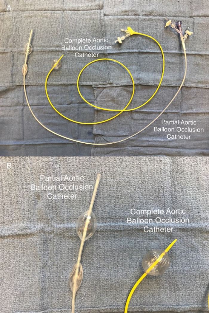
Figure 1: Aortic balloon occlusion catheters. (A) Partial aortic occlusion is achieved using a selective aortic balloon occlusion in trauma (SABOT) catheter, while complete aortic occlusion is achieved using the complete aortic balloon occlusion catheter. (B) The partial aortic balloon occlusion catheter is a two-balloon system that allows an intra-luminal blood flow providing a distal aortic flow. Complete aortic occlusion is provided using a single-balloon system. Please click here to view a larger version of this figure.
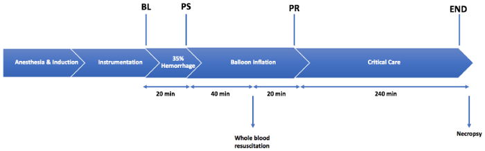
Figure 2: Injury protocol. An injury consisting of a 35% total blood volume hemorrhage is followed by a 1 h period of aortic balloon occlusion. A resuscitation is performed with 20% whole blood over 20 min, after 40 min of balloon occlusion. The animals are monitored in the critical care phase for 4 h following the balloon deflation. BL = the baseline; PS = post-shock; PR = post-resuscitation period. Please click here to view a larger version of this figure.
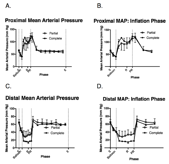
Figure 3: Hemodynamic response to the injury and balloon inflation. These panels show the intraoperative measurements of (A) the proximal pean arterial pressure (MAP), (B) the proximal MAP during the balloon inflation, (C) the distal MAP, and (D) the distal MAP during the balloon inflation. The data are presented as the group mean ± the standard error (SE). S = the shock period (20 min); Balloon = the balloon inflation (60 min); R = the resuscitation (20 min); PR = the post-resuscitation period/balloon deflation; E = the end of the injury phase (5 h following the shock period completion); Complete = the complete aortic balloon occlusion catheter; Partial = the partial aortic balloon occlusion catheter. Please click here to view a larger version of this figure.
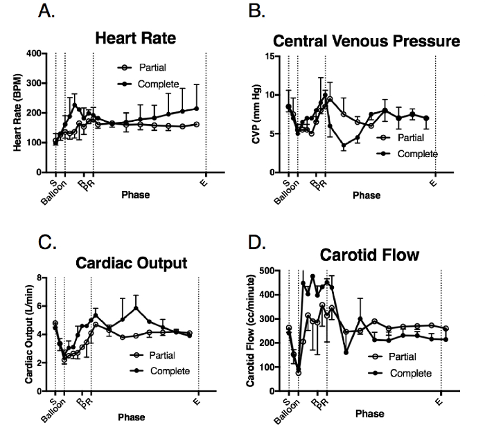
Figure 4: Systemic and physiologic response to the injury and balloon deployment. These panels show the intraoperative measurements of (A) the heart rate (HR), (B) the central venous pressure (CVP), (C) the cardiac output (CO), and (D) the carotid flow (CF). The data are presented as group mean ± SE. S = the shock period (20 min); Balloon = the balloon inflation (60 min); R = the resuscitation (20 min); PR = the post-resuscitation period/balloon deflation; E = the end of the injury phase (5 h following the shock period completion); Complete = the complete aortic balloon occlusion catheter; Partial = the partial aortic balloon occlusion catheter. Please click here to view a larger version of this figure.
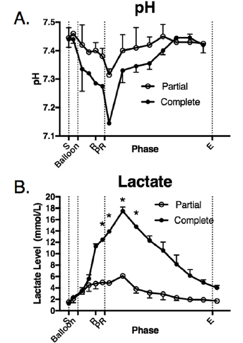
Figure 5: Laboratory parameters in response to the injury and balloon inflation. These panels show the intraoperative measurements of (A) pH and (B) lactate. The data are presented as group mean ± SE. The asterisks indicate the time points that were significantly different (p < 0.05). S = the shock period (20 min); Balloon = the balloon inflation (60 min); R = the resuscitation (20 min); PR = the post-resuscitation period/balloon deflation; E = the end of the injury phase (5 h following the shock period completion). Complete = the complete aortic balloon occlusion catheter; Partial = the partial aortic balloon occlusion catheter. Please click here to view a larger version of this figure.
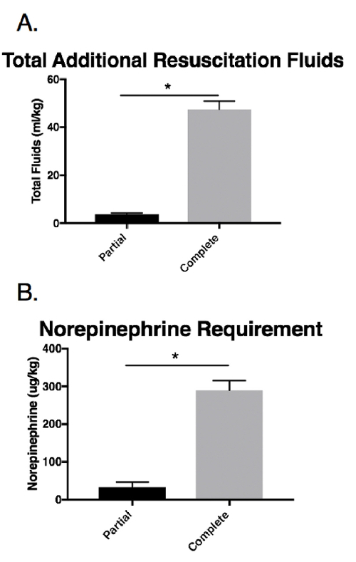
Figure 6: Resuscitation requirements in response to the injury and balloon inflation. These panels show the intraoperative measurements of (A) total additional resuscitation fluids and (B) the norepinephrine use. The data are presented as group mean ± SE. The asterisks indicate significant differences (p < 0.05). Complete = the complete aortic balloon occlusion catheter; Partial = the partial aortic balloon occlusion catheter. Please click here to view a larger version of this figure.
In this protocol, we highlighted a hemorrhagic shock model in swine. This model has been shown to be both reliable and reproducible16,17,18,19. Models similar to this have been employed in several scientific studies investigating the effects of hemorrhagic shock on animal physiology16,20. Furthermore, this model has also been used for testing both pharmacologic and surgical treatment interventions in hemorrhagic shock with marked success12,13,16,19,21.
This model comprises several steps that require great attention to detail. The intubation of a swine is a complex procedure since the animal has a long, beak-like snout and a narrow, long oropharyngeal cavity. Additionally, swine generally have a high tendency to undergo laryngospasm, making orotracheal intubation even more challenging22. An appropriate induction of anesthesia, promoting good muscular relaxation, should be achieved before attempting the intubation. In our experience, having an assistant to use surgical cotton ropes to lift the mandible and tongue and depress the maxilla helps facilitate a wider opening of the oral cavity for easier intubation. If the intubation becomes difficult due to laryngospasm, topical xylocaine spray should be used. Although not utilized in this protocol, a neuromuscular blocker can be administered to promote laryngeal relaxation. Using a neuromuscular blocker, however, requires close supervision by a trained professional. We have also found it helpful to apply lubricating gel to the end of the endotracheal tube, as well as rotating the tube while advancing it through the laryngeal opening. Following the intubation, the endotracheal tube placement should be confirmed with end-tidal CO2. Nevertheless, although pigs can be intubated in various positions, we find intubation in the supine position to be the easiest, especially if the individual performing the intubation has experience with human intubation.
Femoral artery and vein cannulation can be technically challenging. The use of good retraction is important and can be achieved by using self-retaining retractors. An additional retractor, such as an Army-Navy, may be used if needed. Care should be taken while dissecting the neurovascular bundle, since the femoral nerve, which is the lateral-most structure in the bundle, has to be preserved. This is particularly important if the animal needs to survive the experiment. In addition, the cannulation of the right femoral artery is a critical step to the experiment. Following the guidewire cannulation into the vessel, a 14 Fr insertion sheath is inserted. A successful execution of this step requires an initial dilatation with a 10 Fr dilator to upsize the vessel. Also of prime importance is compressing the femoral artery at the site of arteriotomy, following the removal of the 10 Fr dilator, to minimize blood loss. Although not routinely performed in animal studies, gaining proximal and distal control before performing the arteriotomy and venotomy, as demonstrated in this study, can help minimize the bleeding and allow for troubleshooting, should problems arise during the cannulation.
An appropriate positioning and deployment of the aortic balloon occlusion catheters are critical. Caution should be practiced while advancing the catheter inside the aorta, as aggressive manipulation can result in an iatrogenic injury to the femoral artery or the aorta. Although several locations may be targeted for the deployment of the catheter, we chose to position the occlusion balloons in aortic Zone 1, which is the supraceliac aorta. The balloon positioning can be confirmed by manual palpation or fluoroscopy; however, ultrasonography can also be used to easily confirm the balloon placement, which was used for this study. Following the appropriate positioning, the balloon inflation should be conducted with care. In general, balloons should be inflated slowly until no further decrease in the distal MAP is noted. Overinflation of the balloon can potentially cause balloon rupture, which may precipitate an aortic injury. Close attention to the proximal and distal MAP helps to achieve the desired degree of aortic balloon occlusion, whether partial or complete.
Insertion sheaths and aortic balloon occlusion catheters have become smaller in profile in recent years. In this study, we used a 14 Fr insertion sheath before advancing the partial aortic balloon occlusion catheter (i.e., SABOT) into the femoral artery. Currently, this catheter is in phase I of its development, with plans for a future revision involving adjustable balloons and the distal aortic flow, as well as a smaller, low-profile system. Smaller 7 Fr catheters, however, have gained popularity in recent years, as they are associated with fewer ischemic complications. Smaller, low-profile sheaths and aortic balloon occlusion catheters may also be used for deployment in this hemorrhagic shock model, with excellent results.
Several models of hemorrhage are used to test hemorrhagic shock in large animals23,24,25. We employ a fixed-volume model of hemorrhage. In this model, a predetermined hemorrhage volume, which is based on a calculated TBV, is withdrawn from the body over a set period of time. We used a 35% TBV hemorrhage over 20 min, which is commonly used in fixed-volume hemorrhagic shock models26,27,28,29. This model is widely used to investigate shock-induced physiologic changes and compensatory mechanisms, as well as pathophysiologic responses, in hemorrhagic shock. Although this method is highly popular, the degree of shock that is induced as a result of the fixed-volume hemorrhage varies from animal to animal. Furthermore, as the blood-volume-to-body-weight ratio varies, it is important to control for weight in this model in order to achieve reproducible results. Other model types in practice include a fixed-pressure hemorrhage model, an uncontrolled hemorrhage model, and a hemorrhage model with ischemic markers as endpoints. Each of these models, however, has its own set of limitations.
Controlled hemorrhage models have been used to test aortic balloon occlusion catheters with success12. In this study, we used a closed hemorrhage system because this type of hemorrhage model can be employed in a wide variety of experiments. Our goal was to provide readers with the foundation to replicate a hemorrhagic shock model and to deploy aortic balloon occlusion catheters. However, to create the most clinically relevant and meaningful comparison of partial versus complete aortic occlusion, these catheters should ultimately be tested in the setting of an ongoing distal hemorrhage. In combination with other traumatic insults, this model of hemorrhagic shock can be extrapolated to a more clinically realistic model of traumatic injuries16,18.
Resuscitation strategies following traumatic injuries in animal models vary widely. While some are proponents of 'fluid responsiveness'-guiding requirements for ongoing resuscitation28, others propose objective thresholds for administering fluid boluses and vasopressors21,26. In this study, we employed thresholds to determine the fluid bolus administration and vasopressors use for their ease of reproducibility. Although 'fluid responsiveness' replicates clinical practice, objective thresholds for fluid administration and vasopressors may limit a wide variability and the subjectivity of resuscitation requirements in hemorrhagic shock models.
For years, swine have been used in various models of hemorrhagic shock that have provided opportunities to test a wide range of treatment strategies11,12,13,16,17,19,20,21,30. However, it is important to realize that swine are not the perfect animal model and physiologic changes do not exactly translate to humans. For example, some researchers may recommend splenectomy prior to the hemorrhagic shock to better mimic the human physiology, although this is controversial topic31.
In conclusion, this protocol demonstrates the basic foundation for replicating a hemorrhagic shock model in swine and for the deployment of aortic balloon occlusion catheters. The findings of a study that used a similar model of hemorrhagic shock are currently being used in Phase II clinical trials investigating the role of valproic acid (VPA) in traumatic injuries16,19,32,33,34. Also, to be noted is the importance of the role of aortic balloon occlusion catheters in the present era. Aortic balloon occlusion catheters have not only found an application in hemorrhagic shock, they are also being used in cardiac and vascular surgeries, as well as in high-risk elective surgical procedures where a control of the aortic flow is useful in an otherwise devastating circumstance. Overall, we feel that the swine model of hemorrhagic shock described and the aortic balloon occlusion are highly relevant and can be employed in a multitude of experimental studies.
The authors have nothing to disclose.
We would like to acknowledge Rachel O'Connell, and Jessica Lee for their assistance with the animal studies. We would also like to acknowledge Maj. General Harold Timboe, MD, MPH, U.S. Army (Ret.), who has been an advisor and mentor for this project.
Yorkshire-Landrace Swine | Michigan State University Veterinary Farm | ||
Anesthesia: Telazol | Pfizer | Dose: 2-8 mg/kg; IM | |
Anti-cholinergic: Atropine | Pfizer | Dose: 1mg, IM | |
Anesthesia: Isoflurane | Baxter | Dose: 1-5%, INH | |
Betadine | Humco | ||
Alcohol 70% | Humco | NDC 0395-4202-28 | |
Datex-Aespire Anesthesia Machine | GE Healthcare | 7900 | |
Endotracheal tube | DEE Veterinary | 20170518 | Appropriate size for animal (6.5 or 7.0F) |
Laryngoscope | Miller | 85-0045 | |
Stylet | Hudson RCI | 5-151–1 | |
Jelco 20G IV Catheter | Smiths Medical | 4054 | |
Operating Room Monitor (Vital Signs Monitor) | SurgiVet Advisor | V9201 | May require at least 2 |
Surgical Gowns | Kimberly Clark | 90142 | Use appropriate size for surgeon. |
Sterile surgical gloves | Cardinal Health (Allegiance) | 22537-570 | Use appropriate size for surgeon. |
Cautery Pencil | Medline | ESPB 2000 | |
Suction tubing | Medline | DYND50251 | |
Sunction tip: Yankauer | Medline | DYND50130 | |
Bovie Aaron 1250 Electrocautery Unit | Bovie Medical Co. FL | BOV-A1250U | |
Salpel Blade – Size #10 | Cardinal Health (Allegiance) | 32295-010 | |
Scalpel Handle | Martin | 10-295-11 | |
Debakey Forceps | Roboz | RS-7562 | |
Weitlander Retractor | Roboz | RS-8612 | |
Mayo Scissors | Roboz | RS-76870SC | |
Army-navy Retractor | Teleflex | 164715 | |
Mixter Right-angle Forceps | Teleflex | 175073 | |
5F (1.7 mm) 11 cm Insertion Sheath with 0.35" Guidewire | Boston Scientific | 16035-05B | |
8F (2.7 mm) 11 cm Insertion Sheath with 0.35'' Guidewire | Boston Scientific | 16035-08B | |
20G angled Introducer Needle | Arrow | AK-09903-S | |
14F (4.78 mm) 13 cm Insertion Sheath with 10F dilator | Cook Medical | G08024 | |
2-0 Silk 18'' 45 cm | Ethicon | A185H | |
3-0 Vicryl 36'' 90 cm | Ethicon | J344H | |
3-0 Nylon 18'' 45 cm | Ethicon | 663G | |
4-0 Prolene 30'' 75 cm | Ethicon | 8831H | |
20 ml syringe | Metronic/Covidien | 8881512878 | |
3 mL syringe | Metronic/Covidien | 1180300555 | |
6 mL syringe | Metronic/Covidien | 1180600777 | |
1000ml 0.9% Saline | Baxter | 2B1324X | |
Foley Catheter (18F 30 cc) | Bard | 0166V18S | |
Urinary Drainage Bag | Bard | 154002 | |
9F 10 cm Insertion Sheath | Arrow | AK-09903-S | |
Swan-Ganz pulmonary artery catheter (8F) | Edwards Lifesciences co. CA | 746F8 | |
Carotid Flow Probe System | Transonic, Ithaca, NY | 3, 4, or 6 mm probes | |
SABOT catheter | Hayes Inc. | ||
CODA balloon catheter | Cook Medical | 8379144 | |
Ultrasound, M-Turbo | SonoSite | ||
Amplatz Stiff Guidewire (0.035 inch, 260 cm) | Cook Medical | G03460 | |
Arterial Blood Gas Syringes | Smiths Medical | 4041-2 | |
Arterial Blood Gas Analyzer | Nova Biochemical | ABL800 | |
Masterflex Pump | Cole Palmer | HV-77921-75 | |
Blood Collection Bags | Terumo | 1BBD606A | |
Macro IV drip set | Hospira | 12672-28 | |
Pentobarbital | Pfizer | Dose: 100 mg/kg; IV | |
Eppendorf Tubes | Sorenson | 11590 | |
50 cc conical tubes | Falcon | 352097 | |
Formalin | Fisherbrand | 431121 | |
Bair Hugger Normothermia System | Arizant Healthcare, Inc. |