A subscription to JoVE is required to view this content. Sign in or start your free trial.

Method Article

Two-Dimensional Semi-Denaturing Agarose Gel Electrophoresis: A Technique to Separate Polymorphic Amyloids Fibers Based on Size Heterogeneity

2.4K views

⸱

July 8th, 2025

In This Article

Abstract

Source: Hanna-Addams, S., et al. Use of Two Dimensional Semi-denaturing Detergent Agarose Gel Electrophoresis to Confirm Size Heterogeneity of Amyloid or Amyloid-like Fibers. J. Vis. Exp. (2018).

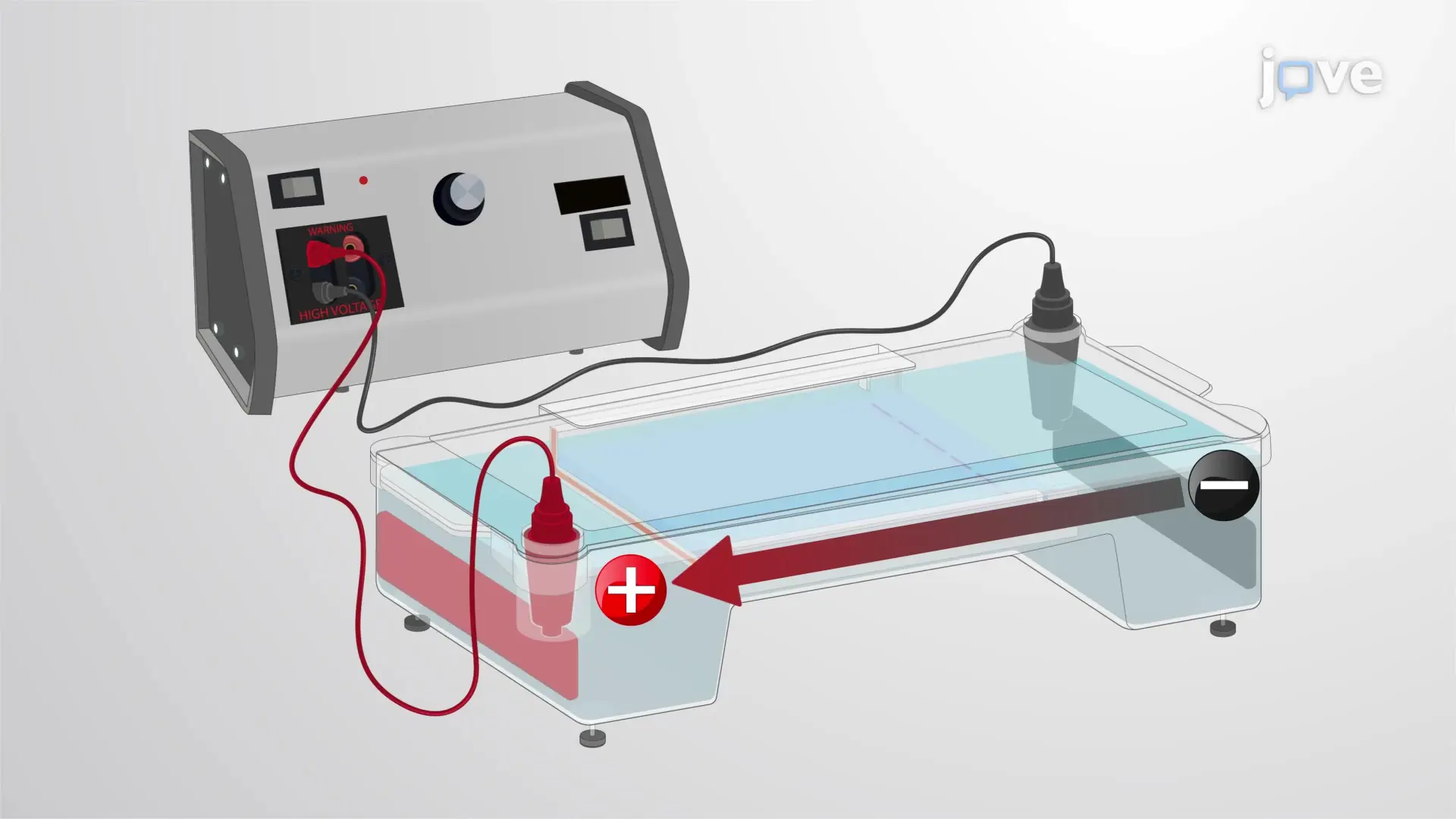
This video demonstrates the two-dimensional semi-denaturing detergent agarose gel electrophoresis-based separation of amyloid fibers. The technique separates the polymorphic amyloid aggregates based on their heterogeneous size.
Protocol

1. Prepare Samples

  1. Culture 2 x 10⁶ amyloid producing HT-29 colon cancer cells in a 10-cm tissue culture dish in 10 mL of Dulbecco's Modified Eagle Medium (DMEM) containing 10% fetal bovine serum and penicillin-streptomycin. Culture cells overnight in a 37 °C incubator with 5% CO2.
    1. After the cells grow to 80% confluency, wash the cells with 10 mL of phosphate-buffered saline (PBS). Add 3 mL of Trypsin solution and incubate at 37 °C for 3 min.
    2. After the cells are totally dissociated from the dish, add 10 mL of culture medium and transfer the cells with a 10-mL pipette to a 15-mL conical ....

Access restricted. Please log in or start a trial to view this content.

Results

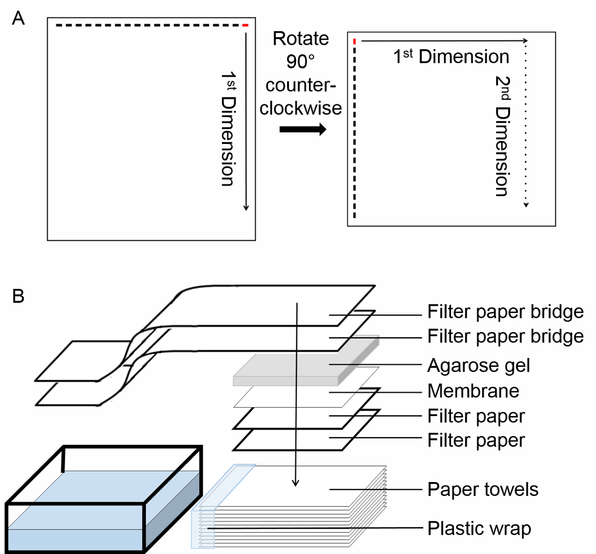
2D electrophoresis diagram; gel rotation, filter paper setup, and transfer process for protein analysis.
Figure 1. Experimental protocol. (A) Schematic of the two-dimensional semi-denaturing detergent agarose gel electrophoresis (2D SDD-AGE). Begin by loading the sample in the right most lane, labeled in red. After termination of the first dimension run,.......

Access restricted. Please log in or start a trial to view this content.

Disclosures

No conflicts of interest declared.

Materials

List of materials used in this article
NameCompanyCatalog NumberComments
gel electrophoresis unitFisherHE99XPROappratus for gel running.
agroseVWR97062-250For agarose gel.
DMEMSigmaD6429for cell culture
fetal bovine serumSigmaF4135for cell culture
penicilin-streptomycinSigmaP4333for cell culture
Trypsin solutionSigmaT4049for cell culture
PBS for tissue cultureSigmaD8662for cell culture
recombinant TNFmade in our labfor inducing necroptosis.
smac-mimeticgift from Dr. Xiaodong Wangfor inducing necroptosis. See reference 11.
ZVAD-FMKApexBioA1902for inducing necroptosis. See reference 11.
Cell CounterBio-Rad1450102Model TC20; for counting cells
Pierce™ BCA Protein Assay KitThermo Scientific23225for measuring protein concentration in cell lysates
Cell lifterFisher07-200-364to remove cells from dish
Lysis Buffer (1 L)20 mL 1 M Tris pH 7.4<br/> 10 mL glycerol<br/> 30 mL 5 M NaCl<br/> 840 mL ddH<sub>2</sub>O<br/> 10 mL Triton-X100<br/> (protease and phosphates inhibitors as desired)
10X TAE (1 L)48.4 g Tris base<br/> 11.42 mL glacial acetic acid<br/> 20 mL 0.5M EDTA pH 8<br/> ddH20 to 1 L
4X SDD-AGE loading buffer (50 mL)5 mL 10X TAE<br/> 10 mL glycerol<br/> 4 mL 20% SDS<br/> 0.5 mL 10% bromophenol blue<br/> 31 mL ddH<sub>2</sub>O
PBST Wash Buffer (1 L)100 mL 10xPBS<br/> 800 mL ddH<sub>2</sub>O<br/> 1 mL Tween20
10X PBS (10 L)800 g NaCl<br/> 20 g KCl<br/> 144 g Na<sub>2</sub>HPO<sub>4</sub>&middot;2H<sub>2</sub>O<br/> 24 g KH<sub>2</sub>PO<sub>4</sub><br/> add ddH<sub>2</sub>O to 10 L

Explore More Articles

Two-Dimensional Gel ElectrophoresisAmyloid Fiber SeparationSize Heterogeneity AnalysisSDS-Containing GelFirst Dimension RunSecond Dimension RunGel Preparation ProtocolTAE Buffer SDSAmyloid Fibril Analysis