1. Preparation of Perfusion and Isolation Solutions
- Prepare the solutions required for the perfusion of the liver piece and the isolation of hepatocytes according to Table 1. Solutions can be stored at 4 °C until use.
- Sterile filter all solutions using a 0.22 μm filter.
- All solutions that come into contact with the liver should be sterile.
2. Preparation of Perfusion Equipment and Solutions
- The equipment for the perfusion of the liver piece should be set up as shown in Figure 1.
- The water bath should be set at an appropriate temperature, which is different in each particular experimental set-up, such that the solutions are at the temperature of 37 °C when they reach the liver piece. In this case, the water bath is set at 41 °C to warm the Solutions 1, 2 and 3 and the jacketed glass condenser. Solution 4 should be warmed up to 37 °C in a separate water bath for use to reduce the loss of collagenase activity.
- Shortly before liver perfusion, turn on the regulator of the gas tank containing 95% O2/5% CO2 to gas the oxygenation apparatus (Figure 1E).
3. Perfusion of the Liver
- A liver piece with as much intact Glisson’s capsule as possible and ideally with only 1 cut surface should be obtained from a pathologist for perfusion.
- Place this liver piece on the Büchner funnel that contains a perforated filter disc (Figure 1B).
- The perfusion system should be primed with Solution 1.
- With a low flow rate, curved irrigation cannulae with olive tips should be inserted into the larger blood vessels on the cut surface of the liver piece. As blood flushes out from the liver, the tissue becomes lighter in areas with good perfusion. The number of cannulae used for various sizes of livers is shown in Figure 2A. The gauge size chosen should result in a snug fit that will hold the cannulae in place. The smaller blood vessels should be left open for the perfusion buffer to drain out of the liver piece.
- Increase the flow rate on the peristaltic pump to between 110-460 ml/min depending on the size of the liver (Figure 2B). This results in an average flow rate of 44±16 ml/min per cannula (Figure 2C). The speed chosen depends on the liver piece and should result in a slight plumping up of the liver piece. In some cases, it may be necessary to clamp shut some of the open vessels with micro vascular clamps to achieve the slight plumping mentioned above. A good perfusion can be observed when the liver piece is a lighter color throughout.
- Keep the liver piece moist during perfusion by covering it with a piece of gauze soaked in saline.
- Perfuse with 1 L of Solution 1 to flush out any remaining blood in the liver piece.
- Change the perfusion fluid to Solution 2 and perfuse for 10 min.
- Switch the perfusion fluid to Solution 3 and perfuse with 0.5 L.
- Change the perfusion fluid to Solution 4, which contains 0.1-0.15% of collagenase (Table 2).
- For this step, perfusion should be carried out in a recirculating manner for 9-12 min or until the liver is sufficiently digested; the liver tissue should appear to break apart slightly under the Glisson’s capsule and feel softened when probed with the blunt side of a scalpel.
4. Isolation of Hepatocytes
- Turn off the peristaltic pump and remove cannulae from the liver piece.
- Place the liver piece in a crystallizing dish containing 100-200 ml of Solution 5.
- Remove the Glisson’s capsule carefully and gently shake out the cells. If there are regions that are not well perfused, a scalpel can be used to cut through these regions to release cells contained within. Add more Solution 5 as needed during the process.
- Add more Solution 5 until a final volume of 500 ml is reached.
- Filter cell suspension twice; first through a 210 μm nylon mesh followed by a 70 μm nylon mesh. Next, pour the cell suspension into 200 ml centrifuge tubes.
- Centrifuge the cell suspension at 72 g for 5 min at 4 °C. Aspirate supernatant and gently resuspend cell pellet gently in 200 ml of Solution 5.
- Repeat the washing step number 4.6 three times. On the final centrifuge step, resuspend cells in cold storage solution (see list of materials). Cells should be approximately 2-5 million hepatocytes per milliliter for assessment of yield and viability using a hemocytometer-based trypan blue exclusion assay.
- To carry out a trypan blue exclusion assay, add 0.1 ml of appropriately diluted cells (≈2-5 million/ml) to a microfuge tube containing 0.5 ml of trypan blue solution (0.4% trypan blue dissolved in phosphate-buffered saline (PBS)) and 0.4 ml of PBS. After mixing the cell suspension thoroughly, load a hemocytometer with the suspension and examine under a microscope at 100X magnification. Under microscopy, the dead cells will be stained blue while the live cells appear unstained. Count the number of live and total cells in each of the 1 mm2 grids marked on the hemocytometer. Viability (%), yield of live cells (million hepatocytes per ml cold storage solution (CSS) or million hepatocytes per g liver) can be calculated using the formulae below.
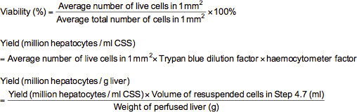
Note: In this case, the trypan blue dilution factor is 10 and the hemocytometer factor is 10,000.
Perfusion Setup
The equipment required for liver perfusion should be set up according to Figure 1.
Viability and Yield of Isolated Human Hepatocytes
The average viability of isolated human hepatocytes was 77±10% and the average yield of hepatocytes was 13±11 million hepatocytes/g liver, with values expressed as means ± standard deviation. The number of hepatocyte isolations carried out to obtain these averages was 648 isolations carried out from January 1999 to December 2012.
Suitable Perfusion Parameters
In order to carry out a successful flushing and perfusion of the liver, the number of cannulae used should vary according to the weight of the liver (Figure 2A). In general, 4-8 cannulae should be used for livers ranging from below 20 g to over 80 g. A suitable rate of perfusion, which is also dependent on liver weight, should be chosen for a successful perfusion of the liver (Figures 2B and C). It has been found that an average perfusion speed of 44 ml/min cannula-1 is ideal across a range of different liver weights and therefore the perfusion speeds should be adjusted appropriately if more cannulae are used. If perfusion is successful, the liver should be pale in color and slightly plumped up.
Purity of Hepatocytes
By means of immunofluorescence, it was found that isolated hepatocytes, which stain positively for albumin, had a purity of 94±1% (N = 4 with 5 replicates each) (Figures 3A and B). Figure 3C is a representative phase contrast image showing the morphological characteristics of hepatocytes such as large cell size and polygonal shaped-cells.
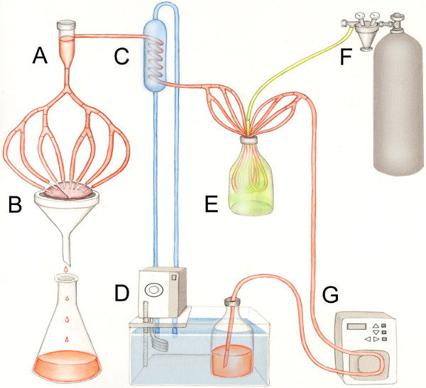
Figure 1. Perfusion setup. (A) bubble trap, (B) liver piece with curved irrigation cannulae with olive tips inserted in blood vessels on a Büchner funnel, (C) glass jacketed condenser, (D) water bath, (E) oxygenation apparatus, (F) 95% O2/5% CO2 gas tank and (G) peristaltic pump.
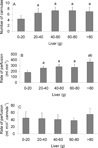
Figure 2. (A) The number of cannulae used, (B) perfusion rate (ml/min), or (C) perfusion rate (ml/min.cannula) for various sizes of liver (g). Values represent means ± standard deviation with N = 25, 41, 18, 14 and 9 for 0-20 g, 20-40 g, 40-60 g, 60-80 g and >80 g liver respectively. aSignificantly different from the 0-20 g condition, P<0.05. abSignificantly different from the 20-40 g, 40-60 g and 60-80 g condition, P<0.05.
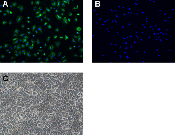
Figure 3. Immunofluorescent images of isolated cells positive for (A) albumin (stained in green) and the corresponding (B) negative control (200X magnification). Nuclei are stained in blue using DAPI. (C) Phase contrast image of isolated cells (100X magnification).
| Solution | Constituent | Final Concentration |
| Solution 1 | Sodium chloride | 154 mM |
| HEPES | 20 mM | |
| Potassium chloride | 5.6 mM | |
| Glucose | 5 mM | |
| Sodium hydrogen carbonate | 25 mM | |
| Solution 2 | Sodium chloride | 152.5 mM |
| HEPES | 19.8 mM | |
| Potassium chloride | 5.5 mM | |
| Glucose | 5.0 mM | |
| Sodium hydrogen carbonate | 24.8 mM | |
| EGTA | 1 mM | |
| To prepare Solution 2, add 10 ml of 100 mM EGTA to 990 ml of Solution 1. | ||
| Solution 3 | Sodium chloride | 152.5 mM |
| HEPES | 19.8 mM | |
| Potassium chloride | 5.5 mM | |
| Glucose | 5.0 mM | |
| Sodium hydrogen carbonate | 24.8 mM | |
| Calcium chloride dihydrate | 5 mM | |
| To prepare Solution 3, add 10 ml of 0.5 M calcium chloride dihydrate to 990 ml of Solution 1. | ||
| Solution 4 | Sodium chloride | 152.5 mM |
| HEPES | 19.8 mM | |
| Potassium chloride | 5.5 mM | |
| Glucose | 5.0 mM | |
| Sodium hydrogen carbonate | 24.8 mM | |
| Calcium chloride dihydrate | 5 mM | |
| Collagenase | See Table 2 | |
| To prepare Solution 4, add appropriate amount of collagenase to Solution 3. | ||
| Solution 5 | Sodium chloride | 120 mM |
| HEPES | 10 mM | |
| Calcium chloride dihydrate | 0.9 mM | |
| Potassium chloride | 6.2 mM | |
| Albumin | 0.1% w/v | |
Table 1. Perfusion and isolation solutions.
| Size of liver piece (g) | Collagenase concentration (%) | Collagenase activity (U/ml) |
| <25 | 0.10 | 250 |
| 25 – 40 | 0.11 | 300 |
| 41 – 80 | 0.13 | 350 |
| >80 | 0.15 | 400 |
Table 2. Collagenase concentrations (%) and activities (U/ml) to be used for various sizes of liver pieces (g).
