1. Generating Elastomeric Stamps
- To generate the PDMS stamp’s silicon masters, design the photomask’s feature patterns using computer-aided design software.
- Design the first pattern as a 20 x 20 array of annuli with 300 μm inner diameter (ID) and 600 μm OD with 1,200 μm center-to-center spacing.
- Design the second pattern as a 20 x 20 array of annuli with 600 μm ID and 900 μm OD with 1,200 μm center-to-center spacing.
- Additionally, place 1 x 1 mm2 square reference marks at all four corners of each array design spaced 1,200 μm center-to-center from the corner pattern at a 45° angle.
- Fabricate the silicon masters for use in this experiment with 1:1 aspect ratios, correlating to a 300 μm feature depth using standard lithography techniques detailed elsewhere15 or in collaboration with a microfluidic foundry.
NOTE: Feature depths lower than 100 μm can lead to abnormal deformation of stamps prior to contact with substrate surfaces.
NOTE: This protocol begins with having already obtained silicon masters with the described patterns of photoresist, which requires specialized equipment and clean rooms to create. It is best to consult with a fabricator or core facility to create these patterned masters.
- Silanize silicon masters O/N by incubation with (tridecafluoro-1,1,2,2-tetrahydroocty) trichlorosilane vapor.
NOTE: Silane vapor is highly toxic and should only be handled in a chemical fume hood. - Create inverse replicas of the silicon masters by curing a 10:1 ratio of PDMS pre-polymer and PDMS curing agent on top of the silanized silicon masters in a petri dish O/N at 60 °C.
- Remove the PDMS stamps from the silicon masters, and bond the stamps to acrylonitrile butadiene styrene (ABS) backings or any other rigid materials using glue.
NOTE: The material of choice need not be transparent or biocompatible. However, it must provide a flat surface for interface with the robotic tooling and higher rigidity than PDMS.
2. Preparing Coverslides
- Rinse microscope coverslides (24 mm x 50 mm #1) in toluene, methanol and sonicate in acetone for 1 min before rinsing with ethanol and drying under a gentle nitrogen stream.
NOTE: Handle toluene and acetone only in a chemical fume hood. - Coat coverslides with ~3.5 nm titanium (Ti) followed by 18.0 nm gold (Au) using a focused electron-beam evaporator.
NOTE: This procedure is compatible with a variety of gold-coated substrates including silicon and polystyrene. While it is possible to generate these on-site, gold-coated substrates are also available via commercial suppliers.
NOTE: Ti layers should be at least 3 nm thick, while Au layers on substrates should be at least 5 nm thick and can range all the way to 1mm in thickness depending on the ideal optical properties of the final substrate16,17. Substrates are not required to be optically clear for fabrication; however, it facilitates microscopic analysis of fabricated substrates. - Rinse gold-coated coverslides with ethanol and dry under gentle nitrogen immediately before use.
3. R-μCP of Outer Annulus
- Before R-μCP with the selectively compliant articulated robot arm (SCARA) system, calibrate the robotic tool effectors and accompanying dual camera systems using the system’s software.
NOTE: These implements are pictured in Figure 1.
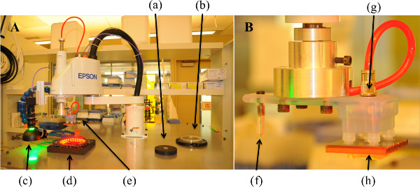
Figure 1. R-μCP System and Robotic Arm Tooling. (A) Large-scale view of R-μCP system with all tooling and fixtures, (a) vacuum chuck, (b) reagent bath, (c) downward facing camera, (d) stamp nesting fixture, (e) robotic tool. (B) Robotic arm tooling depicting (f) diamond-tipped etching tool and (g) pneumatic suction tool holding a (h) ABS-backed PDMS stamp.
- Manually place a PDMS stamp with 600 μm ID and 900 μm OD protruding annuli face down in the stamp nesting fixture. Manually place a freshly cleaned gold-coated coverslide on top of the vacuum chuck, displayed in Figure 1A, and immobilize it using the connected lab vacuum.
- Using the robot controls, align the downward facing camera over the center point of the gold-coated coverslide, as in Figure 2A.
NOTE: This point can initially be defined by visual inspection and does not require great accuracy, as the PDMS stamp is much larger than the gold-coated slide. - Instruct the robotic arm to etch four reference “X” marks at the vertices of a square with dimensions 3.8 mm by 3.8 mm centered on the gold-coated coverslide using the diamond-tipped etching tool attached to the robotic arm. (Depicted in Figure 2B; AUTOMATED PROCESS)
NOTE: This ensures all four reference marks are within one visual frame of the downward facing camera. - Using the robots pneumatic suction tooling, pick-up and hold the PDMS stamp 1 mm above the stamp nesting fixture as in Figure 2C. (AUTOMATED PROCESS)
- Using the upward facing camera and LED illumination ring pictured in Figure 2D and E, visualize the stamp’s square reference marks and identify them with the camera software 10 times in order to determine the stamp’s average X, Y, and angular offset from the center of the robot tooling axis. (AUTOMATED PROCESS)
- As in Figure 2A, use the downward facing camera to visualize and detect the coverslide’s etched reference marks 10 times to determine the coverslide’s average X, Y center locations and angular offset from the center of the robot tooling axis. (AUTOMATED PROCESS).
NOTE: An internal computation is performed by the robotic system’s software to precisely align the stamp’s and coverslip’s X, Y center locations and angular offsets in future R-μCP steps. - 3.8) Place the stamp in a bath of alkanethiol ATRP initiator, ω-mercaptoundecyl bromoisobutyrate (2 mM in ethanol) as depicted in Figure 2F. (AUTOMATED PROCESS)
- Remove the stamp from the alkanethiol solution and place it over nitrogen stream pressurized to 0.48 bar (5 psi) to evaporate the ethanol as in Figure 2G. After 1 min increase the nitrogen stream’s pressure is to 1.03 bar (15 psi) to ensure uniform dryness. (AUTOMATED PROCESS)
NOTE: Incomplete drying will result in total or partial loss of pattern fidelity. - After drying, move the stamp over the calculated center position of the gold-coated slide and lower it at 100 μm increments while monitoring Z-axis motor force as depicted in Figure 2H.
NOTE: This is communicated as torque experienced by the Z-axis motor and displayed in the robot guidance software. (AUTOMATED PROCESS) - Stop lowering the stamp once the predetermined pressure of 79.2 kPa has been achieved, and maintain contact with the gold-coated slide for 15 sec. (AUTOMATED PROCESS)
NOTE: The pressure values here are optimized for the current stamp design and feature height. If the stamp design is changed, specifically for high aspect ratio stamps, tune these accordingly by a trial and error process. - Slowly remove the stamp from the gold-coated slide and place the stamp back in the stamp nesting fixture. (AUTOMATED PROCESS)
4. SI-ATRP of PEGMEMA on Micropatterned Coverslides
- Release vacuum pressure holding the micropatterned coverslide, and transfer it to a 50 ml Schlenk flask. Seal and degas the Schlenk flask using a vacuum pump.
- Add 5.5 ml of ATRP reaction mixture containing the macromonomer PEGMEMA (208.75 mmol), water (34.4 ml), methanol (43.8 ml), copper(II) bromide (1 mmol), and 2’,2-bipyridine (3 mmol) to the Schlenk flask.
- Add 0.5 ml of L-sodium ascorbate (454.3 mM) in water to initiate the reaction, and allow it to continue for 16 hr at RT under inert gas.
NOTE: Following addition of L-sodium ascorbate, reaction color will shift from light green to dark brown as shown in Figure 3A-B.
NOTE: The reaction can continue beyond 16 hr, but it will not significantly increase the length of the PEG brushes.
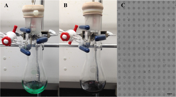
Figure 3. Initiation of SI-ATRP. (A) Initiation of reaction and (B) subsequent color change following addition of L-sodium ascorbate. (C) Microscope image of micropatterned coverslide surface following SI-ATRP procedure. Scale bar 1 mm.
- Remove the micropatterned coverslide from the Schenk flask and rinse with ethanol, water, and ethanol and dry under a gentle nitrogen stream.
NOTE: Following SI-ATRP, surface modifications should be visible to the eye and can be imaged and analyzed under a microscope as in Figure 3C.
NOTE: This is the simplest method to test the precision of the trial as it obviates the need to immunostain the substrates.
5. Azide Functionalization of Micropatterned PEGMEMA Chains
- Place the micropatterned coverslide in a 20 ml glass reaction vial.
- Add 6 ml of N,N-dimethylformamide (DMF) containing 100 mM sodium azide to the reaction vial. Maintain this reaction at 37 °C for 24 hr.
NOTE: Handle DMF only in a chemical fume hood. Exercise caution when weighing out sodium azide. - Upon completion, remove the micropatterned coverslide from the reaction vial and rinse with ethanol then water and dry under a nitrogen stream.
6. Passivation of Bromine Functionalized PEGMEMA Chains
- Place micropatterned coverslide in 20 ml glass reaction vials.
- Add 6 ml of dimethyl sulfoxide (DMSO) containing 100 mM ethanolamine and 300 mM triethylamine to the reaction vial. Hold this reaction at 40 °C for 24 hr.
NOTE: Handle DMSO, ethanolamine, and triethylamine only in a chemical fume hood. - Upon completion remove the micropatterned coverslide from the reaction vial, rinse with ethanol then water and dry under a nitrogen stream.
7. R-μCP of Inner Annulus and SI-ATRP of PEGMEMA
- Carry out R-μCP steps as previously described in Steps 3.2 and 3.6 – 3.12, substituting a PDMS stamp with 300 μm ID and 600 μm OD protruding annuli, such that the annuli with smaller features are placed inside the previously micropatterned annuli.
NOTE: The pressure threshold for this stamp is 132.0 kPa. As previously mentioned, these pressure settings are optimized for the stamp features being used in this experiment. - Carry out SI-ATRP steps as previously described in steps 4.1 – 4.4.
8. Acetylene Functionalization of Micropatterned PEGMEMA Chains
- Place micropatterned coverslide in a 20 ml glass reaction vial.
- Add 6 ml of DMSO containing 100 mM propargylamine to the reaction vial. Maintain this reaction at RT for 24 hr.
NOTE: Handle DMSO and propargylamine only in a chemical fume hood. - Upon completion remove the micropatterned coverslide from the reaction vial and rinse with ethanol then water and dry under a nitrogen stream.
9. Copper-catalyzed “Click” Biotinylation of Acetylene Terminated PEGMEMA Chains
- Place micropatterned coverslide in a 20 ml glass reaction vial.
- Add 6 ml of copper sulfate (15 mM) / Tris[(1-benzyl-1H-1,2,3-triazol-4-yl)methyl]amine (TBTA, 30 mM) (1:1 v/v water/DMF) containing 562 μM azide-PEG4-Biotin conjugate to the reaction vial. Add 1.2 ml of L-ascorbic acid (0.15 mM) in water to the mixture to initialize the reaction.
- Bubble nitrogen through the reaction solution for 10 sec, seal the vial with Parafilm, and react for 24 hr at RT.
- Upon completion remove micropatterned coverslide from the reaction vial, rinse with water and place it in a 12-well polystyrene dish.
10. Immunofluorescent Detection of Biotinylated Acetylene Groups
- Block micropatterned coverslide in DPBS (3% Donkey Serum) for 1 hr at RT. Stain for biotinylated acetylene groups with Streptavidin-546 conjugate (2 μg/ml) in DBPS (3% Donkey Serum in PBS) for 2 hr at RT.
- Rinse micropatterned coverslide 5 times with DPBS for 10 min with gentle agitation.
NOTE: Figure 4B depicts an image of the slide after this step is completed.
11. Copper-free “Click” Biotinylation of Azide Terminated PEGMEMA Chains
NOTE: If desired, this substrate modification step can be performed in situ during cell culture.
- Leave micropatterned coverslide in 12-well polystyrene dish following DBPS rinses.
- Add 2 ml of DBPS (or cell culture media) containing 20 μM DBCO-PEG4-Biotin, and allow this reaction to continue for 24 hr at RT (or at 37 °C in an incubator).
- Upon completion, rinse micropatterned coverslide 5 times with DPBS (or simply perform a media change).
12. Immunofluorescent Detection of Biotinylated Azide Groups
- Stain for biotinylated azide groups with Streptavidin-488 conjugate (2 μg/ml) in 2 ml DPBS (3% Donkey Serum) for 2 hr at RT.
- Rinse micropatterned coverslide 5 times in DPBS for 10 min with gentle agitation.
- Image micropatterned coverslide using confocal fluorescence microscope. Figure 4A-C depicts images of the slide after this step is completed.
NOTE: When using substrates for tissue culture assays, skip sections 10 and 12 of this protocol. After the desired degree of functionalization, sterilize the engineered coverslide by rinsing both sides with 100% ethanol and drying under a nitrogen stream. Transfer the slide to a sterile biological safety cabinet and rinse the slide with PBS five times before proceeding with cell seeding and culture.
The use of manual alignment μCP techniques to engineer culture substrates with arrays of PEG-grafted brushes functionalized with orthogonal “click” chemistries has been demonstrated in previous work6. However, this offers minimal control of pattern orientation and often results in overlap of functionalized areas. Here, a novel R-μCP system is used to overcome this limitation, and its ability to accurately pattern an array of PEG brush annuli with 300 μm ID and 600 μm OD presenting terminal alkyne groups within a separate array of PEG brush annuli with 600 μm ID and 900 μm OD presenting terminal azide groups is demonstrated14. Following the reaction of alkyne presenting PEG brushes with Azide-PEG3-Biotin and the reaction of azide presenting PEG brushes with DBCO-PEG4-Biotin, the substrate was immunostained with fluorescent probes and imaged using a confocal microscope (Figure 4). Analysis of these images using a custom MATLAB program calculated that the two PEG brush arrays were aligned with sub-10 μm accuracy, i.e., X ~ 6.4 μm, Y ~ 1.7 μm, and θ ~ 0.02°, which is at the manufacturer’s cited limit of our SCARA model (i.e., ~10 μm) and indicative of the R-μCP system’s prior performance14. Thus, we believe that the use of higher-end SCARAs in this system would provide even lower, sub-micron resolution. These results demonstrate the versatile substrate engineering capabilities enabled by the combined use of the R-μCP system and sequential nucleophilic substitution reactions. The minimal cross-reactivity evidenced by fluorescent labeling of the orthogonal surface chemistries serves to illustrate the potential of this system for precise immobilization of biochemical cues to generate complex culture substrates for tissue engineering applications.
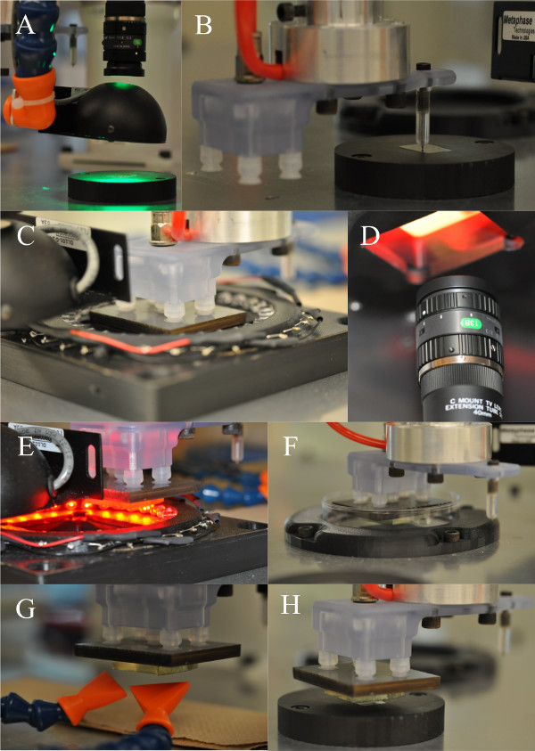
Figure 2. Schematic of R-μCP Process. (A) Downward facing camera visualizing immobilized gold-coated slide, (B) etching reference marks on gold-coated coverslide, (C) removing PDMS stamp from stamp nesting fixture, (D–E) visualizing PDMS stamp reference marks with upward facing camera, (F) placing PDMS stamp in alkanethiol initiator bath, (G) drying alkanethiol initiator solvent over nitrogen streams, and (H) stamping alkanethiol coated PDMS stamp on gold-coated coverslide.
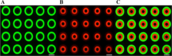
Figure 4. Immunostained Images of MicroPatterned Slide. R-μCP micropatterned coverslide orthogonally functionalized to present terminal (A) azide and (B) alkyne groups, biotinylated using copper-free and copper-mediated click chemistry, and detected with Streptavidin-488 and -546 respectively. (C) Overlay of both fluorescent channels. All scale bars 500 μm.
