Summary

램지 분석의 사용은 유체 분비 및 이온 플럭스 요금을 측정하는<em> 초파리</em> Malpighian하는 Tubule

Published: November 25, 2015
doi:

Summary

이 프로토콜은 초파리에서 격리 Malpighian (신장) 세관에서 유체 분비 속도를 측정 할 수있는 램지 분석의 사용을 설명합니다. 또한, 이온 전극의 특정 사용 transepithelial 이온 플럭스의 계산을 가능하게 분비 유체 나트륨 및 칼륨 농도를 측정하기 위해, 설명한다.

Abstract

신장 상피 이온 수송의 변조는 생물체가 외부 환경 변화에 직면 이온과 삼투압 항상성을 유지할 수 있습니다. 초파리 melanogaster의 Malpighian (신장) 가느 다란 관으로 인해이 생물 및 생리 학적 연구에의 신장 세뇨관의 접근성의 강력한 유전학, 상피 이온 수송의 분자 메커니즘을 연구 할 수있는 최고의 기회를 제공합니다. 여기서는 유체 분비 나트륨 및 칼륨 농도를 측정하는 이온 고유의 전극을 이용하여, 절연 플라이 신 세뇨관에서 유체 분비 속도를 측정하기 램지 분석의 사용을 기술한다. 이 분석은 이온 농도를 측정하는 별도의 장치에 분비 된 유체를 전송할 필요없이, 20 시간에 세관의 transepithelial 유체와 이온 플럭스 ~ 연구를 허용한다. 유전자는 별개의 세뇨관 수송 공정에서 특정 유전자의 역할을 평가하기 위해 분석 될 수있다. 또한, Bathing 식염수 첨가 화학적 특성, 또는 약물 또는 호르몬의 효과를 조사하기 위해 수정 될 수있다. 요약하면,이 기술은 이러한 반송기구의 상피 이온 초파리 세뇨관 운송뿐만 아니라, 규정의 기본 메커니즘의 분자 특성화를 허용한다.

Introduction

신장 상피 이온 수송은 유기체의 iono- 및 삼투의 기초가. 초파리 melanogaster의 Malpighian (신장) 세관 상피 이온 수송의 분자 메커니즘을 연구 할 수있는 최고의 기회를 제공합니다. 이것은 생리 학적 연구에의 신장 세뇨관의 접근성과 짝을 초파리의 강력한 유전학의 조합 때문이다. 기술 1을 개척 연구자의 ​​이름을 따서 명명 램지 분석, 격리 Malpighian 세관에서 유체 분비 속도를 측정하고, 다우와 동료 2에 의해 1994 년에 초파리 년에 설립되었습니다. 이것은 유체 분비 조절 셀 고유 신호 전달 경로를 정의하는, 초파리 등을 GAL4-UAS 시스템 3,4- 유전 도구를 사용하여 추가 연구를위한 기반을 마련. 예는 많은 다른 6,7 사이에 펩티드 호르몬 (5)에 응답하여 칼슘 신호를 포함한다.

비행 세뇨관의 주요 부분에서 염화칼륨이 풍부한 액체의 분비를 통해 발생에 ve_content "> 유전자 기술 및 고전 생리 학적 연구의 조합 뇨 생성을 도시하고있다.이 주로 양이온 평행 transepithelial 분비 통해 이루어 K는 +뿐만 아니라 나트륨 +는 주체 세포, 및 CL를 통해 -. 성상 세포 8-12 내지 분비 별도로 transepithelial K + 및 나트륨 + 플럭스를 측정하는 능력은 유체 분비의 측정보다 반송기구의 상세한 특성화를 허용 홀로. 예를 들어, 자극되지 초파리 세뇨관에서, 나 +가 / K + -ATPase 억제제 인 ouabain 유체 분비 2에 영향을주지 주체 세포 내로의 흡수량은 유기 음이온 수송 체 억제제 타우로 콜산 (13)에 의해 억제 된 경우에도. 그러나, 린톤 및 오도​​넬은 ouabain과는 탈분극 것을 보여 주었다기저 막 잠재력, 그리고 나 + 플럭스 (9)을 증가시킨다. 대표 결과에 도시 된 바와 같이, 우리는 이러한 발견을 복제하고, 14 병용 감소 K + 플럭스가 있음을 보여 주었다; 증가 나 + 플럭스 및 K 감소 + 플럭스 분비없는 순 변화의 결과로 유체 분비에 반대하는 효과를 가지고있다. . 따라서, "ouabain과 모순", 즉 두 가지 해결책이 존재하며, 초파리 세뇨관 유체 분비에 영향을주지 ouabain과 초기 관찰 : 제 자극 세뇨관에서 유체 분비 ouabain과 효과는 명백하지 않다 인한 유기 음이온 수송 (13)에 의해 그 흡수; 둘째, 자극되지 세관에서, ouabain과는 (대표 결과와 심판을 참조하십시오. 9) 유체 분비없는 순 변화의 결과로, transepithelial 나 +와 K + 플럭스에 영향을 반대하고있다. 따라서, 나 +의 주요 역할 / K + -ATP비자극 세뇨관에서 ASE는 나트륨 +는 기저 외측 막에 걸쳐 전송 프로세스 -coupled위한 유리한 농도 기울기를 생성하는 세포 나 + 농도를 낮출 수있다. 실제로, 별도로 나 +와 K + 플럭스를 측정하여, 우리는 세관이 비행 나트륨 – 칼륨 – 2 – 염화 송체 (NKCC)를 부족하지 ouabain과 첨가 한 후 더 이상의 감소, transepithelial K + 플럭스 감소했으며, transepithelial의 변화가 있음을 보여 주었다 나 + 14 플럭스. 이러한 연구 결과는 NKCC를 통해 세포를 입력 나 +가+ / K + -ATPase를 통해 재활용 우리의 결론을지지했다. 다른 예에서, Ianowski는 외. mM의 10 mM 내지 6 욕 K + 농도를 낮추면 유체 분비없는 순 변화와 Rhodnius의 prolixus에서 세뇨관에서 transepithelial의 K + 플럭스 증가 transepithelial 나 + 플럭스 감소 관찰 <sup> 15. 애벌레 세관에서 나 + 플럭스 및 K + 플럭스에 차등 효과는 염분 (17) 양육에 대한 응답으로 다양한 소금 다이어트 (16)와 두 모기 종에 대한 응답으로 초파리 세관에서 관찰되었다.

램지 분석 준비 transepithelial 이온 플럭스의 측정에서 가장 큰 도전은 분비 된 유체 내 이온 농도의 판정이다. 이 문제는 불꽃 photometery (18), 방사성 이온 (19)의 사용, 전자 프로브 파장 분산 분광기 (20)를 포함하여 다양한 솔루션을 만났다되었습니다. 이러한 기술은 이온 농도의 측정을위한 도구로 분비되는 액체 방울의 전송을 필요로한다. 비자극 초파리 세뇨관 분비 유체의 부피가 작기 때문에, 일반적으로 ~ 0.5 NL / 분, 이것은 기술적 도전을 제기하고 분비 된 유체의 일부가 오류가있는 경우에도 소개이전에 잃었다. 대조적으로, 특정 이온 전극의 사용은 인 시츄 (이온 농도가 계산 될 수있는) 이온 활성의 측정을 허용한다. 현재 프로토콜은 K + 이온 운반체 (21)로 발리 노마 이신 (Valinomycin) 사용 Rhodnius의 세뇨관 걸쳐 transepithelial의 K + 플럭스를 측정하도록 Maddrell 동료가 사용하는 각색 및 또한 4- 급의 -butylcalix [4] 아렌 – 테트라 아세트산의 사용을 설명했다 테트라 에틸 에스테르 계 나은 Messerli 특징으로 특이 이온 특정 전극을 +. 등. (22). 이온 특정 전극 성인 9,23과 애벌레 (16) 초파리에서 램지 분석에 Malpighian 세관에 의해 분비되는 유체의 이온 농도를 측정하는 데 사용 된, 뉴질랜드 알파인 웨타 (Hemideina 마오리어) (24) 모기 (17).

여기서는 상세히 램지의 사용 등을 서술유체 분비 초파리 melanogaster의 발 Malpighian 세뇨관의 속도뿐만 아니라 분비 유체 내 K + 및 나트륨 +의 농도를 결정하는 이온 고유의 전극을 사용하므로 transepithelial 이온 플럭스의 계산을 측정하기 말한다. 분석의 개요는도 1에 제공된다.

그림 1

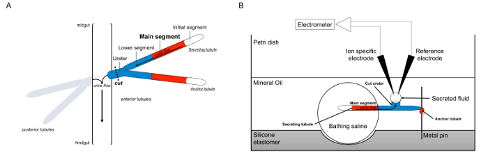
Malpighian 세관의 그림 1. 도식과 이온 농도를 측정하기 위해 이온 특정 전극의 사용과 램지 분석. 이 그림은 램지 분석에 대한 설정을 보여줍니다. (A) 각 플라이는 네 세뇨관, 전방 세뇨관의 쌍 및 후방 세뇨관의 쌍에 의해 둘러싸인 체액 복강 그 플로트있다. 각 쌍에서 두 세관은 중장 hindgu과의 교차점에 뇨를 비운다 요관에서 합류티. 세관은 블라인드 종료됩니다. 소변 (빨간색으로 표시) 유체 분비 주요 세그먼트에 의해 발생하고, 장에 요관으로 밖으로 흐른다. 절개 후 세관 쌍 요관을 직감에 병행하여 해리된다. (B)은 세뇨관의 쌍은 분석 접시의 웰 내에서 입욕 식염수 방울로 전달된다. 여기에 언급 된 두 세뇨관의 일 "앵커 세관은,"금속 핀 감싸 불활성이다. 다른 세관은 분비의 가느 다란 관이다. 초기 세그먼트 (유체 분비하지 않는)과 분비 세관의 주요 부분은 목욕 식염수의 액적 내에 남아있다. 생체 내에서 발생하는 것에 이온과 목욕 식염수에서 주 세그먼트의 세관 루멘에 물 이동, 그리고, 요관으로 이동합니다. 낮은 세그먼트 (파란색) 목욕 식염수 때문에 불활성 밖에 있습니다. 요관이 절단되기 때문에, 유체는 분비 요관의 절단 단부로부터 액적으로 나온다. 티그 분비는 계속 유체 액적 시간 경과 확대 분비, 그 직경은 접안 마이크로 미터를 사용하여 측정된다. 미네랄 오일 층은 분비 된 유체의 증발을 방지한다. 참조 및 이온 특정 전극이 분비 유체의 이온 농도를 측정한다. 이 그림의 더 큰 버전을 보려면 여기를 클릭하십시오.

Protocol

1. 준비 해부, 교정 및 분석 요리 참고 :이 단계에서, 실리콘 엘라스토머 늘어선 세 개의 플라스틱 페트리 접시를 준비한다 : 램지 분석 ( "분석 요리")을 수행하기위한 해부 용 하나, 하나, 교정을 수행하기위한 하나를. 이 요리는 실험 실험에서 다시 사용된다, 따라서이 단계는 접시가 깨진 경우 반복 될 필요가있다. 분석 접시의 그림은 그림 2에 표시됩?…

Representative Results

도 7 및도 8은 유전 학적 및 약리학 적으로 별개의 K + 및 나트륨 + 플럭스만으로는 유체 분비 속도를 측정함으로써 캡쳐되지 않은 정보를 구분할 수있는 K + 및 나트륨 + 농도를 측정하기 위하여 이온 – 특정 전극 램지 분석의 사용을 보여준다.도를 나트륨 + 플럭스 (도 7b)이 변경되지 않은 상태 NKCC에서 동형 널 돌연변이…

Discussion

함께 특정한 이온 전극 램지 분석의 사용은, 절연 곤충 Malpighian (신장)에서 유체 세뇨관 분비 속도 및 이온 플럭스를 측정 할 수있다. 스물 이상의 세관은 체외 microperfused 세뇨관에서 개별의 분석법에 비해 더 높은 처리량을 가능하게 한 번에 분석 될 수있다. 또한, 이온 전극을 특정 제 2 장치로의 유체의 소량의 전사에 도입 될 수있는 오류를 제한 시츄 분비 유체 내 이온 농도?…

Divulgazioni

The authors have nothing to disclose.

Acknowledgements

The authors wish to thank Drs. Sung-wan An and Mike O’Donnell for practical advice on establishing this assay, Dr. Chih-Jen Cheng for helpful discussions on the use of ion-specific electrodes, and Dr. Chou-Long Huang for his mentorship and support. This work was supported by the National Institutes of Health (K08DK091316 to ARR) and the American Society of Nephrology Gottschalk Award to ARR.

Materials

Sylgard 184 Silicone Elastomer Kit Ellsworth Adhesives http://www.ellsworth.com/dow-corning-sylgard-184-silicone-encapsulant-0-5kg-kit-clear/ May be purchased from multiple distributors
Petri dish, polystyrene, 100 mm x 15 mm Fisher FB0875712 Specific brand is not important
Petri dish, polystyrene, 35 mm x 10 mm Corning Life Sciences Fisher 08-757-100A Specific brand is not important
Scalpel Handle #3 Fine Science Tools 10003-12 Specific brand is not important
Scalpel Blades #1 Fine Science Tools 10011-00 Specific brand is not important; use appropriate sharps precautions
Needle, 30G x 1/2 Becton Dickinson 305106 Use appropriate sharps precautions
Minutien pins, black anodized, 0.15 mm Fine Science Tools 26002-15
Stereomicroscope with ocular micrometer Nikon SMZ800 Specific brand is not important; this is given as an example
Sheet of black stained glass, 3 mm (1/8 inch) thick Hobby shop Example includes Spectrum Black Opal by Spectrum Glass (http://www.delphiglass.com/spectrum-glass/opalescent/spectrum-black-opal)
Glass cutting tools (glass cutter, glass cutting pliers) Hobby shop Examples include the Studio Pro Lightweight Running Pliers by Diamond Tech (http://www.delphiglass.com/glass-cutters-tools/pliers-nippers/studio-pro-lightweight-running-pliers) and the Studio Pro Brass Glass Cutter by Diamond Tech (http://www.delphiglass.com/glass-cutters-tools/glass-cutters/studio-pro-brass-glass-cutter). Use appropriate safety precautions when cutting glass
Borosilicate glass capillary tube, unfilamented, GC120-10, OD 1.2 mm, ID 0.69 mm, length 10 cm Warner Instruments 30-0042
Borosilicate glass capillary tube, filamented, GC120F-10, OD 1.2 mm, ID 0.69 mm, length 10 cm Warner Instruments 30-0044
Nitric acid, 70% Sigma 438073 CAUTION: see Material Data Safety Sheet for appropriate storage and handling guidelines. Specific brand is not important
Cimarec 7 in x 7 in hotplate Fisher 11675911Q Specific brand is not important; caution when heated
Selectophore dichlorodimethylsilane Sigma 40136-1ML CAUTION: see Material Data Safety Sheet for appropriate storage and handling guidelines
Two-step vertical pipet puller Narishige PC-10 Other pipet pullers can be used; this is given as an example
Glass petri dish, 150 mm diameter x 15 mm height Fisher 08-748E Specific brand is not important; only one dish needed
World Precision Instruments E210 1 mm micropipette storage jar Fisher 50-821-852 May be available from other distributors. Useful to have two jars. Note that although this jar is specified for 1 mm pipets, and the pipets used here are 1.2 mm, in our experience the 1 mm jar works best for the 1.2 mm pipets.
Silica Gel, Tel-Tale Desiccant, indicating, 10-18 mesh Fisher S161-500 Indicating silica useful for determining whether silica gel retains desiccating ability
World Precision Instruments MicroFil, 34G Fisher 50-821-914 May be available from other distributors.
1 ml syringe with luer lock Becton Dickinson 309659 May be available from other distributors.
3 ml syringe with luer lock Becton Dickinson 309657 May be available from other distributors.
D300 3-way stopcock with female luer lock inlet port, male luer outlet port with rotating collar and guard Cole-Parmer UX-30600-02 Specific brand is not important
Female Luer Locking Connector 4 Medical Solutions ADC 9873-10 Specific brand is not important; barbed end is ~4 mm at narrowest point and ~7 mm at widest point.
Silicone Tubing I.D. x O.D. x Wall: 1/16 x 1/8 x 1/32 in. (1.59 x 3.18 x 0.79 mm) Fisher 14-179-110 Specific brand is not important
E-3603 tubing, I.D. x O.D.: 1/32 x 3/32 in Fisher 14171208 Specific brand is not important
Modeling clay Specific brand is not important
Selectophore potassium ionophore I, cocktail B Sigma 99373 CAUTION: see Material Data Safety Sheet for appropriate storage and handling guidelines
Selectophore sodium ionophore X Sigma 71747 Sodium ionphore X = 4-tert-butylcalix[4]arene-tetraacetic acid tetraethylester
Selectophore 2-nitrophenyl octyl ether Sigma 73732
Selectophore sodium tetraphenylborate Sigma 72018
Schneider's Drosophila medium Life Technologies 21720024
High impedance electrometer World Precision Instruments FD223a
Microelectrode holder 1 mm with 45° body, vented, with handle Warner Instruments 64-1051
Microelectrode holder 1 mm with straight body, vented Warner Instruments 64-1007
Silver wire Warner Instruments 64-1318
Micromanipulators, pair Leitz Various brands/models will work; this is an example
Faraday cage Technical Manufacturing Corporation 81-334-03 This is an example; any Faraday cage will work
Single gooseneck fiberoptic light Nikon Specific brand is not important
mineral oil Fisher BP-2629 Specific brand is not important
forceps, Dumont #5 with Biologie tip Fine Science Tool 11295-10 May be available from other distributors.

Riferimenti

  1. Ramsay, J. A. Active Transport of Water by the Malpighian Tubules of the Stick Insect, Dixippus-Morosus (Orthoptera, Phasmidae). J Exp Biol. 31, 104-113 (1954).
  2. Dow, J. A., et al. The malpighian tubules of Drosophila melanogaster: a novel phenotype for studies of fluid secretion and its control. J Exp Biol. 197, 421-428 (1994).
  3. Brand, A. H., Perrimon, N. Targeted gene expression as a means of altering cell fates and generating dominant phenotypes. Development. 118, 401-415 (1993).
  4. Sozen, M. A., Armstrong, J. D., Yang, M., Kaiser, K., Dow, J. A. Functional domains are specified to single-cell resolution in a Drosophila epithelium. Proc Natl Acad Sci U S A. 94, 5207-5212 (1997).
  5. Rosay, P., et al. Cell-type specific calcium signalling in a Drosophila epithelium. J Cell Sci. 110 (15), 1683-1692 (1997).
  6. Dow, J. T., Davies, S. A. Integrative physiology and functional genomics of epithelial function in a genetic model organism. Physiol Rev. 83, 687-729 (2003).
  7. Beyenbach, K. W., Skaer, H., Dow, J. A. The developmental, molecular, and transport biology of Malpighian tubules. Annu Rev Entomol. 55, 351-374 (2010).
  8. Donnell, M. J., et al. Hormonally controlled chloride movement across Drosophila tubules is via ion channels in stellate cells. Am J Physiol. 274, 1039-1049 (1998).
  9. Linton, S. M., O’Donnell, M. J. Contributions of K+:Cl- cotransport and Na+/K+-ATPase to basolateral ion transport in malpighian tubules of Drosophila melanogaster. J Exp Biol. 202, 1561-1570 (1999).
  10. Rheault, M. R., O’Donnell, M. J. Analysis of epithelial K(+) transport in Malpighian tubules of Drosophila melanogaster: evidence for spatial and temporal heterogeneity. J Exp Biol. 204, 2289-2299 (2001).
  11. Donnell, M. J., Dow, J. A., Huesmann, G. R., Tublitz, N. J., Maddrell, S. H. Separate control of anion and cation transport in malpighian tubules of Drosophila Melanogaster. J Exp Biol. 199, 1163-1175 (1996).
  12. Cabrero, P., et al. Chloride channels in stellate cells are essential for uniquely high secretion rates in neuropeptide-stimulated Drosophila diuresis. Proc Natl Acad Sci U S A. 111, 14301-14306 (2014).
  13. Torrie, L. S., et al. Resolution of the insect ouabain paradox. Proc Natl Acad Sci U S A. 101, 13689-13693 (2004).
  14. Rodan, A. R., Baum, M., Huang, C. L. The Drosophila NKCC Ncc69 is required for normal renal tubule function. Am J Physiol Cell Physiol. 303, 883-894 (2012).
  15. Ianowski, J. P., Christensen, R. J., O’Donnell, M. J. Na+ competes with K+ in bumetanide-sensitive transport by Malpighian tubules of Rhodnius prolixus. J Exp Biol. 207, 3707-3716 (2004).
  16. Naikkhwah, W., O’Donnell, M. J. Salt stress alters fluid and ion transport by Malpighian tubules of Drosophila melanogaster: evidence for phenotypic plasticity. J Exp Biol. 214, 3443-3454 (2011).
  17. Donini, A., et al. Secretion of water and ions by malpighian tubules of larval mosquitoes: effects of diuretic factors, second messengers, and salinity. Physiol Biochem Zool. 79, 645-655 (2006).
  18. Maddrell, S. H. Secretion by Malpighian Tubules of Rhodnius movements of Ions and Water. J Exp Biol. 51, 71-97 (1969).
  19. Maddrell, S. H., Overton, J. A. Stimulation of sodium transport and fluid secretion by ouabain in an insect malpighian tubule. J Exp Biol. 137, 265-276 (1988).
  20. Williams, J. C., Beyenbach, K. W. Differential effects of secretagogues on Na and K secretion in the Malpighian tubules of Aedes Aegypti (L). J Comp Physiol. 149, 511-517 (1983).
  21. Maddrell, S. H., O’Donnell, M. J., Caffrey, R. The regulation of haemolymph potassium activity during initiation and maintenance of diuresis in fed Rhodnius prolixus. J Exp Biol. 177, 273-285 (1993).
  22. Messerli, M. A., Kurtz, I., Smith, P. J. Characterization of optimized Na+ and Cl- liquid membranes for use with extracellular, self-referencing microelectrodes. Anal Bioanal Chem. 390, 1355-1359 (2008).
  23. Ianowski, J. P., O’Donnell, M. J. Basolateral ion transport mechanisms during fluid secretion by Drosophila Malpighian tubules: Na+ recycling, Na+:K+:2Cl- cotransport and Cl- conductance. J Exp Biol. 207, 2599-2609 (2004).
  24. Neufeld, D. S., Leader, J. P. Electrochemical characteristics of ion secretion in malpighian tubules of the New Zealand alpine weta (Hemideina maori). J Insect Physiol. 44, 39-48 (1997).
  25. Greenspan, R. J. . Fly Pushing: The Theory and Practice of Drosophila Genetics. , (1997).
  26. Jayakannan, M., Babourina, O., Rengel, Z. Improved measurements of Na+ fluxes in plants using calixarene-based microelectrodes. J Plant Physiol. 168, 1045-1051 (2011).
  27. Wu, Y., Schellinger, J. N., Huang, C. L., Rodan, A. R. Hypotonicity Stimulates Potassium Flux through the WNK-SPAK/OSR1 Kinase Cascade and the Ncc69 Sodium-Potassium-2-Chloride Cotransporter in the Drosophila Renal Tubule. J Biol Chem. 289, 26131-26142 (2014).
  28. Blumenthal, E. M. Modulation of tyramine signaling by osmolality in an insect secretory epithelium. Am J Physiol Cell Physiol. 289, 1261-1267 (2005).
  29. Dow, J. A., Maddrell, S. H., Davies, S. A., Skaer, N. J., Kaiser, K. A novel role for the nitric oxide-cGMP signaling pathway: the control of epithelial function in Drosophila. Am J Physiol. 266, 1716-1719 (1994).
  30. Dube, K., McDonald, D. G., O’Donnell, M. J. Calcium transport by isolated anterior and posterior Malpighian tubules of Drosophila melanogaster: roles of sequestration and secretion. J Insect Physiol. 46, 1449-1460 (2000).
  31. Efetova, M., et al. Separate roles of PKA and EPAC in renal function unraveled by the optogenetic control of cAMP levels in vivo. J Cell Sci. 126, 778-788 (2013).
  32. Rheault, M. R., O’Donnell, M. J. Organic cation transport by Malpighian tubules of Drosophila melanogaster: application of two novel electrophysiological methods. J Exp Biol. 207, 2173-2184 (2004).
  33. Donnell, M. J. Too much of a good thing: how insects cope with excess ions or toxins in the diet. J Exp Biol. 212, 363-372 (2009).
  34. Cheng, C. J., Truong, T., Baum, M., Huang, C. L. Kidney-specific WNK1 inhibits sodium reabsorption in the cortical thick ascending limb. Am J Physiol Renal Physiol. 303, 667-673 (2012).
  35. Cheng, C. J., Yoon, J., Baum, M., Huang, C. L. STE20/SPS1-related Proline/alanine-rich Kinase (SPAK) is Critical for Sodium Reabsorption in Isolated Perfused Thick Ascending Limb. Am J Physiol Renal Physiol. , (2014).
check_url/it/53144?article_type=t

Play Video

Citazione di questo articolo
Schellinger, J. N., Rodan, A. R. Use of the Ramsay Assay to Measure Fluid Secretion and Ion Flux Rates in the Drosophila melanogaster Malpighian Tubule. J. Vis. Exp. (105), e53144, doi:10.3791/53144 (2015).

View Video