Summary

問題のあるプラント、卵菌類、および真菌サンプルのための電子顕微鏡(SEM)プロトコルをスキャン

Published: February 03, 2017
doi:

Summary

Problems in the processing of biological samples for scanning electron microscopy observation include cell collapse, treatment of samples from wet microenvironments and cell destruction. Low-cost and relatively rapid protocols suited for preparing challenging samples such as floral meristems, oomycete cysts, and fungi (Agaricales) are compiled and detailed here.

Abstract

走査型電子顕微鏡(SEM)による観察のための生物学的サンプルの処理における一般的な問題は、細胞崩壊、湿式微環境からのサンプルを処理し、細胞の破壊を含みます。 SEM下の画像キャプチャのための試料処理における主な課題のいくつかを克服するため、ここで説明されているデリケートなサンプルを処理するために、特定のプロトコルを若い花の組織、卵菌類嚢胞、および例などの真菌胞子(ハラタケ目)を使用します。

花分裂組織FAA(ホルマリン酢酸 – アルコール)で固定し、臨界点乾燥機(CPD)で処理が崩壊セルラー壁や歪ん臓器が表示されませんでした。これらの結果は、花の開発の復興のために非常に重要です。このようなグルタルアルデヒド固定卵菌類嚢胞等の湿式微小環境からのサンプルの同様のCPDに基づく治療、suコマンドの異なる種類の診断特性( 例えば、嚢胞棘)の差動成長をテストすることが最適ですbstrates。カビの胞子に取り付けられたナース細胞の破壊を再水和、脱水、及びCPD処理、これらの細胞のさらなる機能研究のための重要なステップの後に回避されました。

ここでの詳細なプロトコルは、成長プロセスを再構築し、診断特性を研究するために、良好な品質の画像を取得するための低コストで迅速な代替物を表します。

Introduction

生物学では、走査型電子顕微鏡(SEM)の使用は、構造進化の研究、比較形態、器官の発達、及び集団又は種1の特徴に拡張されています。微細構造体の、その2次元図では、このような微細形態や系統などの分野は、20 世紀後半以降のSEM技術の進歩から利益を得ました。例えば、1970年代にスパッタコーティング方法論の導入は、このような非導電性組織2、3のイメージングを高める茎頂や花などの繊細な素材の可能な観察を行いました。 SEMは、高真空環境4の形状を再現するために、試料の表面から放出された電子を使用します。

SEMを含む研究は、構造的な文字の推論とgrowtの復興の両方に重点を置いています時間プロセス。広範囲の生物の分類と系統に関連する新しい構造的な文字は、SEM観察から発見されています。例えば、このような木材5、柱頭の多様性6、蜜腺と花の形態7、8の衣をまとったピットなどの種の診断またはsupraspecific分類ごとに使用される植物の形質、トライコームの詳細9、および花粉粒10、11 、適切にせずに可視化することはできませんSEM。従来のSEMで成功観察はまた、長時間のホルマリン固定生物12と植物植物標本13のために達成されています。

一方、SEMを用いた成長プロセスの再構成の研究は、このような臓器の発達14、infeなどのトピックの広い範囲を含みます細菌15、植物の根生理学16、寄生虫ホスト接続機構17、18、寄生虫19、mycoparasitismおよび抗生20、21、成長奇形22、野生および変異個体23の比較開発、およびライフサイクル全体を上薬の効果によって誘発されるctions 24。環境走査型電子顕微鏡(ESEM)25は、成長プロセスにおいて湿潤生体試料の観察に重要な利点を有していてもよいが、繊細な材料が依然としてもESEM)の低真空状態で妥協し、損失を回避するために適切に処理する必要があることができます貴重な形態学的観察。

本論文では、3差分のSEM観察のための特定のプロトコルのレビューではサンプルのerent種類が提示されている:花の分裂組織、卵菌類( ミズカビ )、および真菌物質を。これらのプロトコルは、特定の困難や代替案が見出されている我々の以前のSEMベースの研究26、27、28、29、30、31、32、33、の経験をコンパイルします。植物の比較発達と構造研究の場合には、SEMの使用は、1970年代34、35で開始して以来、研究者は、特定の花の特徴は、以前に36を考えられていたよりも不安定であることを発見しました。花の開発の復興は、若い花の分裂組織と開花間のすべての段階の捕獲を伴います。この目的を達するためには、ESSEあります試料のトポグラフィー及び細胞壁の完全性は、固定およびその後の脱水後に損なわれないことをntial。若い花分裂組織の細胞壁の崩壊に対して特に脆弱である( 図1A、1B)。同様に、蜜腺、花弁、柱頭および胞子嚢のような繊細な構造が効果的とundamagingプロトコルを必要とします。このレビューは、SEMイメージングのためそのまま若くて繊細な組織を維持するための最適なプロトコルをまとめたものです。

卵菌類の微生物や植物からの無脊椎動物と脊椎動物37に至るまでのホストと寄生虫の最も多様かつ広範なグループの(ストラメノパイル) -オンの場合、 -成長し、湿潤環境での開発胞子があります。胞子は、標準的なSEMプロトコルには適していない適切な基板を必要とするので、この条件は、SEM観察のための課題です。彼らはcaのため卵菌類のうち、 サプロレグニアの種は特に重要ですnは水産養殖、漁業、および両生類の個体数38で深刻な減少を引き起こします。そのような嚢胞のフック棘などの微細形態の特徴は、感染の制御および潜在的な治療法39を確立すること基本であるサプロレグニア、の種を同定するのに有用であることが見出されていますここでは、異なる基板上の嚢胞の脊椎の成長パターンを比較し、臨界点乾燥(CPD)の調製とその後のSEM観察用の試料を操作するための実験プロトコルがあります。

第三のケースでは、真菌のPhelloriniaのherculanea fの胞子の検査後に思い付いた興味深い調査結果があります。 stellataの F。新星(ハラタケ目)31。一緒に胞子で、予想外の保育園の細胞のグループがSEMで確認されました。以前の伝統的なプロトコルおよび未処理材料で、ナース細胞は、OUを来ましたtは完全に( 図1c)を崩壊しました。胞子に関連する特定の組織についてのさらなる推論は、ここで説明する( 図1D)は、標準的なアプローチにシンプルだが非常に重要な変更を加えて行うことができます。

このレビューでは、このような細胞の崩壊と分裂組織の縮小、嚢胞棘の非最適な成長、および破壊など、さまざまな被子植物におけるSEM観察に関連する問題、卵菌類、およびハラタケ目に対処するために使用することができ、詳細なSEMプロトコルが存在し、それぞれはかない組織。

図1
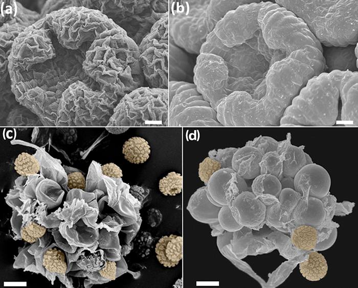
図1(C)なしで(B、D)プロトコルFAAエタノール-CPDで処理したサンプルの比較。 ( – b)は Anacyclusのclavatus、ミッド開発の花蕾。バドは<四酸化オスミウム46で処理されました/ SUP>(a)およびFAA-CPDプロトコル(B)で処理した出芽。 (CD)Phelloriniaのherculanea fの胞子とナース細胞。 stellata。任意の治療(C)なしで、ここハラタケ目(d)に記載のプロトコルを用いてサンプルを乾燥させました。オレンジ色で胞子。スケール:(AB)が100μm、(CD)50μmです。写真はY.ルイス・レオンによって撮影されました。 この図の拡大版をご覧になるにはこちらをクリックしてください。

Protocol

注:このプロトコルは、すべての(4-6)に一般的な手順を記述した特定の生物(セクション1-3)、および三から三献身的な6つの主要なセクションが含まれています。アスタリスク(*)は、実験者によって変更された手順を示しています。 1.開発の研究と完全に形成されたプラントの構造 コレクションと固定 植物材料は、ドラフトチャンバー?…

Representative Results

花の開発と開発の固定と完全に形成されたプラントの構造ここで説明FAA-CPDのプロトコルを使用して、若い成熟した植物組織を最適に固定され、SEMイメージングのために脱水されています。芽の地形や形状が収縮細胞( 図1B、1D、図4A-F)によって歪まされていないので、このような花の発達等の処理を再構成す?…

Discussion

標準的なSEMプロトコルに関しては、ここで紹介する手順は、比較的、迅速な追随しやすく、かつ低コストの方法論を含みます。試料の量と処理の容易さに応じて、良好な品質の画像を取得するために4〜5日かかります。 CPDとSEMの操作のための適切な安全措置を含め、手続きは、取り扱いが容易です。特に注意が(1.1.3およびプロトコルの2.1.5へのステップ1.1.1を参照)、ホルマリンやグルタル?…

Divulgazioni

The authors have nothing to disclose.

Acknowledgements

このプロジェクトは、このマニュアルでは、唯一の著者の見解を反映し、欧州委員会は、情報を挙げることができる任意の使用のための責任を負うことはできません付与契約番号634429.の下で、欧州連合(EU)のホライゾン2020年研究と技術革新プログラムから資金提供を受けていますその中に含まれます。我々はまた、レアルハルディンボタニコ、CSIC製の資金拠出を認めます。 SRはサプロレグニアで彼女の研究をサポートするため、欧州連合(EU)[ITN-SAPRO-238550]に感謝しています。我々はまた、親切Phelloriniaのherculanea画像を提供し、処理サンプルについてB.プエヨ(図5)のためのフランシスコカロンジュに感謝したいと思います。すべての画像はマドリードでレアルハルディンボタニコ-CSICのSEMサービスによって撮影されました。

Materials

Acetic acid No specific supplier Skin irritation, eye irritation
aluminium stubs Ted Pella, Inc. 16221 www.tedpella.com
Centrifuge tubes No specific supplier
Critical Point Dryer Polaron Quatum Technologies CPD7501
D (+) Glucose Merck 1,083,421,000
Double sided sellotape No specific supplier
Ethanol absolute No specific supplier. Flammable
European bacteriological agar Conda 1800.00 www.condalab.com
Filter paper No specific supplier
Forceps No specific supplier
Formalin 4% No specific supplier. Harmful, acute toxicity, skin sensitisation, carcinogenicity. Flammable
Glass cover slips No specific supplier
Glass hermetic container  No specific supplier
Glutaraldehyde 25% DC 253857.1611  (L) Dismadel S.L. 3336 www.dismadel.com
Mycological peptone Conda 1922.00 www.condalab.com
needles No specific supplier
Petri dishes No specific supplier
Plastic containers No specific supplier
Sample holder with lid  for the critical point dryer  Ted Pella, Inc. 4591 www.tedpella.com
scalpels No specific supplier
Scanning Electron Microscope Hitachi S3000N
Software for SEM
Solution A: NaH2PO4
Solution B: Na2HPO4
Specimen holders No specific supplier
Sputter coater Balzers SCD 004
Stereomicroscope No specific supplier
Transmission Electron Microscope (TEM) grids Electron Microscopy Sciences G200 (Square Mesh) www.emsdiassum.com
Tweezers No specific supplier

Riferimenti

  1. Endress, P. K., Baas, P., Gregory, M. Systematic plant morphology and anatomy: 50 years of progress. Taxon. 49 (3), 401-434 (2000).
  2. Falk, R. H., Gifford, E. M., Cutter, E. G. Scanning electron microscopy of developing plant organs. Science. 168 (3938), 1471-1474 (1970).
  3. Damblon, F. Sputtering, a new method of coating pollen grains in scanning electron microscopy. Grana. 15 (3), 137-144 (1975).
  4. Everhart, T. E., Thornley, R. F. M. Wide-band detector for micro-microampere low-energy electron currents. J. Sci. Instrum. 37 (7), 37246-37248 (1960).
  5. Collins, S. P., et al. Advantages of environmental scanning electron microscopy in studies of microorganisms. Microsc. Res. Techniq. 25 (5-6), 398-405 (1993).
  6. Fannes, W., Vanhove, M. P. M., Huyse, T., Paladini, G. A scanning electron microscope technique for studying the sclerites of Cichlidogyrus. Parasitol. Res. 114 (5), 2031-2034 (2015).
  7. Erbar, C., Leins, P. Portioned pollen release and the syndromes of secondary pollen presentation in the Campanulales-Asterales complex. Flora. 190 (4), 323-338 (1995).
  8. Jansen, S., Smets, E., Baas, P. Vestures in woody plants: a review. IAWA Journal. 19 (4), 347-382 (1998).
  9. Bortolin Costa, M. F., et al. Stigma diversity in tropical legumes with considerations on stigma classification. Bot. Rev. 80 (1), 1-29 (2014).
  10. Almeida, O. J. G., Cota-Sánchez, J. H., Paoli, A. A. S. The systematic significance of floral morphology, nectaries, and nectar concentration in epiphytic cacti of tribes Hylocereeae and Rhipsalideae (Cactaceae). Perspect. Plant Ecol. 15 (5), 255-268 (2013).
  11. Konarska, A. Comparison of the structure of floral nectaries in two Euonymus L. species (Celastraceae). Protoplasma. 252 (3), 901-910 (2015).
  12. Giuliani, C., Maleci Bini, L. Insight into the structure and chemistry of glandular trichomes of Labiatae, with emphasis on subfamily Lamioideae. Plant Syst. Evol. 276 (3-4), 199-208 (2008).
  13. Li, K., Zheng, B., Wang, Y., Zhou, L. L.Breeding system and pollination biology of Paeonia delavayi (Paeoniaceae), an endangered plant in the Southwest of China. Pak. J. Bot. 46 (5), 1631-1642 (2014).
  14. García, L., Rivero, M., Droppelmann, F. Descripción morfológica y viabilidad del polen de Nothofagus nervosa (Nothofagaceae). Bosque. 36 (3), 487-496 (2015).
  15. Prenner, G., Klitgaard, B. B. Towards unlocking the deep nodes of Leguminosae: floral development and morphology of the enigmatic Duparquetia orchidacea (Leguminosae, Caesalpinioideae). Am. J. Bot. 95 (11), 1349-1365 (2008).
  16. Ratnayake, K., Joyce, D. C., Webb, R. I. A convenient sample preparation protocol for scanning electron microscope examination of xylem-occluding bacterial biofilm on cut flowers and foliage. Sci. Hortic-Amsterdam. 140 (1), 12-18 (2012).
  17. Çolak, G., Celalettin Baykul, M., Gürler, R., Çatak, E., Caner, N. Investigation of the effects of aluminium stress on some macro and micro-nutrient contents of the seedlings of Lycopersicon esculentum Mill. by using scanning electron microscope. Pak. J. Bot. 46 (1), 147-160 (2014).
  18. Arafa, S. Z. Scanning electron microscope observations on the monogenean parasite Paraquadriacanthus nasalis from the nasal cavities of the freshwater fish Clarias gariepinus in Egypt with a note on some surface features of its microhabitat. Parasitol. Res. 110 (5), 1687-1693 (2012).
  19. Uppalapatia, S. R., Kerwinb, J. L., Fujitac, Y. Epifluorescence and scanning electron microscopy of host-pathogen interactions between Pythium porphyrae (Peronosporales, Oomycota)and Porphyra yezoensis (Bangiales, Rhodophyta). Bot. Mar. 44 (2), 139-145 (2001).
  20. Meaney, M., Haughey, S., Brennan, G. P., Fairweather, I. A scanning electron microscope study on the route of entry of clorsulon into the liver fluke, Fasciola hepatica. Parasitol. Res. 95 (2), 117-128 (2005).
  21. Sundarasekar, J., Sahgal, G., Subramaniam, S. Anti-candida activity by Hymenocallis littoralis extracts for opportunistic oral and genital infection Candida albicans. Bangladesh J. Pharmacol. 7 (3), 211-216 (2012).
  22. Benhamou, N., Rey, P., Picard, K., Tirilly, Y. Ultrastructural and cytochemical aspects of the interaction between the mycoparasite Pythium oligandrum and soilborne plant pathogens. Phytopathology. 89 (6), 506-517 (1999).
  23. Singh, A., et al. First evidence of putrescine involvement in mitigating the floral malformation in mangoes: A scanning electron microscope study. Protoplasma. 251 (5), 1255-1261 (2014).
  24. Xiang, C., et al. Fine mapping of a palea defective 1 (pd1), a locus associated with palea and stamen development in rice. Plant Cell Rep. 34 (12), 2151-2159 (2015).
  25. Mendoza, L., Hernandez, F., Ajello, L. Life cycle of the human and animal oomycete pathogen Pythium insidiosum. J. Clin. Microbiol. 31 (11), 2967-2973 (1993).
  26. Bello, M. A., Rudall, P. J., González, F., Fernández, J. L. Floral morphology and development in Aragoa (Plantaginaceae) andrelated members of the order Lamiales. Int. J. Plant Sci. 165 (5), 723-738 (2004).
  27. Bello, M. A., Hawkins, J. A., Rudall, P. J. Floral morphology and development in Quillajaceae and Surianaceae (Fabales), the species-poor relatives of Leguminosae and Polygalaceae. Ann. Bot. 100 (4), 1491-1505 (2007).
  28. Bello, M. A., Hawkins, J. A., Rudall, P. J. Floral ontogeny in Polygalaceae and its bearing on the homologies of keeled flowers in Fabales. Int. J. Plant Sci. 171 (5), 482-498 (2010).
  29. Bello, M. A., Alvarez, I., Torices, R., Fuertes-Aguilar, J. Floral development and evolution of capitulum structure in Anacyclus (Anthemideae, Asteraceae). Ann. Bot. 112 (8), 1597-1612 (2013).
  30. Bello, M. A., Martínez-Asperilla, A., Fuertes-Aguilar, J. Floral development of Lavatera trimestris and Malva hispanica reveals the nature of the epicalyx in the Malva generic alliance. Bot. J. Linn. Soc. 181 (1), 84-98 (2016).
  31. Calonge, F. D., Martínez, A. J., Falcó, I., Samper, L. E. Phellorinia herculanea f. stellata f. nova encontrada en España. Bol. Soc. Micol.Madrid. 35 (1), 65-70 (2011).
  32. Liu, Y., et al. Deciphering microbial landscapes of fish eggs to mitigate emerging diseases. ISME J. 8 (10), 2002-2014 (2014).
  33. Sandoval-Sierra, J. V., Diéguez-Uribeondo, J. A comprehensive protocol for improving the description of Saprolegniales (Oomycota): two practical examples (Saprolegnia aenigmatica sp. nov. and Saprolegnia racemosa sp. nov.). PLOS one. , (2015).
  34. Endress, P. K. Zur vergleichenden Entwicklungsmorphologie, Embryologie und Systematik bei Laurales. Bot. Jahrb. Syst. 92 (2), 331-428 (1972).
  35. Tucker, S. Floral development in Saururus cernuus (Saururaceae):1. Floral initiation and stamen development. Am. J. Bot. 62 (3), 993-1005 (1975).
  36. Endress, P. K., Matthews, M. L. Progress and problems in the assessment of flower morphology in higher-level systematics. Plant Syst. Evol. 298 (2), 257-276 (2012).
  37. Beakes, G. W., Glockling, S. L., Sekimoto, S. The evolutionary phylogeny of the oomycete "fungi&#34. Protoplasma. 249 (1), 3-19 (2012).
  38. Romansic, J. M., et al. Effects of the pathogenic water mold Saprolegnia ferax on survival of amphibian larvae. Dis. Aquat. Organ. 83 (3), 187-193 (2009).
  39. van West, P. Saprolegnia parasitica, an oomycete pathogen with a fishy appetite: new challengues for an old problem. Mycologist. 20 (3), 99-104 (2006).
  40. Johansen, D. A. . Plant microtechnique. , (1940).
  41. Unestam, T. Studies on the crayfish plague fungus Aphanomyces astaci. Some factors affecting growth in vitro. Physiol. Plantarum. 18 (2), 483-505 (1965).
  42. Cerenius, L., Söderhäll, K. Repeated zoospore emergence from isolated spore cysts of Aphanomyces astaci. Exp. Mycol. 8 (4), 370-377 (1984).
  43. Diéguez-Uribeondo, J., Cerenius, L., Söderhäll, K. Repeated zoospore emergence in Saprolegnia parasitica. Mycol. Res. 98 (7), 810-815 (1994).
  44. Söderhäll, K., Svensson, E., Unestam, T. Chitinase and protease activities in germinating zoospore cysts of a parasitic fungus, Aphanomyces astaci, Oomycetes. Mycopathologia. 64 (1), 9-11 (1978).
  45. Echlin, P. . Handbook of sample preparation for scanning electron microscopy and X-Ray Microanalysis. , (2009).
  46. Osumi, M., et al. Preparation for observation of fine structure of biological specimens by high-resolution SEM. Microscopy. 32 (4), 321-330 (1983).
  47. Rezinciuc, S. . The Saprolegniales morpho-molecular puzzle: an insight into markers identifying specific and subspecific levels in main parasites. , (2013).
check_url/it/55031?article_type=t

Play Video

Citazione di questo articolo
Bello, M. A., Ruiz-León, Y., Sandoval-Sierra, J. V., Rezinciuc, S., Diéguez-Uribeondo, J. Scanning Electron Microscopy (SEM) Protocols for Problematic Plant, Oomycete, and Fungal Samples. J. Vis. Exp. (120), e55031, doi:10.3791/55031 (2017).

View Video