Summary

幻影的多模态3D打印模拟生物组织

Published: January 11, 2020
doi:

Summary

旋转涂层、多喷流打印和熔融沉积建模集成在一起,可生成多层异构幻象,模拟生物组织的结构和功能特性。

Abstract

生物医学光学成像在诊断和治疗各种疾病中起着重要的作用。然而,光学成像器件的精度和可重复性受其元件性能特性、测试环境和操作的影响很大。因此,有必要通过可追溯的幻象标准校准这些设备。然而,目前可用的大多数幻象都是同质幻象,无法模拟生物组织的多模态和动态特性。在这里,我们展示了使用集成旋转涂层模块、多喷流模块、熔融沉积建模 (FDM) 模块和自动控制框架的生产线进行异构组织模拟幻象的制造。”数字光学幻象”的结构信息和光学参数在原型文件中定义,导入生产线,并逐层在不同打印方式之间进行顺序切换。这种生产线的技术能力体现在皮肤模拟幻象的自动打印,包括表皮、真皮、皮下组织和嵌入肿瘤。

Introduction

生物医学光学成像代表一系列医疗成像工具,根据光与生物组织相互作用检测疾病和组织异常。与其他成像模式(如磁共振成像 (MRI) 和计算机断层扫描 (CT) 相比,生物医学光学成像利用低成本和便携式设备1、2、3、4等非侵入性测量组织结构、功能和分子特性。然而,尽管光学成像在成本和便携性方面具有优势,但临床诊断和治疗指导并未被广泛接受,部分原因在于其可重复性差,光学和生物参数之间缺乏定量映射。造成这种限制的主要原因是生物医学光学成像设备缺乏可追溯的定量校准和验证标准。

过去,各种组织模拟幻象被开发用于各种组织类型的生物医学光学成像研究,如脑5、6、7、皮肤8、9、10、11、12、膀胱13和乳房组织14、15、16、17。这些幻象主要由以下制造工艺之一生产:1) 旋转涂层10,18 (用于模拟均质和薄层组织);2)成型19(用于模拟具有几何特征的笨重组织);和3)三维(3D)打印20,21,22(用于模拟多层异构组织)。成型产生的皮肤幻象能够模拟皮肤组织的体积光学特性,但不能模拟横向光学异质性19。本茨等人采用双通道FDM3D打印方法模拟生物组织的不同光学特性23。然而,使用两种材料不能充分模拟组织光学异质性和各向异性。Lurie等人通过结合3D打印和旋转涂层13,为光学相干断层扫描(OCT)和膀胱镜片创建了一个膀胱幻象。然而,幻象的异构特征,如血管,必须手工绘制。

在上述幻象制造工艺中,3D 打印为模拟生物组织的结构和功能异质性提供了最大的灵活性。然而,许多生物组织类型(如皮肤组织)由多层和多尺度的成分组成,无法通过单个 3D 打印过程进行有效复制。因此,必须集成多个制造工艺。我们推荐一条3D打印生产线,集成了多种制造工艺,用于自动生产多层和多尺度组织模拟幻像,作为生物医学光学成像的可追溯标准(图1)。尽管旋转涂层、多喷头打印和 FDM 在我们的 3D 打印生产线中是自动化的,但每种模式都保留了与既定工艺相同的功能特性。因此,本文为生产多尺度、多层、异构组织模拟幻象提供了一般指南,无需在单个仪器中物理集成多个过程。
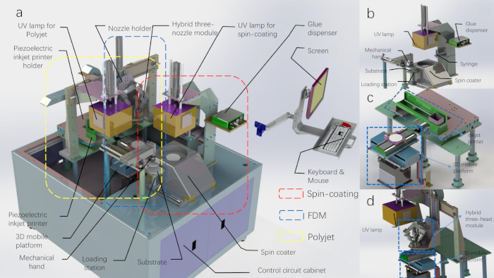
Figure 1
图1:3D打印生产线的CAD图。A) 3D 打印生产线,并拆下顶部外壳。(B) 旋转涂层模块和机械手模块的示意图。(C) 多喷打印模块的示意图。(D) FDM 打印模块的示意图(UV 灯属于多喷头打印模块)。请点击此处查看此图的较大版本。

Protocol

1. 准备 3D 打印材料 注:我们的光学幻象生产线使用各种印刷材料来模拟生物组织的结构和功能异质性。打印材料的选择也取决于制造工艺。 用于旋转涂层印刷的材料准备 将100毫克二氧化钛(TiO2)粉末加入含有100mL立体光刻(SLA)光聚合物树脂的烧杯中。 在磁性搅拌器上搅拌烧杯中的混合物 30 分钟。 用锡箔密封烧杯,并在超声波机中?…

Representative Results

旋转涂层制造的幻影旋转涂层通过旋转转盘均匀地将液滴均匀地分布在基材上,固化后将制造原始主体的单层。基板的旋转速度和旋转时间不仅影响幻象的表面质量,而且决定幻像每一层的厚度。不同厚度的幻影可以通过逐层重复的旋转涂层制造。幻象的光学参数可以通过改变散射和吸收材料的比例来确定,如我们上一份出版物24所述。增加光固化树脂中的TiO…

Discussion

在多层幻象的制造中,用于旋转涂层的材料是一种轻固化材料,而不是PDMS。中间层采用多喷喷印刷方法印刷,采用光固化树脂为原料。虽然在添加三丁基醇后,可通过旋转涂层进行薄 PDMS 幻像,但在多喷流打印过程中,PDMS 层无法有效地与光固化材料结合。因此,我们选择了轻固化树脂作为旋转涂层。

目前,只有两种材料可用于多喷头打印。在光固化材料中加入TiO2?…

Divulgazioni

The authors have nothing to disclose.

Acknowledgements

这项工作得到了国家自然科学基金(第11002139号)和中央大学基础研究基金的支持。我们感谢科技大学的Zachary J. Smith提供音频画外音。

Materials

2-Hydroxy-2-methylpropiophenone aladdin H110280-500g Light initiator
http://www.aladdin-e.com/
3D printing control system USTC USTC-3DPrinter_control1.0 custom-made
github:
https://github.com/macanzhen/
3D printing system USTC USTC-3DPrinter1.0 custom-made
AcroRip color Human Plus AcroRip v8.2.6
All-in-one nozzle slicing script Shenzhen CBD Technology Co.,Ltd. github:
https://github.com/macanzhen/
Chinese Red Dye Juents Oil-soluble
Cura Ultimaker Cura_15.04.6
Gel Wax Shanghai Lida Industry Co.,ltd. LP melting point: 56 °C
Graphite aladdin G103922-100g Change object optical absorption parameters
http://www.aladdin-e.com/
PDMS Dow Corning 184
Titanium dioxide ALDRICH 24858-100G 347 nm
Triethylene glycol dimethacrylate aladdin T101642-250ml Photocured monomer
http://www.aladdin-e.com/
UV ink SLA Photopolymer Resin time80s RESIN-A http://www.time80s.com/zlxz

Riferimenti

  1. Lu, G., Fei, B. Medical hyperspectral imaging: a review. Journal of Biomedical Optics. 19 (1), 010901 (2014).
  2. Wang, K., et al. Development of a non-uniform discrete Fourier transform based high speed spectral domain optical coherence tomography system. Optics Express. 17 (14), 12121-12131 (2009).
  3. Zhao, H., Gao, F., Tanikawa, Y., Homma, K., Yamada, Y. Time-resolved diffuse optical tomographic imaging for the provision of both anatomical and functional information about biological tissue. Applied Optics. 44 (10), 1905-1916 (2005).
  4. Ding, Z., Ren, H., Zhao, Y., Nelson, J. S., Chen, Z. High-resolution optical coherence tomography over a large depth range with an axicon lens. Optics Letters. 27 (4), 243-245 (2002).
  5. Iida, H., et al. Three-dimensional brain phantom containing bone and grey matter structures with a realistic head contour. Annals of Nuclear Medicine. 27 (1), 25-36 (2013).
  6. Mobashsher, A. T., Abbosh, A. Three-dimensional human head phantom with realistic electrical properties and anatomy. IEEE Antennas and Wireless Propagation Letters. 13, 1401-1404 (2014).
  7. Li, J. B., et al. A new head phantom with realistic shape and spatially varying skull resistivity distribution. IEEE Transactions on Biomedical Engineering. 61 (2), 254-263 (2013).
  8. Bykov, A., et al. Multilayer tissue phantoms with embedded capillary system for OCT and DOCT imaging. Life Sciences. (International Society for Optics and Photonics). , 73760 (2011).
  9. Bykov, A. V., Popov, A. P., Priezzhev, A. V., Myllylä, R. Skin phantoms with realistic vessel structure for OCT measurements in Laser Applications. European Conference on Biomedical Optics. , 80911 (2010).
  10. Park, J., et al. Fabrication of double layer optical tissue phantom by spin coating method: mimicking epidermal and dermal layer. Design and Performance Validation of Phantoms Used in Conjunction with Optical Measurement of Tissue V. , 85830 (2013).
  11. Wróbel, M. S., et al. Use of optical skin phantoms for preclinical evaluation of laser efficiency for skin lesion therapy. Journal of Biomedical Optics. 20 (8), 085003 (2015).
  12. Sheng, S., Wu, Q., Han, Y., Dong, E., Xu, R. Fabricating optical phantoms to simulate skin tissue properties and microvasculature. Design and Performance Validation of Phantoms Used in Conjunction with Optical Measurement of Tissue Vii. , 932507 (2015).
  13. Lurie, K. L., Smith, G. T., Khan, S. A., Liao, J. C., Ellerbee, A. K. Three-dimensional, distendable bladder phantom for optical coherence tomography and white light cystoscopy. Journal of Biomedical Optics. 19 (3), 36009 (2014).
  14. Hahn, C., Noghanian, S. Heterogeneous breast phantom development for microwave imaging using regression models. Journal of Biomedical Imaging. 2012, 6 (2012).
  15. Ansari, M. A., Mohajerani, E. Estimation of optical abnormalities in breast phantom by diffuse equation. Optik-International Journal for Light and Electron Optics. 125 (20), 5978-5981 (2014).
  16. Roman, M., Gonzalez, J., Carrasquilla, J., Erickson, S. J., Godavarty, A. A Gen-2 Hand-Held Optical Imager: Phantom and Preliminary in-vivo Breast Imaging Studies. 29th Southern Biomedical Engineering Conference. , 103-104 (2013).
  17. Michaelsen, K. E., et al. Anthropomorphic breast phantoms with physiological water, lipid, and hemoglobin content for near-infrared spectral tomography. Journal of Biomedical Optics. 19 (2), 026012 (2014).
  18. Park, J., et al. Optical tissue phantoms based on spin coating method. Design and Performance Validation of Phantoms Used in Conjunction with Optical Measurement of Tissue VII. , 93250 (2015).
  19. Mustari, A., et al. Agarose-based tissue mimicking optical phantoms for diffuse reflectance spectroscopy. Journal of Visualized Experiments. (138), e57578 (2018).
  20. Luciano, N. J., et al. Utilizing 3D printing technology to merge MRI with histology: A protocol for brain sectioning. Journal of Visualized Experiments. (118), e54780 (2016).
  21. Dong, E., et al. Three-dimensional fuse deposition modeling of tissue-simulating phantom for biomedical optical imaging. Journal of Biomedical Optics. 20 (12), 121311 (2015).
  22. Beltrame, E. D. V., et al. 3D Printing of Biomolecular Models for Research and Pedagogy. Journal of Visualized Experiments. (121), e55427 (2017).
  23. Bentz, B. Z., Chavan, A. V., Lin, D., Tsai, E. H., Webb, K. J. Fabrication and application of heterogeneous printed mouse phantoms for whole animal optical imaging. Applied Optics. 55 (2), 280-287 (2016).
  24. Liu, G., et al. Fabrication of a multilayer tissue-mimicking phantom with tunable optical properties to simulate vascular oxygenation and perfusion for optical imaging technology. Applied Optics. 57 (23), 6772-6780 (2018).
check_url/it/60563?article_type=t

Play Video

Citazione di questo articolo
Ma, C., Shen, S., Liu, G., Guo, S., Guo, B., Li, J., Huang, K., Zheng, Y., Shao, P., Dong, E., Chu, J., Xu, R. X. Multimodal 3D Printing of Phantoms to Simulate Biological Tissue. J. Vis. Exp. (155), e60563, doi:10.3791/60563 (2020).

View Video