Summary

생물학적 조직을 시뮬레이션하기 위한 팬텀의 멀티모달 3D 프린팅

Published: January 11, 2020
doi:

Summary

스핀 코팅, 폴리젯 프린팅 및 융합 증착 모델링은 생물학적 조직의 구조적 및 기능적 특성을 시뮬레이션하는 다층 이기종 팬텀을 생성하기 위해 통합됩니다.

Abstract

생물 의학 광학 화상 진찰은 각종 질병의 진단 그리고 처리에 있는 중요한 역할을 하고 있습니다. 그러나 광학 이미징 장치의 정확성과 재현성은 구성 요소의 성능 특성, 테스트 환경 및 작업의 영향을 크게 받습니다. 따라서 추적 가능한 팬텀 표준에 따라 이러한 장치를 교정해야 합니다. 그러나, 현재 유효한 팬텀의 대부분은 생물학 조직의 다중 모달 및 동적 특성을 시뮬레이션할 수 없는 균질한 유령입니다. 여기서, 우리는 스핀 코팅 모듈, 폴리젯 모듈, 융합 증착 모델링 (FDM) 모듈 및 자동 제어 프레임 워크를 통합 생산 라인을 사용하여 이기종 조직 시뮬레이션 유령의 제조를 보여줍니다. “디지털 광학 팬텀”의 구조 정보와 광학 파라미터는 프로토타입 파일에 정의되어 생산 라인으로 가져오고 서로 다른 인쇄 양식 간의 순차적 전환을 통해 제작된 레이어별 레이어별로 정의됩니다. 이러한 생산 라인의 기술적 능력은 표피, 진피, 피하 조직 및 임베디드 종양을 포함하는 피부 시뮬레이션 유령의 자동 인쇄에 의해 예시된다.

Introduction

생물 의학 광학 화상 진찰은 생물학 조직과의 가벼운 상호 작용에 근거를 둔 질병 및 조직 이상을 검출하는 의학 화상 진찰 공구의 가족을 나타냅니다. 자기 공명 영상(MRI) 및 컴퓨터 단층 촬영(CT)과 같은 다른 영상 양식과 비교하여, 생체 의학 광학 이미징은 저비용 휴대용 장치를 사용하여 조직 구조, 기능 및 분자 특성의 비침습적 측정을 활용합니다1,2,3,4. 그러나, 비용과 휴대성의 우수성에도 불구하고, 광학 화상 진찰은 광학과 생물학 매개변수 사이 그것의 나쁜 재현성과 양적 매핑의 부족 때문에 부분적으로 임상 진단 및 치료 지도를 위해 넓게 받아들여지지 않았습니다. 이러한 제한의 주된 이유는 생체 의학 광학 이미징 장치의 정량적 교정 및 검증을 위한 추적 가능한 표준이 없기 때문입니다.

과거에는5,6,7,피부8,9,10,11,12,방광 13, 유방 조직14,15,16,17과 같은 다양한 조직 유형에서 생물 의학 광학 이미징 연구를 위해 다양한 조직 시뮬레이션 팬텀이 개발되었습니다. 이러한 팬텀은 주로 다음 제조 공정 중 하나에 의해 생산된다 : 1) 스핀 코팅10,18 (균질및 얇은 층 조직을 시뮬레이션하기위한); 2) 성형19 (기하학적 특징으로 부피가 큰 조직을 시뮬레이션하기위한); 및 3) 3 차원 (3D) 인쇄20,21,22 (다층 이종 조직 시뮬레이션). 성형에 의해 생성된 피부 유령은 피부 조직의 대량 광학 적 특성을 모방할 수 있지만 횡측 광학이질성(19)을시뮬레이션할 수는 없다. Bentz 외. 생물학적조직(23)의상이한 광학적 특성을 모방하기 위해 2채널 FDM 3D 프린팅 방법을 사용했다. 그러나, 두 물질을 사용하면 조직 광학 이질성과 이방성 등을 충분히 시뮬레이션할 수 없다. Lurie 등은 3D 프린팅과 스핀 코팅13을결합하여 광학 일관성 단층 촬영 (OCT) 및 방광경 검사를위한 방광 팬텀을 만들었습니다. 그러나 혈관과 같은 유령의 이질적인 특징은 손으로 그려야했습니다.

위의 팬텀 제조 공정 중에서 3D 프린팅은 생물학적 조직의 구조적 및 기능적 이질성을 시뮬레이션하는 데 가장 많은 유연성을 제공합니다. 그러나 피부 조직과 같은 많은 생물학적 조직 유형은 단일 3D 프린팅 프로세스로 효과적으로 복제할 수 없는 다층 및 다중 스케일 구성 요소로 구성됩니다. 따라서 여러 제조 공정을 통합해야 합니다. 우리는 생물 의학 광학 화상 진찰을 위한 추적 가능한 표준으로 유령을 시뮬레이션하는 다층 및 다중 스케일 조직의 자동 생산을 위한 다중 제조 공정을 통합하는 3D 인쇄 생산 라인을 제안합니다(그림 1). 스핀 코팅, 폴리젯 프린팅 및 FDM은 3D 프린팅 생산 라인에서 자동화되어 있지만 각 양식은 기존 공정과 동일한 기능적 특성을 유지합니다. 따라서 이 백서는 단일 장치에서 여러 공정을 물리적으로 통합할 필요 없이 다중 스케일, 다층 및 이기종 조직 시뮬레이션 팬텀을 생산하기 위한 일반적인 지침을 제공합니다.

Figure 1
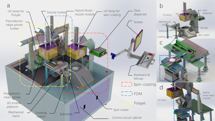
그림 1: 3D 프린팅 생산 라인의 CAD 다이어그램입니다. (A)상단 쉘이 제거된 3D 프린팅 생산 라인입니다. (B)스핀 코팅 모듈과 기계식 핸드 모듈의 회로도. (C)폴리젯 인쇄 모듈의 회로도입니다. (D)FDM 인쇄 모듈의 회로도(UV 램프는 폴리젯 인쇄 모듈에 속합니다). 이 그림의 더 큰 버전을 보려면 여기를 클릭하십시오.

Protocol

1. 3D 프린팅을 위한 자료 준비 참고 : 우리의 광학 팬텀 생산 라인은 생물학적 조직의 구조적 및 기능적 이질성을 시뮬레이션하기 위해 다양한 인쇄 재료를 사용합니다. 인쇄 재료의 선택은 제조 공정에 따라 달라집니다. 스핀 코팅 인쇄를 위한 재료 준비 100 mg의 이산화 티타늄 (TiO2)분말을 100 mL의 스테레오 리소그래피 (SLA) 광중합체 수지가 들어있는 비커…

Representative Results

스핀 코팅으로 제작된 팬텀스핀 코팅은 턴테이블을 회전시켜 기판에 방울을 고르게 분배하고, 원래 본체의 단일 층은 경화 후 제작된다. 기판의 회전 속도와 회전 시간은 팬텀의 표면 품질에 영향을 줄 뿐만 아니라 팬텀의 각 층의 두께를 결정합니다. 서로 다른 두께의 팬텀은 반복적 인 스핀 코팅 층별로 제작 될 수 있습니다. 팬텀의 광학 파라미터는 우리의 이전 간행물<sup class="x…

Discussion

다층 팬텀의 제조에서 스핀 코팅에 사용되는 재료는 PDMS 대신 경화 가능한 재료의 일종입니다. 중간 층은 원료로 광 경화 수지를 사용하는 폴리젯 인쇄 방법으로 인쇄됩니다. 얇은 PDMS 팬텀은 테르트 부틸 알코올을 첨가 한 후 스핀 코팅으로 만들 수 있지만 PDMS 층은 폴리젯 인쇄 중에 경화 성 물질에 효과적으로 결합 할 수 없습니다. 따라서 스핀 코팅용 경화 수지(light-curable)를 선택했습니다.

<p…

Divulgazioni

The authors have nothing to disclose.

Acknowledgements

이 작품은 중국 국립 자연 과학 재단 (그랜트 번호 11002139 및 81327803)과 중앙 대학의 기초 연구 기금에 의해 지원되었다. 우리는 오디오 음성 해설을 제공하는 과학 기술 대학의 재커리 J. 스미스에게 감사드립니다.

Materials

2-Hydroxy-2-methylpropiophenone aladdin H110280-500g Light initiator
http://www.aladdin-e.com/
3D printing control system USTC USTC-3DPrinter_control1.0 custom-made
github:
https://github.com/macanzhen/
3D printing system USTC USTC-3DPrinter1.0 custom-made
AcroRip color Human Plus AcroRip v8.2.6
All-in-one nozzle slicing script Shenzhen CBD Technology Co.,Ltd. github:
https://github.com/macanzhen/
Chinese Red Dye Juents Oil-soluble
Cura Ultimaker Cura_15.04.6
Gel Wax Shanghai Lida Industry Co.,ltd. LP melting point: 56 °C
Graphite aladdin G103922-100g Change object optical absorption parameters
http://www.aladdin-e.com/
PDMS Dow Corning 184
Titanium dioxide ALDRICH 24858-100G 347 nm
Triethylene glycol dimethacrylate aladdin T101642-250ml Photocured monomer
http://www.aladdin-e.com/
UV ink SLA Photopolymer Resin time80s RESIN-A http://www.time80s.com/zlxz

Riferimenti

  1. Lu, G., Fei, B. Medical hyperspectral imaging: a review. Journal of Biomedical Optics. 19 (1), 010901 (2014).
  2. Wang, K., et al. Development of a non-uniform discrete Fourier transform based high speed spectral domain optical coherence tomography system. Optics Express. 17 (14), 12121-12131 (2009).
  3. Zhao, H., Gao, F., Tanikawa, Y., Homma, K., Yamada, Y. Time-resolved diffuse optical tomographic imaging for the provision of both anatomical and functional information about biological tissue. Applied Optics. 44 (10), 1905-1916 (2005).
  4. Ding, Z., Ren, H., Zhao, Y., Nelson, J. S., Chen, Z. High-resolution optical coherence tomography over a large depth range with an axicon lens. Optics Letters. 27 (4), 243-245 (2002).
  5. Iida, H., et al. Three-dimensional brain phantom containing bone and grey matter structures with a realistic head contour. Annals of Nuclear Medicine. 27 (1), 25-36 (2013).
  6. Mobashsher, A. T., Abbosh, A. Three-dimensional human head phantom with realistic electrical properties and anatomy. IEEE Antennas and Wireless Propagation Letters. 13, 1401-1404 (2014).
  7. Li, J. B., et al. A new head phantom with realistic shape and spatially varying skull resistivity distribution. IEEE Transactions on Biomedical Engineering. 61 (2), 254-263 (2013).
  8. Bykov, A., et al. Multilayer tissue phantoms with embedded capillary system for OCT and DOCT imaging. Life Sciences. (International Society for Optics and Photonics). , 73760 (2011).
  9. Bykov, A. V., Popov, A. P., Priezzhev, A. V., Myllylä, R. Skin phantoms with realistic vessel structure for OCT measurements in Laser Applications. European Conference on Biomedical Optics. , 80911 (2010).
  10. Park, J., et al. Fabrication of double layer optical tissue phantom by spin coating method: mimicking epidermal and dermal layer. Design and Performance Validation of Phantoms Used in Conjunction with Optical Measurement of Tissue V. , 85830 (2013).
  11. Wróbel, M. S., et al. Use of optical skin phantoms for preclinical evaluation of laser efficiency for skin lesion therapy. Journal of Biomedical Optics. 20 (8), 085003 (2015).
  12. Sheng, S., Wu, Q., Han, Y., Dong, E., Xu, R. Fabricating optical phantoms to simulate skin tissue properties and microvasculature. Design and Performance Validation of Phantoms Used in Conjunction with Optical Measurement of Tissue Vii. , 932507 (2015).
  13. Lurie, K. L., Smith, G. T., Khan, S. A., Liao, J. C., Ellerbee, A. K. Three-dimensional, distendable bladder phantom for optical coherence tomography and white light cystoscopy. Journal of Biomedical Optics. 19 (3), 36009 (2014).
  14. Hahn, C., Noghanian, S. Heterogeneous breast phantom development for microwave imaging using regression models. Journal of Biomedical Imaging. 2012, 6 (2012).
  15. Ansari, M. A., Mohajerani, E. Estimation of optical abnormalities in breast phantom by diffuse equation. Optik-International Journal for Light and Electron Optics. 125 (20), 5978-5981 (2014).
  16. Roman, M., Gonzalez, J., Carrasquilla, J., Erickson, S. J., Godavarty, A. A Gen-2 Hand-Held Optical Imager: Phantom and Preliminary in-vivo Breast Imaging Studies. 29th Southern Biomedical Engineering Conference. , 103-104 (2013).
  17. Michaelsen, K. E., et al. Anthropomorphic breast phantoms with physiological water, lipid, and hemoglobin content for near-infrared spectral tomography. Journal of Biomedical Optics. 19 (2), 026012 (2014).
  18. Park, J., et al. Optical tissue phantoms based on spin coating method. Design and Performance Validation of Phantoms Used in Conjunction with Optical Measurement of Tissue VII. , 93250 (2015).
  19. Mustari, A., et al. Agarose-based tissue mimicking optical phantoms for diffuse reflectance spectroscopy. Journal of Visualized Experiments. (138), e57578 (2018).
  20. Luciano, N. J., et al. Utilizing 3D printing technology to merge MRI with histology: A protocol for brain sectioning. Journal of Visualized Experiments. (118), e54780 (2016).
  21. Dong, E., et al. Three-dimensional fuse deposition modeling of tissue-simulating phantom for biomedical optical imaging. Journal of Biomedical Optics. 20 (12), 121311 (2015).
  22. Beltrame, E. D. V., et al. 3D Printing of Biomolecular Models for Research and Pedagogy. Journal of Visualized Experiments. (121), e55427 (2017).
  23. Bentz, B. Z., Chavan, A. V., Lin, D., Tsai, E. H., Webb, K. J. Fabrication and application of heterogeneous printed mouse phantoms for whole animal optical imaging. Applied Optics. 55 (2), 280-287 (2016).
  24. Liu, G., et al. Fabrication of a multilayer tissue-mimicking phantom with tunable optical properties to simulate vascular oxygenation and perfusion for optical imaging technology. Applied Optics. 57 (23), 6772-6780 (2018).
check_url/it/60563?article_type=t

Play Video

Citazione di questo articolo
Ma, C., Shen, S., Liu, G., Guo, S., Guo, B., Li, J., Huang, K., Zheng, Y., Shao, P., Dong, E., Chu, J., Xu, R. X. Multimodal 3D Printing of Phantoms to Simulate Biological Tissue. J. Vis. Exp. (155), e60563, doi:10.3791/60563 (2020).

View Video