This protocol describes a juvenile porcine model of orthotopic left lung allotransplantation designed for use with ESLP research. Focus is made on anesthetic and surgical techniques, as well as critical steps and troubleshooting.
Lung transplantation is the gold-standard treatment for end-stage lung disease, with over 4,600 lung transplantations performed worldwide annually. However, lung transplantation is limited by a shortage of available donor organs. As such, there is high waitlist mortality. Ex situ lung perfusion (ESLP) has increased donor lung utilization rates in some centers by 15%-20%. ESLP has been applied as a method to assess and recondition marginal donor lungs and has demonstrated acceptable short- and long-term outcomes following transplantation of extended criteria donor (ECD) lungs. Large animal (in vivo) transplantation models are required to validate ongoing in vitro research findings. Anatomic and physiologic differences between humans and pigs pose significant technical and anesthetic challenges. An easily reproducible transplant model would permit the in vivo validation of current ESLP strategies and the preclinical evaluation of various interventions designed to improve donor lung function. This protocol describes a porcine model of orthotopic left lung allotransplantation. This includes anesthetic and surgical techniques, a customized surgical checklist, troubleshooting, modifications, and the benefits and limitations of the approach.
Lung transplantation is the preeminent long-term treatment for end-stage lung disease. Over 4,600 lung transplantations are performed worldwide annually1. However, lung transplantation currently has significant limitations. For one, the necessity for organs continues to eclipse available donors. Despite rates of lung transplantation increasing every year since 2012 due to the combined effects of more candidates being listed for transplant, an increase in the number of donors, and improved use of recovered organs, the transplant waitlist mortality has not decreased significantly2. Organ quality concerns represent another major limitation, with reported organ utilization rates as low as 20%-30%3,4,5. Finally, the trends in the post-operative outcomes of lung transplantation are less than satisfactory, with long-term graft and patient outcomes still lagging that of other solid organ transplantations2.
An emerging technology, ex situ lung perfusion (ESLP), has the potential to mitigate these limitations. ESLP has been increasingly applied as a method to assess and recondition marginal donor lungs and has demonstrated acceptable short- and long-term outcomes following transplantation of extended criteria donor (ECD) lungs6,7,8,9,10. Consequently, ESLP has increased utilization rates in some centers by 15%-20%6,7,8,9,10,11.
Proper ESLP research requires the in vivo validation of in vitro findings; however, there is limited literature on porcine lung transplantation models for ESLP12,13,14,15. Furthermore, available literature provides inadequate details regarding anesthetic management of Yorkshire pigs for lung transplantation, which can be highly unstable hemodynamically12,13,14,15. Establishing an easily reproducible model would permit the in vivo validation of current ESLP strategies and the preclinical evaluation of various interventions to reduce lung ischemia-reperfusion injury. The objective of the present study is to describe a porcine model of orthotopic left lung allotransplantation for use with ESLP. The protocol includes descriptions of the anesthetic and surgical techniques, a custom surgical checklist, and details regarding the troubleshooting experience and protocol modifications. The limitations and benefits of the left lung porcine transplantation model have also been discussed in this work. This manuscript does not outline the retrieval process of porcine lungs in 35-50 kg Yorkshire pigs, nor does it cover the establishment and termination of ESLP. This protocol exclusively addresses the recipient transplantation operation.
All the procedures were performed in compliance with the guidelines of the Canadian Council on Animal Care and the guide for the care and use of laboratory animals. The protocols were approved by the institutional animal care committee of the University of Alberta. This protocol has been applied in female juvenile Yorkshire pigs between 35-50 kg. Pigs are pathogen-free, food-grade specimens. They are purchased from the Swine Research and Technology Centre in Edmonton, AB, Canada (https://srtc.ualberta.ca). All individuals involved in ESLP procedures had received proper biosafety training.
1. Pre-surgical preparations and anesthesia
NOTE: Pigs are fasted overnight prior to surgery for a maximum duration of 12 h.
2. Insertion of central venous and arterial lines
3. Left lung procurement
4. Termination of ESLP, division of left lung, and flushing with electrolyte solution
5. Left lung transplantation
6. Isolated Left Lung Assessment
All of the results are in the context of 4 h of reperfusion following 12 h of NPV-ESLP16. During lung explant, there are several clinical outcomes to anticipate (Figure 3). Typically, the pig will remain hemodynamically stable following a successful left lung explantation but may require a low dose infusion of phenylephrine (dose range: 2-10 mg/h) due to a vasodilatory response to surgery. Heart rate should target approximately 100-120 bpm, respiratory rate (RR) 8-30 for SpO2 > 90%, mean arterial pressure (MAP) > 60 mmHg, normothermic (38 °C), and tidal volumes (TVs) are targeted at 5 mL/kg while on one-lung ventilation with peak pressures of 20-24 cm H2O. During one-lung ventilation, the ventilation volumes were reduced by half to protect the left lung from overinflation. The respiratory rate was increased to target a physiologic end-tidal carbon dioxide level (Figure 3). Thus, Figure 3 displays typical hemodynamic and ventilatory parameters during critical points of the transplant.
During lung implant, the following results are typical. The left lung will have absorbed fluid during the ESLP run and appears heavier and larger than the explanted lung. For this reason, the recipient should be slightly larger than the donor (2-4 kg), so the thorax can accommodate the somewhat edematous lung. The lung will require gentle pressure to insert into the chest through the thoracotomy. It is easier to insert the lower lobe first, followed by the upper lobe. The bronchus is a direct end-to-end anastomosis and should be performed first. 4-0 prolene on a TF needle is recommended. The LA cuffs are highly friable but not too difficult to sew due to the redundancy and pliability of the tissue. 6-0 prolene on BV-1 needles work well for the LA anastomoses. The PA is the last anastomosis performed. This vessel can tear easily with little traction. If it tears, it is possible to open the pericardium and move the clamp proximally toward healthy tissue for sewing. Again, a 6-0 prolene on BV-1 needles works well for this anastomosis.
At the time of reperfusion, the following trends were noticed. Once the bronchus is unclamped and TVs are increased back to 10 mL/kg, the left lung will begin to inflate. Although the target was 10 mL/kg for tidal volumes, generally 6-8 mL/kg was attained, which is achieved gradually over the first 2-3 h of reperfusion, depending on the ESLP protocol used and the quality of the implanted lung. Rarely, there can be a small air leak, and this can be remedied with a simple stitch on the anterior wall. The posterior wall is more difficult to repair and will require packing. Great effort should be made to avoid air leaks from the bronchial anastomosis. Upon bronchoscopy, the right lung appears normal, and the left lung is typically edematous. The suture line is inspected, and approximately 50-100 mL of clear fluid is suctioned from the airways. The TV will drop significantly during suctioning from 300 s to 20 s, so this action should be performed quickly to allow the pig to recover. If arterial saturation drops below 90%, the bronchoscopy should be terminated, and the pig is allowed to recover over 1-2 min of ventilation. The first arterial blood gas (ABG) is typically normal because the right lung is functioning well as the left lung recovers.
The proactive administration of furosemide, dextrose, and insulin at the time of reperfusion serves to mitigate a dramatic rise in potassium through intracellular shifting. The potassium will predictably rise during 60-120 min of reperfusion (Table 1). Table 1 demonstrates a sample of ABGs over transplantation with 4 h reperfusion following 12 h of normothermic negative pressure ventilation (NPV) ESLP. Approximately two to four shifts are required during 4 h reperfusion to keep potassium < 5 mmol/L. If the trend is upward and appears as a rapid change between two gases drawn at 30 min intervals, the target is K+< 4.5 mmol/L. Shifts include 40 mg of furosemide, 100 mL of 25% dextrose (D25), and 10 units of regular insulin administered as IV push via the central line. Occasionally, the pig will require a low dose dobutamine infusion (1.5-5 mcg/kg/min) along with phenylephrine (2-10 mg/h) after 30-60 min of reperfusion to treat a developing vasoplegic response. It is preferable to use phenylephrine in this situation exclusively. However, dobutamine can be a useful supplemental inotrope to maintain a mean arterial pressure greater than 60 mmHg, particularly if the heart rate is bradycardic.
Upon thoracotomy closure and turning the pig prone, an improvement in ventilation and hemodynamics is demonstrated. The modification can be drastic and occur over 5-10 min, but occasionally the response takes 1 h. Tidal volumes increase as pressure/weight is taken off the right lung, and the left lung continues to ventilate with improved compliance and recruitment. A repeat bronchoscopy can be performed further to clear the airway after a change in position. Over the following 4 h, phenylephrine requirements decrease, TVs approach the target 10 mL/kg, and ABGs stabilize (Table 1). To reiterate, if TVs of 10 mL/kg are targetted, typically TVs in the range of 6-8 mL/kg are achieved (Figure 3).
At the time of the final isolated left lung assessment, a stable pattern of behavior has been observed. The pig is less tolerant hemodynamically in the supine position for sternotomy and may require additional vasopressor support. Inspection of the left lung reveals variable degrees of mild hyperemia from ischemic reperfusion injury (IRI). The right lung appears normal. Upon clamping the right hilum, the pig becomes sinus tachycardic (120-140 bpm), and 100% of the cardiac output is diverted to the left lung. Targeted tidal volumes are not decreased at this time as the entire process takes 10 min. The pig remains stable up to the 5 min mark, but the heart may develop ventricular fibrillation between 5-10 min and manual cardiac massage is potentially required to continue perfusing the left lung. The left lung is explanted, weighed, and the anastomoses are inspected for patency. The pig expires rapidly at the time of exsanguination, which coincides with the explantation of the previously transplanted lung.
A successful transplant has predictable findings after the experiment (Table 1 and Figure 4). Figure 4 displays typical P:F ratio changes and edema formation during the transplant protocol. Typically, the left lung will experience an approximate 35% (+/-15%) weight gain; however, residual blood in the circulation contributes to this weight. PF ratios drop by approximately 100 at reperfusion as the left lung is not immediately effective at oxygenation, but this discrepancy improves over 2-3 h. Upon isolated left lung assessment at 4 h, the PF ratio will remain stable or decline slightly. Generally, the isolated left lung gas at 10 min will be similar to the final gas analysis post 12 h ESLP (Table 1). However, this is entirely dependent on the ESLP protocol employed, and the extent of IRI incurred. An unsuccessful transplant can be caused by clotting of the LPA, which results in an infarcted lung that does not oxygenate. Likewise, the duration of the transplant surgery can affect the quality of the reperfused lung function. An implantation surgery should take between 30-60 min. Longer operations expose the donor lung to damaging warm ischemic time that exacerbates ischemic reperfusion injury and can confound the results of the experimental ESLP protocol. The specific ESLP protocol of a given experiment may produce a non-functioning lung that fails to oxygenate after transplantation despite patent anastomoses. Such isolated left lung gases will be very dark in color (deoxygenated) with a low partial pressure of oxygen (PaO2).
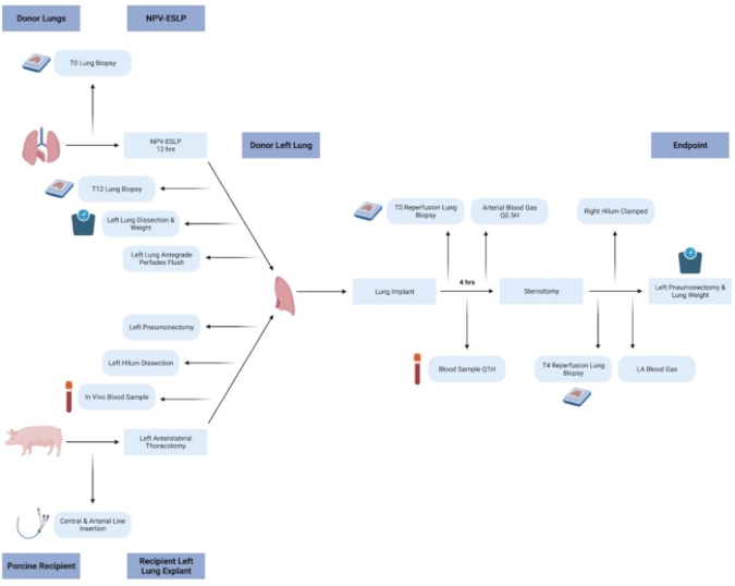
Figure 1: Schematic of porcine left lung transplant protocol. Schematic representation of 12 h NPV-ESLP run followed by left lung transplantation in a Yorkshire pig. Please click here to view a larger version of this figure.
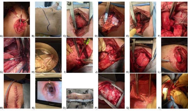
Figure 2: Photos of porcine left lung transplant surgery protocol. (A) Internal jugular and common carotid line placement. (B) Thoracotomy incision. (C) Thoracotomy. (D) Left Hemi-azygous vein. (E) Ligated Left Hemi-azygous vein. (F) Isolation of pulmonary veins. (G) Clamped left atrial cuff, left bronchus, and left pulmonary artery. (H) Left donor lung with pulmonary vein, bronchial and PA cuffs. (I) Pulmonary artery anastomosis. (J) Left lung transplanted and unclamped. (K) Lung repositioned. (L) Chest tube positioned. (M) Thoracotomy closure. (N) Bronchial anastomosis. (O) Pig in prone position. (P) Sternotomy. (Q) Accessory lobe clamped (Right lung clamped, but not shown). (R) Left pulmonary vein blood samples were drawn from pulmonary vein anastomosis (bleeding from prior puncture site). Please click here to view a larger version of this figure.
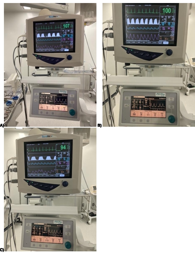
Figure 3: Monitoring and ventilation parameters for porcine left lung transplant surgery. (A) Typical parameters for recipient pre-transplant. (B) Typical parameters at recipient left lung explant. (C) Typical parameters 4 h post left lung donor transplant. Please click here to view a larger version of this figure.
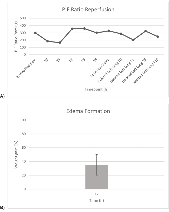
Figure 4: P:F ratio and weight gain pre-and post-transplant. (A) PaO2:FiO2 ratios throughout the transplant. (B) Weight gain of left lung throughout transplant after 12 h of NPV-ESLP. Please click here to view a larger version of this figure.
Arterial Blood Gases (100% FiO2) | In vivo Recipient | T0 Reperfusion | T1 Reperfusion | T2 Reperfusion | T3 Reperfusion | T4 Reperfusion | Isolated Left Lung Pre-clamp | Isolated Left Lung Post-clamp (0 min) | Isolated Left Lung Post-clamp (1 min) | Isolated Left Lung Post-clamp (5 min) | Isolated Left Lung Post-clamp (10 min) |
Blood Gas Values | |||||||||||
pH | 7.402 | 7.327 | 7.284 | 7.402 | 7.421 | 7.479 | 7.504 | 7.399 | 7.371 | 7.423 | 7.435 |
pCO2 (mmHg) | 47.7 | 57.3 | 56.4 | 36.9 | 35.3 | 35.6 | 34.2 | 45.6 | 48.1 | 40.6 | 36.6 |
pO2 (mmHg) | 299 | 184 | 165 | 355 | 358 | 300 | 327 | 287 | 207 | 335 | 249 |
Oximetry Values | |||||||||||
Hb (g/dL) | 11.2 | 12.5 | 11.3 | 11.6 | 10.3 | – | 17.1 | 11.7 | 13.5 | 16.3 | 13.8 |
sO2 (%) | 100.1 | 99.2 | 99 | 99.8 | 99.8 | – | 99.9 | 100.2 | 99.7 | 99.8 | 99.9 |
Electrolyte Values | |||||||||||
K+ (mmol/L) | 4.5 | 6.2 | 4.4 | 4 | 4.1 | 4.6 | 5.2 | 5.4 | 5.3 | 6.9 | 7.4 |
Na+ (mmol/L) | 141 | 143 | 140 | 245 | 145 | 144 | 140 | 141 | 139 | 137 | 136 |
Ca2+ (mmol/L) | 0.99 | 0.88 | 0.81 | 0.74 | 0.66 | 0.61 | 0.36 | 0.98 | 0.42 | 0.36 | 0.38 |
Cl– (mmol/L) | 97 | 97 | 95 | 101 | 100 | 96 | 91 | 102 | 94 | 91 | 94 |
Osm (mmol/kg) | 287 | 287.9 | 293.7 | 292.4 | 297.5 | 293.5 | 284.7 | 287.1 | 282.9 | 278.2 | 277.1 |
Metabolite values | |||||||||||
Glucose (mmol/L) | 4,2 | 2.7 | 13.4 | 2.8 | 8.3 | 5 | 5.1 | 4.9 | 4.5 | 4.6 | 4.2 |
Lactate (mmol/L) | 1.2 | 1.3 | 3.8 | 2.5 | 1.3 | 1.2 | 1.4 | 1.8 | 1.4 | 1.9 | 2.7 |
Acid Base status | |||||||||||
HCO-3 (mmol/L) | 29 | 29.1 | 25.9 | 22.4 | 22.5 | 26.1 | 26.7 | 27.6 | 27.1 | 26.1 | 24.1 |
Table 1: Blood gas analysis performed following left lung transplant post 12 h of ESLP. Ca+, calcium ion; Cl–, chloride ion; Hb, hemoglobin; HCO3–, bicarbonate ion; K+, potassium ion; Na+, sodium ion; Osm, osmolarity; paCO2, arterial partial pressure of carbon dioxide; PaO2, arterial partial pressure of oxygen; sO2, oxygen saturation; isolated left lung pre-clamp, right hilum open; Isolated left lung post-clamp, 1 min after right hilum clamped.
Supplementary File 1: Surgical safety checklist for left lung transplantation. Please click here to download this File.
Several critical surgical steps are involved in this protocol, and troubleshooting is needed to ensure successful transplantation and lung assessment. Juvenile porcine lungs are incredibly delicate compared to adult human lungs, so the operating surgeon must be cautious when handling porcine lungs. This is especially true after a 12 h run of ESLP as the organ will have taken on fluid volume and be susceptible to injury from excessive manipulation. Any undue pressure will cause atelectasis or trauma to the experimental lung that will affect assessment results. Likewise, the vascular structures are very delicate in the juvenile pig. It is critical to avoid torsion of the PA clamp as this can cause a tear or dissection of the tissue layers. A tear in the PA will necessitate opening the pericardium to access a more proximal portion of the left PA that can be anastomosed to the implanting lung. A DeBakey vascular clamp has a low profile that fits well in the surgical field, but this instrument can cause injury to the delicate PA if the surgeon is not careful. It is helpful to secure the clamp in position using a silk tie that is snapped to the drapes to prevent dislodgement or torsion. Bronchoscopy of the transplanted lung after unclamping of the bronchial anastomosis is also critical. There is often fluid within the donor lung airway after 12 h of ESLP and transplant. Suctioning this fluid is vital to ensure optimal recovery of left lung function and thereby assessment after 4 h of reperfusion. After bronchoscopy and the first ABG has returned with satisfactory potassium levels, it is critical to insert a chest tube, close the incision, and prone the pig. The pig's hemodynamics and ventilation are considerably more stable in the prone position, with the ribcage reapproximated. Elevated potassium > 5.5 mmol/L at this stage risks bradycardic arrest and will require emergent re-opening and manual cardiac massage to support perfusion, which is best avoided. Due to the significant risk of hyperkalemia and bradycardic arrest upon reperfusion, it is critical to perform serial ABGs beginning at reperfusion and recurring every 30 min until the 4 h exsanguination. ABGs give essential readings of oxygenation, partial pressure of carbon dioxide (PCO2), potassium, and glucose. Monitoring these four components closely and treating them appropriately is vital to a successful experiment. A continuous telemetry reading is also critical to monitor for peaked T waves associated with hyperkalemia and the anticipation of bradycardia. At the final stages of the experiment, it is crucial to clamp the right lung hilum and the accessory lobe before drawing final blood samples from the LA anastomosis. The right hilum supplies blood to the accessory lung lobe, and the accessory lobe drains adjacent to the left inferior pulmonary vein, often via a common trunk. The right hilum and accessory lobe need to be clamped separately to ensure no right lung function contributes to the sample LA gases through blood mixing. Drawing the left lung ABG sample from the PV anastomosis or just beyond it is suggested.
Several modifications have been made to this protocol along with significant troubleshooting of the described methods. Initially, it was attempted to perform the implantation via median sternotomy; however, the exposure was suboptimal due to the orientation of the pig PA, bronchus, and LA. The approach was successfully performed, but a thoracotomy was attempted on subsequent surgeries for improved exposure. This proved to be a superior surgical approach from visualization and technical perspective. Another essential modification was developing and implementing a surgical safety/protocol checklist (Supplementary File 1). There was a significant learning curve for all the team members involved, and these experiments are resource-intensive. A checklist was developed to guide the communication and document protocol development (Supplementary File 1). The checklist allowed to systemize and simplify the protocol for faster learning. The heparinization protocol was also modified. Two of the first ten transplants performed suffered from left lung ischemia due to clot formation in the left PA. Initially, 5000 units of heparin IV was administered 5 min before PA clamping and an additional 5000 units was administered 5 min before PA unclamping. Dosing frequency was increased to include 5000 units every hour after PA unclamping, and there have not been any issues with bleeding or PA clotting since adopting this approach. A strategy that utilizes less heparin was developed to control expenses, with a dose of 5000 units IV heparin 5 min before PA clamping and 5 min before partial PA unclamping. This is followed by 1000 unit IV heparin boluses every hour for the remainder of the case. There was no access to ACT analysis, which would be the most accurate means of accessing adequacy of heparinization.
The unclamping of the PA was also modified from a sudden unclamping to an approach that gradually reintroduces full flow to the transplanted lung over 10 min. The left inferior PV and LA cuff remain clamped upon PA unclamping to allow for antegrade de-airing. Full PA flow produced significant pressure on the delicate LA suture lines and considerable pressure within the lung vasculature, which appeared damaging. Prolonged PA unclamping allows for the antegrade de-airing of the LA with a gradual increase in flow as opposed to sudden unclamping and a sudden increase in flow. Prolonged unclamping protects the suture lines and lung endothelium from sudden increase in pressure. Even with ESLP, an ischemic insult to the transplanted lung and cell death contributes to a significant release of potassium into the pig's circulation following ischemic-reperfusion. For managing hyperkalemia proactively, the protocol was modified to pre-emptively shift potassium at the time of reperfusion by administering furosemide 40 mg IV, 100 mL of 25% dextrose (D25), and 10 units of regular insulin. This maintains target potassium on the ABGs within the first hour of reperfusion, and the pig can be safely proned earlier in the experiment, which helps with graft function. From a hemodynamic perspective, the protocol is modified to use phenylephrine as the predominant vasopressor support. Vasopressin was found to be less effective. A low dose drip of dobutamine was occasionally run to increase cardiac output, along with a phenylephrine infusion to maintain blood pressure. Still, dobutamine is used sparingly due to its arrhythmogenic properties. Finally, the assessment of the isolated left lung was modified. After clamping the right lung hilum, the LA gases were initially drawn from the body of the LA after lifting the heart cephalad; however, gas mixing from the accessory lobe drainage into the LA produced falsely high PaO2 readings. Now, samples are drawn distal to the LA anastomosis line after clamping the right lung and the accessory lobe individually. These samples are taken at 0, 1, 2, 5, and 10 min after clamping the right hilum and are a more accurate representation of the isolated left lung function. Manual cardiac massage may be required between the 5-10 min mark. The most recent protocol improvement pertains to the superior pulmonary vein (SPV) anastomoses. Initially, the recipient SPVs were oversewn due to their small caliber and propensity to clot. Still, the donor's upper lobe occasionally suffered congestion as collateral drainage was variable and inadequate between pigs. To remedy this, the donor SPV and IPV were incorporated into the recipient's IPV/LA anastomosis, eliminating any issue with venous drainage and lung congestion. This protocol will continue to benefit from further modification as experience grows.
There are several limitations with this method of left lung transplantation. The model has only been assessed with a 4 h period, which only considers the transplanted lung function in the acute post-operative period following 12 h of ESLP. This protocol was designed with the animal's recovery in mind; however, it has yet to be tested in that capacity. The technical operation requires considerable surgical skill and necessitates a trained surgeon or highly independent surgical trainee to perform. There are many opportunities for fatal errors to occur that would compromise the entire experiment, and proper surgical technique is needed to avoid or correct such hazards. The only true assessment of the transplanted lung occurs at the very end of reperfusion. The native right lung is capable of meeting the oxygen requirements of the pig and producing satisfactory ABGs. When the right lung is completely clamped at the hilum, it is prevented from receiving fresh oxygen, fresh deoxygenated blood supply, and oxygenated blood drainage. This is a pivotal moment to determine the transplanted left lung function as 100% of cardiac output is redirected toward the transplanted lung, which becomes solely responsible for systemic oxygenation.
There are multiple benefits of this method concerning existing/alternative methods. After reviewing the literature12,13,14,15, this method is the most detailed and reproducible after an initial learning curve of 1 or 2 pigs in the hands of a junior cardiac surgical trainee or fully qualified surgeon. The operation is straightforward; however, the hemodynamics of the pig (including its susceptibility for lethal arrhythmias) creates a learning opportunity for those accustomed to operating on adult humans, which are more robust from a cardiopulmonary perspective. The methods for isolated left lung functional assessment, although brief, are easy to perform and highly reproducible. In particular, this methodology provides more details about anesthetic management than is currently available in the literature.
In vivo transplantation is essential for ESLP and lung transplantation research. ESLP is the most crucial development in lung transplantation since the introduction of antirejection medication, with some centers already benefitting from the increased organ utilization rates afforded by this technology6,7,8,9,10,11,12. Further advancement in this field of research is needed to decrease waitlist mortality and expand the accessibility of ESLP platforms. In vitro analysis with ESLP benefits from the in vivo assessment and confirmation of a large animal model. Large animal models that confirm in vitro findings are often necessary for clinical research trial approval for developing labs. This method provides a reliable and relatively straightforward transplant method for labs performing ESLP research.
The authors have nothing to disclose.
This research is funded on behalf of the University Hospital Foundation.
ABL 800 FLEX Blood Gas Analyzer | Radiometer | 989-963 | |
Adult-Pediatric Electrostatic Filter HME – Small | Covidien | 352/5877 | |
Allison Lung Retractor | Pilling | 341679 | |
Arterial Filter | SORIN GROUP | 01706/03 | |
Backhaus Towel Clamp | Pilling | 454300 | |
Bovine Serum Albumin | MP biomedicals | 218057791 | |
Biomedicus Pump | Maquet | BPX-80 | |
Bronchoscope | |||
Cable Ties – White 12” | HUASU International | HS4830001 | |
Calcium Chloride | Fisher Scientific | C69-500G | |
Cooley Sternal Retractor | Pilling | 341162 | |
CUSHING Gutschdressing Forceps | Pilling | 466200 | |
Debakey-Metzenbaum Dissecting | Pilling | 342202 | |
Scissors | Pilling | 342202 | |
DeBakey Peripheral Vascular Clamp | Pilling | 353535 | |
Debakey Straight Vascular Tissue Forceps | Pilling | 351808 | |
D-glucose | Sigma-Aldrich | G5767-500G | |
Drop sucker | |||
Endotracheal Tube 9.0mm CUFD | Mallinckrodt | 9590E | |
Flow Transducer | BIO-PROBE | TX 40 | |
Infusion Pump | Baxter | AS50 | |
Inspire 7 M Hollow Fiber Membrane Oxygenator | SORIN GROUP | K190690 | |
Intercept Tubing Connector 3/8" x 1/2" | Medtronic | 6013 | |
Intercept Tubing 1/4" x 1/16" x 8' | Medtronic | 3108 | |
Intercept Tubing 3/8" x 3/32" x 6' | Medtronic | 3506 | |
Laryngoscope | N/A | N/A | Custom-made with 10-inch blade |
Metzenbaum Dissecting Scissors | Pilling | 460420 | |
Medical Carbon Dioxide Tank | Praxair | 5823115 | |
Medical Oxygen Tank | Praxair | 2014408 | |
Medical Nitrogen Tank | Praxair | NI M-K | |
Mosquito Clamp | Pilling | 181816 | |
Harken Auricle Clamp | |||
Organ Chamber | Tevosol | ||
PlasmaLyte A | Baxter | TB2544 | |
Poole Suction Tube | Pilling | 162212 | |
Potassium Phosphate | Fischer Scientific | P285-500G | |
PERFADEX Plus | XVIVO | 19811 | |
Satinsky Clamp | Pilling | 354002 | |
Scale | TANITA | KD4063611 | |
Silicon Support Membrane | Tevosol | ||
Sodium Bicarbonate | Sigma-Aldrich | 792519-1KG | |
Sodium Chloride 0.9% | Baxter | JB1324 | |
Sorin XTRA Cell Saver | SORIN GROUP | 75221 | |
Sternal Saw | Stryker | 6207 | |
Surgical Electrocautery Device | Kls Martin | ME411 | |
TruWave Pressure Transducer | Edwards | VSYPX272 | |
Two-Lumen Central Venous Catheter 7fr X2 | Arrowg+ard | CS-12702-E | |
Vorse Tubing Clamp | Pilling | 351377 | |
Willauer-Deaver Retractor | Pilling | 341720 | |
Yankauer Suction Tube | Pilling | 162300 | |
0 ETHIBOND Green 1X36" Endo Loop 0 | ETHICON | D8573 | |
0 PDS II CP-1 2×27” | ETHICON | Z467H | |
1 VICRYL MO-4 1×18” | ETHICON | J702D | |
2-0 SILK Black 12" x 18" Strands | ETHICON | SA77G | |
4-0 PROLENE Blue TF 1×24” | ETHICON | 8204H | |
6-0 PROLENE Blue BV 2×30” | ETHICON | M8776 | |
21-Gauge Needle |