Summary

כלים מיקרופלואידיים לחקר אינטראקציות פטרייתיות-מיקרוביאליות ברמה התאית

Published: June 23, 2022
doi:

Summary

בשל האטימות של האדמה, לא ניתן לדמיין בקלות אינטראקציות בין המיקרובים המרכיבים אותה עם רזולוציה תאית. כאן מוצגים שני כלים מיקרופלואידיים, המציעים הזדמנויות חדשות לחקר אינטראקציות פטרייתיות-מיקרוביאליות. המכשירים הם רב-תכליתיים ופשוטים לשימוש, ומאפשרים שליטה מרחבית-טמפורלית גבוהה והדמיה ברזולוציה גבוהה ברמה התאית.

Abstract

פטריות נימה הן תושבות מוצלחות של הקרקע וממלאות תפקיד מרכזי במערכות אקולוגיות בקרקע, כגון בפירוק חומר אורגני ואנאורגני, כמו גם בוויסות רמות חומרי ההזנה. שם הם גם מוצאים הזדמנויות רבות לקיים אינטראקציה עם מגוון של מיקרובים אחרים כגון חיידקים או פטריות אחרות. עם זאת, חקר אינטראקציות פטרייתיות ברמה התאית יכול להיות מאתגר בשל האופי דמוי הקופסה השחורה של האדמה. כלים מיקרופלואידיים חדשים מפותחים לחקר אינטראקציות פטרייתיות; מודגשות שתי פלטפורמות שנועדו לחקור אינטראקציות חיידקיות-פטרייתיות ופטרייתיות-פטרייתיות. בתוך מיקרו-ערוצים אלה, ניתן לנטר אינטראקציות פטרייתיות-מיקרוביאליות בסביבות פיסיקליות-כימיות מבוקרות ברזולוציה טמפורלית ומרחבית גבוהה יותר ממה שהיה אפשרי בעבר. היישום של כלים אלה הניב תובנות ביולוגיות חדשניות רבות, כגון תצפית על התקשרות קוטבית חיידקית להיפאים או חשיפת אנטגוניזמים פטרייתיים-פטרייתיים לא אופייניים. מאפיין מרכזי של מתודולוגיות אלה מתייחס לקלות השימוש בכלי זה על ידי אנשים שאינם מומחים, מה שמניב טכנולוגיות הניתנות לתרגום גבוה לשימוש במעבדות מיקרוביולוגיה.

Introduction

אדמה היא סביבה מגוונת במיוחד המכילה שפע של מיקרואורגניזמים המסייעים למחזורי פחמן וזרחן 1,2. פטריות נימה הן מרכיב מרכזי במערכות אקולוגיות רבות כמפרקים של חומר אורגני ואנאורגני ויכולות לשפר את התזונה של צמחים באמצעות יצירת יחסים סימביוטיים 3,4. בתוך האדמה, פטריות מתקשרות באופן דינמי עם מספר רב של מיקרובים כגון פטריות אחרות5, חיידקים6, וירוסים7 ונמטודות8. לאינטראקציות אלה יש השלכות משמעותיות על בריאות הקרקע והצמחים. עם זאת, בשל היעדר מערכות ניסיוניות מתאימות המסוגלות להדמיית מיקרואורגניזמים מתקשרים עם מיקרואורגניזמים ברזולוציה גבוהה, רבים נותרו בלתי מוגדרים.

לחקירות הנוגעות לאינטראקציות בין חיידקים לפטריות (BFIs) ולאינטראקציות פטרייתיות-פטרייתיות (FFIs) יש יישומים בעלי ערך במגוון תחומים, כולל חומרים אנטי-מיקרוביאליים ברפואה וחומרי הדברה ביולוגית בחקלאות. לדוגמה, הפטרייה Coprinopsis cinerea מייצרת את הפפטיד קופסין, אשר הוכח כי הוא מפגין פעילות אנטיבקטריאלית נגד הפתוגן האנושי ליסטריה מונוציטוגנים9. באופן דומה, התרכובת שמקורה בפטריות, griseofulvin, נמצאת בשימוש נרחב כטיפול בזיהומים פטרייתיים אנושיים והיא גם מסוגלת לעכב את הצמיחה של הפטרייה הפתוגנית הצמחית Alternaria solani10,11. מספר זנים של החיידק השוכן בקרקע, Bacillus subtilis, הוכחו גם כחומרים ביו-מבוקרים יעילים של הפתוגן הצמחי הפטרייתי Rhizoctonia solani 12,13. עם זאת, בשל מגבלות הקשורות למתודולוגיות מסורתיות, BFIs ו- FFIs אינם מובנים היטב ברמה של תאים בודדים.

מחקרים קונבנציונליים חוקרים בדרך כלל BFIs ו-FFIs על המאקרו-קנה מידה באמצעות לוחות אגר עם שני מינים או יותר שנמצאים בעימות. האינטראקציה ביניהם נבחנת על ידי מדידת קצבי גדילה וייצור מטבוליטים של המינים המתעמתים 14,15,16; עם זאת, מתודולוגיה זו נפתרת רק לרמת המושבה. כדי לחקור אינטראקציות ברמה התאית, ניתן לטפח אינוקולנטים חיידקיים ופטרייתיים על גבי שקופיות מיקרוסקופ זכוכית מצופות באגר שמצולמות לאחר מכן תחת מיקרוסקופ17. עם זאת, זה יכול להיות קשה לעקוב אחר hypha יחיד באמצעות שקופיות מיקרוסקופ בשל חוסר כליאה, כלומר תמונות קיטועי זמן קשה יותר להשיג. יתר על כן, ההזדמנות להגביל מרחבית מיקרואורגניזמים אחרים בתוך אזורים מוגדרים של התפטיר הפטרייתי או ליצור סביבות כימיות מוגדרות שניתן להפריע להן, למשל, אינה אפשרית במערכים כאלה. אופי “הקופסה השחורה” של האדמה מוסיף גם למורכבות של חקר אינטראקציות פטרייתיות-מיקרוביאליות ברמה של תאים בודדים18. על ידי התבוננות במינים מתקשרים הרחק מהמגוון המדהים של המיקרוביום הקרקעי, ניתן להעריך את האופן המדויק שבו חברים בודדים מתקשרים. לפיכך, קיים צורך מתמשך בפלטפורמות רב-תכליתיות המאפשרות הדמיה חד-תאית ברזולוציה גבוהה של BFIs ו-FFIs.

טכנולוגיות מיקרופלואידיות, מה שמכונה מערכות מעבדה על שבב, מספקות פלטפורמה אידיאלית לחקר BFIs ו- FFIs ברמה של תאים בודדים. תחום המיקרופלואידיקה, שמקורו בטכנולוגיות שפותחו לצורך אנליזה כימית ומיקרו-אלקטרוניקה, אומץ על ידי מדעי הביולוגיה19. טכנולוגיות מיקרופלואידיות מווסתות כמויות קטנות של נוזלים בתוך רשת מותאמת אישית של תעלות ממוזערות, בעלות ממד אחד לפחות בסולם המיקרומטר, והשימוש בהן במחקר ביולוגי מתרחבל-20. בפרט, התקנים מיקרופלואידיים פותחו כדי לבחון את הצמיחה של פטריות נימה 21,22,23,23,24,25,26,27,28,29,30. אחד היתרונות של שימוש בטכנולוגיה זו הוא שכליאה של hyphae והתפלגות חומרי המזון בתוך מיקרו-ערוצים דומה יותר למבנה סביבת הקרקע מאשר שיטות אגר קונבנציונליות31. לאחרונה, פלטפורמות מיקרופלואידיות שימשו כדי לחקור אינטראקציות בין נויטרופילים אנושיים לבין פתוגנים פטרייתיים32, חיידקים ושורשי צמחים33, כמו גם פטריות ונמטודות34,35.

אחד היתרונות הרבים של שימוש במיקרופלואידיקה לחקר אינטראקציות מיקרוביאליות כולל את הבקרה הספציפית של סביבת המיקרו-ערוצים. לדוגמה, ניתן לנצל משטרי זרימה למינריים כדי ליצור גרדיאנטים מוגדרים של ריכוז, וזה שימושי במיוחד כאשר בוחנים את הכימוטקסיס החיידקי36. יתרון נוסף הוא שהאופי השקוף של פולי(דימתילסילוקסן) (PDMS), פולימר אלסטומרי זול וניתן להתאמה ביולוגית הנפוץ בייצור התקנים מיקרופלואידיים, מאפשר הדמיה ברזולוציה גבוהה של תאים בודדים באמצעות מיקרוסקופיה פלואורסצנטית של שדה בהיר ופלואורסצנטי37. באופן דומה, כליאה של מיקרובים בתוך מיקרו-ערוצים פירושה שניתן לבצע ניסויים בהילוך מהיר העוקב אחר תאים בודדים, מה שמאפשר לרשום ולכמת תגובות תאיות בודדות37. לבסוף, מכיוון שניתן לתכנן התקנים מיקרופלואידיים כך שיהיו ידידותיים למשתמש, הם יכולים להיות מועסקים בקלות על ידי אנשים שאינם מומחים38.

קידום הידע על יחסי הגומלין בין מיקרואורגניזמים שוכני קרקע חשוב לשיפור שיטות ניהול אקולוגיות בנות קיימא השומרות על המגוון הביולוגי ולמיתון ההשפעה של שינויי האקלים על סביבות יבשתיות39. לפיכך, פיתוח כלים מיקרופלואידיים חדשניים הוא בסיסי להרחבת ההבנה של פטריות והאינטראקציות שלהן ברמה התאית. הפרוטוקול כאן יתמקד בשני התקנים מיקרופלואידיים שיוצרו עבור המחקר של BFIs40 ו-FFIs41 כפי שהם מיוצגים באיור 1.

Figure 1
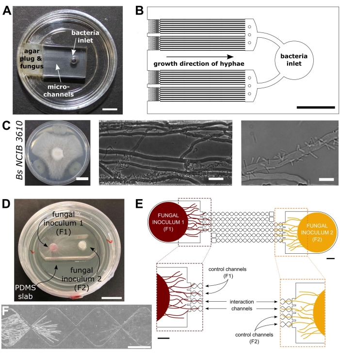
איור 1: ייצוג חזותי וסכמטי של התקני האינטראקציה החיידקית-פטרייתית (BFI) והתקני האינטראקציה הפטרייתית-פטרייתית (FFI). תקע תפטיר ממוקם בכניסה לקצה אחד של המיקרו-ערוצים כדי לאפשר צמיחת היפל לתוך המכשיר. המפרצון החיידקי נמצא בקצה הנגדי. סרגל קנה מידה = 5 מ”מ. (B) סקירה סכמטית של התקן ה- BFI, המתארת את המיקום של מפרצוני החיידקים ואת כיוון הצמיחה ההיפלית באמצעות מיקרו-ערוצי האינטראקציה. הערוצים הם בעומק של 10 מיקרומטר, ברוחב של 100 מיקרומטר ובאורך של 7 מ”מ, עם 28 ערוצי תצפית בסך הכל. (C) מבחן עימות על לוח אגר בין Coprinopsis cinerea ו Bacillus subtilis NCIB 3610, סרגל קנה מידה = 20 מ”מ (משמאל). תמונות מיקרוסקופיה מראות את האינטראקציה בין C. cinerea ו– B. subtilis NCIB 3610 בתוך המיקרו-ערוץ (האמצעי והימין), כלומר חיבור קוטבי של חיידקים להיפאה פטרייתית. סרגל קנה מידה = 25 מיקרומטר (באמצע) ו- 10 מיקרומטר (מימין). (D) תמונה של מכשיר ה-FFI המחובר לצלחת פטרי בעלת תחתית זכוכית, מחוסנת פעמיים בתקעים תפטיריים. סרגל קנה מידה = 1 ס”מ. (E) סקירה סכמטית של התקן ה- FFI. שני תקעי אינוקולנט פטרייתיים מוכנסים לתוך המפרצים בשני קצות המכשיר, ומאפשרים חקירה היפלית של המיקרו-ערוצים. ערוצי הבקרה מחוברים לכניסה פטרייתית אחת בלבד ויש להם ערוץ ללא מוצא, מה שמונע אינטראקציות בין פטריות הבדיקה. ערוצי אינטראקציה מחברים בין שני המפרצים הפטרייתיים ומאפשרים אינטראקציות היפאליות בין הנבדקים בתוך המיקרו-ערוץ. כל תעלת אינטראקציה מורכבת מ-18 מקטעים בצורת יהלום, באורך כולל של 8.8 מ”מ (490 x 430 מיקרומטר ליהלום), בעומק של 10 מיקרומטר ובבעל אזור חיבור בין כל יהלום של 20 מיקרומטר. סוגי הערוצים משוכפלים, סרגלי קנה מידה = 1 מ”מ. (F) אזור אינטראקציה בין שתי חזיתות היפל מתקרבות, הגדלות מקצוות מנוגדים של ערוץ האינטראקציה המחובר. תמונת מיקרוסקופיה של ניגודיות פאזה, סרגל קנה מידה = 250 מיקרומטר. הלוחות באיור זה שונו מ-Stanley et al., 2014 (A-C)40 ו-Gimeno et al., 2021 (D-F)41. אנא לחץ כאן כדי להציג גרסה גדולה יותר של נתון זה.

Protocol

הערה: סיכום של ההליכים המתוארים בפרוטוקול זה מתואר באופן חזותי באיור 2. איור 2: ייצוג סכמטי של המתודולוגיה המוצגת המורכב מחמישה חלקים עיקריים המפורטים בפרוטוקול זה.</str…

Representative Results

תוצאות מייצגות מוצגות ממכשירי BFI40 ו- FFI41 למופת. ניתן להשיג בקלות מדידות קצב גדילה של Hyphal באמצעות מכשירים אלה בשילוב עם טכניקות מיקרוסקופיה בסיסיות. איור 3A-B ממחיש את האינטראקציות בין חיידקים לפטריות בין C. cinerea hyphae לבין B. subti…

Discussion

מאמר זה מציג פרוטוקול לחקר אינטראקציות פטרייתיות-מיקרוביאליות באמצעות מיקרופלואידיקה של ערוצים. המחברים שואפים להדגים את הרבגוניות של מכשירים אלה ולעודד הסתגלות שתתאים לתחומי העניין של החוקר. באמצעות מכשירי BFI ו-FFI למופת, ניתן לחקור אינטראקציות פטרייתיות-מיקרוביאליות בפירוט רב יותר מאש…

Divulgazioni

The authors have nothing to disclose.

Acknowledgements

אנו מכירים בתמיכה כספית מהמחלקה לביו-הנדסה באימפריאל קולג’ בלונדון ומקרן Leverhulme (הפניה למענק מחקר: RPG-2020-352).

Materials

Agar Difco Laboratories 214010 Used to solidify culture medium for bacterial and fungal cultivation within Petri dishes
Aluminum foil Fisher Scientific Ltd 11759408
AutoCAD 2021 Autodesk, USA
Autoclave (VX-75) Systec
Centrifuge (5810R) Eppendorf
Chlorotrimethysilane Merck Life Sciences 386529 CAUTION: Chlorotrimethylsilane is a hazardous substance. Wear appropriate PPE and handle with care. Avoid contact with skin and eyes and prevent inhalation. Keep away from sources of ignition and use in a well-ventilated area.
Cork borer SLS COR1000
Developer solution (mr-Dev 600) Microresist Technologies CAUTION: mr-Dev 600 developer solution is flammable
Erlenmeyer flasks VWR 214-1108 e.g. 200 mL; choose size to suit your exact needs
Ethanol (70% v/v)  Fisher Scientific Ltd E/0650DF/15 Diluted from 99.8% (Analytical Reagent Grade)
Fiji ImageJ Exemplar software package for imaging processing
Filtered, compressed air Available as standard in most labs. Altervatively, an oil-free compressor with air regulator can be used.
Flat-headed wafer tweezers SLS INS5026
Forceps Fisher Scientific Ltd 10008051 Bent, sharp
Glass bottom petri dish World Precision Instruments FD35-100 35 mm
Glass bottom petri dish World Precision Instruments FD5040-100 50 mm
Glass crystallisation dishes VWR 216-1865 Used for washing of PDMS slabs
Glass crystallisation dishes VWR 216-1866 Used in the development of master moulds
Glass media bottles Fisher Scientific Ltd 15456113 e.g. 250 mL; choose size to suit your exact needs
Glass syringe (Hamilton) Fisher Scientific Ltd 10625251 Used for dispensing chlorotrimethylsilane
Hot plate (HP 160 III BM) SAWATEC
Inoculation loop VWR COPA175CS01
Isopropyl alcohol Sigma-Aldrich W292907
Laminar flow hood Air Science (PCR) Exemplar laminar flow hood used for device fabrication
LB medium Fisher Scientific Ltd BP9723-500 Exemplar nutrient broth for bacterial overnight culture
Light emitting diode light engine (LedHUB) Omicron-Laserage Laserprodukte GmbH Exemplar light source that can be used for imaging fungal-microbial interactions (fluorescence)
MA6 Ultraviolet mask aligner Suss Microtec
Malt extract VWR 84618 Used to make exemplar fungal culture medium (Malt extract agar)
Mask Writer Applied Materials 4700DP Example of a mask writer which can be used to print photo-mask for photolithography
Master mould plastic mount 3D-printed bespoke holder manufactured in-house
Microbiological safety cabinet  (BioMat2) Contained Air Solutions Exemplar MSC used for microbial culture and device inoculation
Milli-Q purified water Available as standard in biology labs. 
NaOH Fisher Scientific Ltd BP359-500
NIS-Elements Advanced Research imaging software Nikon Exemplar software package for image acquisition
NIS-Elements Free Viewer Nikon Exemplar software package for viewing acquired images
Oven (Binder BD115) Fisher Scientific Ltd 15602126 Used for curing poly(dimethylsiloxane)(PDMS)
Oven (CLO-2AH-S) KOYO Used for preparing silicon wafers
Parafilm Bemis HS234526B transparent film
Petri dishes, square sterile Fisher Scientific Ltd 11708573 120.5 mm
Petri dishes, sterile Fisher Scientific Ltd 15370366 90 mm 
Photolithography mask Micro Lithography Services Ltd. UK
Plasma cleaner (Zepto) Diener Electronic 100012601
Plastic cup Semadeni 8323
Plastic spatula Semadeni 3340
Portable precision balance (OHAUS Scout) Fisher Scientific Ltd 15519631 Used for weighing PDMS, media components etc.
Precision cutter Syneo HS1251135P1183 Cutting edge diameter: 3.18 mm
Precision cutter  Syneo HS1871730P1183S Cutting edge diameter: 4.75 mm
Profilometer  Bruker Dektak XT-stylus
Razor blades Häberle Labortechnik 9156110
Refridgerator Haden 4-6 °C
Retiga R1 CCD camera Qimaging Exemplar camera that can be used for imaging fungal-microbial interactions
Scotch magic tape Office Depot 3969954 19 mm invisible tape; clear tape
Shaking incubator (Cole-Parmer SI500) Fisher Scientific Ltd 10257954
Silicon wafer Inseto 100 mm
Soda lime glass plate Inseto 125 mm x 125 mm x 2 mm. Used to hold photolithography mask in mask aligner
Sodium chloride Sigma-Aldrich S7653
Spincoater  SAWATEC SM-180-BM
SU-8 2010 photoresist MicroChem CAUTION: SU-8 photoresist is hazardous, take care when handling and prevent inhalation and contact with skin. Flammable, potentially carcinogenic and toxic to the environment. 
Sylgard 184 elastomer kit VWR 634165S Used for the preparation of poly(dimethylsiloxane)(PDMS) devices
Temperature controlled incubator Okolab Exemplar incubator that can be used for imaging fungal-microbial interactions
Ti2-E inverted epifluorescence microscope Nikon MEA54000 Exemplar microscope that can be used for imaging fungal-microbial interactions
Ultrasonic cleaner S-Line Fisher Scientific Ltd FB15050
Vacuum desiccator Fisher Scientific Ltd 10528861 Silianisation and PDMS degassing should be conducted in separate desiccators
x10/0.3 NA CFI Plan Fluor DL objective lens Nikon MRH20105 Exemplar objective lens that can be used for imaging fungal-microbial interactions
x20/0.45 NA CFI Plan Fluor DL objective lens Nikon MRH48230 Exemplar objective lens that can be used for imaging fungal-microbial interactions

Riferimenti

  1. Zhu, Y. -. G., Miller, R. M. Carbon cycling by arbuscular mycorrhizal fungi in soil-plant systems. Trends in Plant Science. 8 (9), 407-409 (2003).
  2. Dai, Z., et al. Long-term nutrient inputs shift soil microbial functional profiles of phosphorus cycling in diverse agroecosystems. The ISME Journal. 14 (3), 757-770 (2020).
  3. Op De Beeck, M., et al. Regulation of fungal decomposition at single-cell level. The ISME Journal. 14 (4), 896-905 (2020).
  4. Bender, S. F., et al. Symbiotic relationships between soil fungi and plants reduce N2O emissions from soil. The ISME Journal. 8 (6), 1336-1345 (2014).
  5. Dullah, S., et al. Melanin production and laccase mediated oxidative stress alleviation during fungal-fungal interaction among basidiomycete fungi. IMA Fungus. 12 (1), 33 (2021).
  6. Deveau, A., et al. Bacterial-fungal interactions: ecology, mechanisms and challenges. FEMS Microbiology Reviews. 42 (3), 335-352 (2018).
  7. Bian, R., et al. Facilitative and synergistic interactions between fungal and plant viruses. Proceedings of the National Academy of Sciences of the United States of America. 117 (7), 3779-3788 (2020).
  8. Jiang, X., Xiang, M., Liu, X. Nematode-trapping fungi. Microbiology Spectrum. 5 (1), (2017).
  9. Essig, A., et al. a novel peptide-based fungal antibiotic interfering with the peptidoglycan synthesis. Journal of Biological Chemistry. 289 (50), 34953-34964 (2014).
  10. Tang, H. -. Y., Zhang, Q., Li, H., Gao, J. -. M. Antimicrobial and allelopathic metabolites produced by Penicillium brasilianum. Natural Product Research. 29 (4), 345-348 (2015).
  11. Bai, Y. -. B., et al. Antifungal activity of griseofulvin derivatives against phytopathogenic fungi In vitro and In vivo and three-dimensional quantitative structure-activity relationship analysis. Journal of Agricultural and Food Chemistry. 67 (22), 6125-6132 (2019).
  12. Solanki, M. K., et al. Characterization of antagonistic-potential of two Bacillus strains and their biocontrol activity against Rhizoctonia solani in tomato. Journal of Basic Microbiology. 55 (1), 82-90 (2015).
  13. Jamali, H., Sharma, A., Srivastava, A. K. Biocontrol potential of Bacillus subtilis RH5 against sheath blight of rice caused by Rhizoctonia solani. Journal of Basic Microbiology. 60 (3), 268-280 (2020).
  14. Válková, H., Novotný, &. #. 2. 6. 8. ;., Malachová, K., Šlosarčíková, P., Fojtík, J. Effect of bacteria on the degradation ability of Pleurotus ostreatus. Science of The Total Environment. 584-585, 1114-1120 (2017).
  15. Leyva-Rojas, J. A., Coy-Barrera, E., Hampp, R. Interaction with soil bacteria affects the growth and amino acid content of Piriformospora indica. Molecules. 25 (3), 572 (2020).
  16. Dullah, S., et al. Fungal interactions induce changes in hyphal morphology and enzyme production. Mycology. 12 (4), 279-295 (2021).
  17. Marfetán, J. A., Romero, A. I., Folgarait, P. J. Pathogenic interaction between Escovopsis weberi and Leucoagaricus sp.: mechanisms involved and virulence levels. Fungal Ecology. 17, 52-61 (2015).
  18. Cortois, R., De Deyn, G. B. The curse of the black box. Plant and Soil. 350 (1), 27-33 (2012).
  19. Whitesides, G. M. The origins and the future of microfluidics. Nature. 442 (7101), 368-373 (2006).
  20. Sackmann, E. K., Fulton, A. L., Beebe, D. J. The present and future role of microfluidics in biomedical research. Nature. 507 (7491), 181-189 (2014).
  21. Hanson, K. L., et al. Fungi use efficient algorithms for the exploration of microfluidic networks. Small. 2 (10), 1212-1220 (2006).
  22. Held, M., Edwards, C., Nicolau, D. V. Probing the growth dynamics of Neurospora crassa with microfluidic structures. Fungal Biology. 115 (6), 493-505 (2011).
  23. Thomson, D. D., et al. Contact-induced apical asymmetry drives the thigmotropic responses of Candida albicans hyphae. Cellular Microbiology. 17 (3), 342-354 (2015).
  24. Lee, K. K., Labiscsak, L., Ahn, C. H., Hong, C. I. Spiral-based microfluidic device for long-term time course imaging of Neurospora crassa with single nucleus resolution. Fungal Genetics and Biology. 94, 11-14 (2016).
  25. Asenova, E., Lin, H. Y., Fu, E., Nicolau, D. V., Nicolau, D. V. Optimal fungal space searching algorithms. IEEE Transactions on NanoBioscience. 15 (7), 613-618 (2016).
  26. Soufan, R., et al. Pore-scale monitoring of the effect of microarchitecture on fungal growth in a two-dimensional soil-like micromodel. Frontiers in Environmental Science. 6, (2018).
  27. Uehling, J. K., et al. Microfluidics and metabolomics reveal symbiotic bacterial-fungal interactions between Mortierella elongata and Burkholderia include metabolite exchange. Frontiers in Microbiology. 10, 2163 (2019).
  28. Millet, L. J., et al. Increasing access to microfluidics for studying fungi and other branched biological structures. Fungal Biology and Biotechnology. 6 (8), 1-14 (2019).
  29. Baranger, C., Fayeulle, A., Le Goff, A. Microfluidic monitoring of the growth of individual hyphae in confined environments. Royal Society Open Science. 7 (8), 191535 (2020).
  30. Aleklett, K., Ohlsson, P., Bengtsson, M., Hammer, E. C. Fungal foraging behaviour and hyphal space exploration in micro-structured Soil Chips. The ISME Journal. 15 (6), 1782-1793 (2021).
  31. Aleklett, K., et al. Build your own soil: exploring microfluidics to create microbial habitat structures. The ISME Journal. 12 (2), 312-319 (2018).
  32. Ellett, F., Jorgensen, J., Frydman, G. H., Jones, C. N., Irimia, D. Neutrophil interactions stimulate evasive hyphal branching by Aspergillus fumigatus. PLOS Pathogens. 13 (1), 1006154 (2017).
  33. Massalha, H., Korenblum, E., Malitsky, S., Shapiro, O. H., Aharoni, A. Live imaging of root-bacteria interactions in a microfluidics setup. Proceedings of the National Academy of Sciences of the United States of America. 114 (17), 4549-4554 (2017).
  34. Schmieder, S. S., et al. Bidirectional propagation of signals and nutrients in fungal networks via specialized hyphae. Current Biology. 29 (2), 217-228 (2019).
  35. Tayyrov, A., Stanley, C. E., Azevedo, S., Künzler, M. Combining microfluidics and RNA-sequencing to assess the inducible defensome of a mushroom against nematodes. BMC Genomics. 20 (1), 243 (2019).
  36. Stanley, C. E., Grossmann, G., Casadevall i Solvas, X., deMello, A. J. Soil-on-a-Chip: microfluidic platforms for environmental organismal studies. Lab on a Chip. 16 (2), 228-241 (2016).
  37. Stanley, C. E., vander Heijden, M. G. A. Microbiome-on-a-Chip: new frontiers in plant-microbiota research. Trends in Microbiology. 25 (8), 610-613 (2017).
  38. Ortseifen, V., Viefhues, M., Wobbe, L., Grünberger, A. Microfluidics for biotechnology: bridging gaps to foster microfluidic applications. Frontiers in Bioengineering & Biotechnology. 8, 589074 (2020).
  39. Jansson, J. K., Hofmockel, K. S. The soil microbiome-from metagenomics to metaphenomics. Current Opinion in Microbiology. 43, 162-168 (2018).
  40. Stanley, C. E., et al. Probing bacterial-fungal interactions at the single cell level. Integrative Biology (Camb). 6 (10), 935-945 (2014).
  41. Gimeno, A., et al. A versatile microfluidic platform measures hyphal interactions between Fusarium graminearum and Clonostachys rosea in real-time. Communications Biology. 4 (1), 262 (2021).
  42. Duffy, D. C., McDonald, J. C., Schueller, O. J. A., Whitesides, G. M. Rapid prototyping of microfluidic systems in poly(dimethylsiloxane). Analytical Chemistry. 70 (23), 4974-4984 (1998).
  43. Stanley, C. E., et al. Fabrication and use of the dual-flow-RootChip for the imaging of Arabidopsis roots in asymmetric microenvironments. Bio-protocol. 8 (18), 3010 (2018).
  44. Choi, C. -. H., Lee, H., Weitz, D. A. Rapid patterning of PDMS microfluidic device wettability using syringe-vacuum-induced segmented flow in nonplanar geometry. ACS Applied Materials & Interfaces. 10 (4), 3170-3174 (2018).
  45. Sanders, E. R. Aseptic laboratory techniques: plating methods. Journal of Visualized Experiments. (63), e3064 (2012).
  46. Schindelin, J., et al. Fiji: an open-source platform for biological-image analysis. Nature Methods. 9 (7), 676-682 (2012).
  47. Harting, R., et al. Pseudomonas strains induce transcriptional and morphological changes and reduce root colonization of Verticillium spp. Frontiers in Microbiology. 12, 652468 (2021).
  48. Boenisch, M. J. . Structural and molecular characterisation of the penetration process of Fusarium graminearum during Fusarium head blight infection. , (2013).
  49. Eynck, C., Koopmann, B., Grunewaldt-Stoecker, G., Karlovsky, P., von Tiedemann, A. Differential interactions of Verticillium longisporum and V. dahliae with Brassica napus detected with molecular and histological techniques. European Journal of Plant Pathology. 118 (3), 259-274 (2007).
  50. Ghanem, N., Stanley, C. E., Harms, H., Chatzinotas, A., Wick, L. Y. Mycelial effects on phage retention during transport in a microfluidic platform. Environmental Science & Technology. 53 (20), 11755-11763 (2019).
  51. Alrifaiy, A., Lindahl, O. A., Ramser, K. Polymer-based microfluidic devices for pharmacy, biology and tissue engineering. Polymers. 4 (3), 1349-1398 (2012).
  52. Duncombe, T. A., Tentori, A. M., Herr, A. E. Microfluidics: reframing biological enquiry. Nature Reviews Molecular Cell Biology. 16 (9), 554-567 (2015).
  53. Hoelzle, D., et al. Microfluidic device design, fabrication, and testing protocols. Protocol Exchange. , (2015).

Play Video

Citazione di questo articolo
Masters-Clark, E., Clark, A. J., Stanley, C. E. Microfluidic Tools for Probing Fungal-Microbial Interactions at the Cellular Level. J. Vis. Exp. (184), e63917, doi:10.3791/63917 (2022).

View Video