This protocol was approved by the Institutional Animal Care and Use Committee (IACUC) at the University of Florida and is in compliance with the “Principle of Laboratory Animal Care” (NIH publication No. 86-23, revised 1985).
1. Materials
- Animals: Male C57BL/6 mice (Harlan Laboratories, Indianapolis, IN, USA) weighing 20–26 grams at the time of surgery.
- Anesthesia:
- Anesthesia machine: VetEquip inhalation anesthesia system (VetEquip Inc., Pleasanton, CA, USA).
- Inhalation anesthetic: Isoflurane (Baxter Pharmaceutics, Deerfield, IL).
- Surgical tools: SuperCut Iris scissors, straight (World Precision Instruments, Inc., Sarasota, FL, USA); Vannas micro-scissors, straight (World Precision Instruments, Inc., Sarasota, FL, USA); Dumont forceps (fine tip, 45° bent tips, World Precision Instruments, Inc., Sarasota, FL, USA); Bipolar coagulation unit (High Frequency Desiccator 900, World Precision Instruments, Inc., Sarasota, FL, USA); micro vessel clip (World Precision Instruments, Inc., Sarasota, FL, USA)
- Surgical supplies: Sterile cotton tips; Wipes; Gauze sponges; Suture (7-0 Silk suture, Ethicon, Inc., Somerville, NJ, USA); Tissue adhesive (Vetbond, 3M Company, St Paul, MN, USA); Surgical scrub: Bethadine, 70% ethanol, and sterile 0.9% saline; Eye ointment (Puralube Vet Ointment, Pharmaderm, Melville, NY, USA).
- Occlusion suture preparation for MCAO:
- Filament for MCA occlusion: 7-0 surgical Nylon monofilament suture (Ethicon, Inc., Somerville, NJ, USA).
- Heat source for blunting suture tip (Thermal Cautery Unit, World Precision Instruments, Inc., Sarasota, FL).
- Liquid silicone rubber (Silicone RTV adhesive, World Precision Instruments, Inc., Sarasota, FL, USA).
- Surgical/Dissecting microscope (Leica Stereo Microscope MZ6).
- Fiber optic illuminator (Fiber Optic illuminator FO1–150, TechniQuip Corp., Livermore, CA, USA).
- Laser doppler flowmetry (PeriFlux System 5000, Perimed, Stockholm, Sweden).
- Flexible microtip (MT B500-0 Straight Microtip, Perimed, Stockholm, Sweden)
- Temperature parameters:
- Rectal temperatures monitor (BAT-12, World Precision Instruments, Inc., Sarasota, FL).
- Thermal blanket (T/PUMP, TP600, Gaymar, NY, USA).
- Staining and quantitative measurement of volume infarct:
- 2% solution 2,3,5-triphenyltetrazolium chloride (2% TTC, Sigma-Aldrich Co., St Louis, MO, USA).
- Mice brain matrix slicer (Zivic-Miller Lab., Inc., Allison Park, PA, USA).
- Digital camera (V1253, Kodak Co., Rochester, NY, USA).
- Image J software (ImageJ 1.42q software, U.S. National Institutes of Health, Bethesda, MA, USA).
2. Pre-surgical steps
- Prior to surgery, the mice are housed under a 12:12 light/dark cycle with free access to water and rodent chow.
- Sutures for MCAO are prepared by blunting 1-2mm of the suture tip with heat and coating 3-4mm of suture shaft from its tip with liquid silicone rubber, allowing it to dry (7-0; tip diameter, 0.20-0.25mm; coating length, 3-4mm; suture length 11-13mm). Sterility should be preserved in all of the aforementioned steps.
- After weighing the mouse, anesthesia is induced with 4% isoflurane in an induction chamber and maintained during the procedure with 1.5-2% isoflurane in 30% O2 and 70% N2O gas mixture via a facemask.
- The mouse is placed in a prone position on a gauze pad lying on a temperature-controlled operating surface (thermal blanket); the head fur is shaved.
- Lubricant ophthalmic ointment is applied to both eyes, and eyelids are closed to prevent eye desiccation during the surgical process.
- A midline incision (1 cm) is made on skin overlying the calvarium from superior nuchal line to the nasion and the skin is pulled laterally to affix a flexible microtip (MT B500-0 Straight Microtip; 0.5mm diameter). The microtip is placed perpendicular to the surface of the right parietal skull (1 mm posterior and 5 mm lateral to the bregma) to monitor blood flow in the MCA territory by laser doppler flowmetry.
- Make sure that the flexible microtip is attached to skull firmly before the animal is turned to a supine position. The animal’s neck is placed on a soft pad and its nose is held in nose cone of the anesthetic apparatus. This will result in having enough space for the flexible microtip.
- A rectal temperature probe is inserted to maintain a constant animal core temperature of 37±0.5°C.
- Both forepaws and hindpaws are taped to the operating platform surface, and the tail is taped to the rectal tube.
3. MCA occlusion surgery:
- Under the operating microscope, a midline neck incision is made with scissors between the manubrium and the jaw by gently lifting the loose skin.
- The paired underlying submandibular glands are bluntly divided, with the left gland left in situ. The right submandibular gland is retracted cranially and secured along with the sternocleidomastoid muscle.
- The exposed division of the omohyoid muscle covering the carotid sheath is carefully divided with pickups.
- The right common carotid artery (CCA) is carefully separated and isolated from the vagus nerve and its sheath (located lateral to CCA).
- Hereafter, the 2 surgical methods that have been used frequently to perform MCA occlusion diverge:
Koizumi’s4 method
- Three loose collar sutures (7-0 silk) are prepared around the right CCA.
- The external carotid artery (ECA) is identified, isolated, and cauterized (2-3mm distal to bifurcation). It can subsequently be divided with micro-scissors. To avoid inadvertent peripheral damages at the cauterization site, the power should be set at 1 using the power knob. This is consistent for all cauterizations.
- Sufficient tension is applied to the most proximal collar suture on the CCA to occlude the blood flow (permanent ligation). The flow occlusion is verified by a ~50% drop of baseline cortical perfusion values.
- Then a vessel clip is placed just before the CCA bifurcation to avoid retrograde flow at the time of arteriotomy.
- An arteriotomy is performed between the proximal ligated suture and the vessel clip on the CCA with micro-scissors (closer to ligation).
- The heat-blunted silicone-coated 7-0 nylon suture is introduced via the arteriotomy and advanced till it reaches the vessel clip. The 2 remaining loose collar sutures are gently tightened around the CCA between the arteriotomy and the vessel clip to avoid back flow leakage through CCA, with caution not to traumatize the arterial wall; then the vessel clip is withdrawn.
Longa’s3 method
- The ECA is identified, dissected as much as possible (near its bifurcation into lingual and maxillary arteries), cauterized (with power set to 1), and divided with micro-scissors.
- Two loose collar sutures (7-0 silk) are prepared around the proximal ICA just above the CCA bifurcation.
- The CCA is temporarily closed with a vessel clip. The flow occlusion is verified by a ~50% drop of baseline cortical perfusion values.
- Then the ICA is temporarily closed with a vessel clip above the two loose collar sutures to avoid retrograde flow at the time of arteriotomy.
- An arteriotomy is performed using micro-scissors in the reflected ECA close to the stump.
- The heat-blunted silicone-coated 7-0 nylon suture is introduced via the arteriotomy in ECA and advanced till it reaches the applied vessel clip in ICA. The 2 loose collar sutures in proximal ICA are gently tightened around the proximal ICA to avoid reflux blood flow through ICA, with enough attention not to traumatize the arterial wall; then the vessel clip is withdrawn.
The rest of the procedure is largely convergent:
- The occluding suture is slowly advanced through internal carotid artery (ICA) toward the cranial base until a mild resistance is felt. A concurrent drop in cerebral blood flow value as a percentage (= 80%) relative to baseline is confirmed by laser doppler flowmetry. The 2 collar sutures are tightened around the inserted filament.
- For transient cerebral ischemia experiments, the occluding suture is withdrawn gently after 45 minutes. This time point can be altered for various experimental aims.
- After withdrawing the suture filament, the arteriotomy site is closed to avoid post-surgical bleeding. In Koizumi’s method, this involves securely tightening the collar sutures around the CCA. In Longa’s technique, the arteriotomy site at the base of ECA is cauterized, then the collar sutures at the proximal ICA are loosened and removed to restore the carotid flow.
- To close the incision, the submandibular gland and SCM are laid over the operative field. The wound edges are then approximated with tissue adhesive in cervical and scalp areas. Alternatively, an absorbable suture or metal staple could be used to close the wound especially when long-term survival of the animal is intended.
- After incision closure, the animal receives 1 ml warm saline subcutaneously as volume replenishment. A dose of appropriate analgesics (i.e. Buprenorphine at 0.05-0.1 mg/kg) should be used to minimize animal’s pain and discomfort during recovery period. The animal is removed from the surgical suite and placed in a warm recovery area/cage to prevent hypothermia, with free, easy access to soft food and water.
4. Post-Op neurological evaluation
After the animal regains total consciousness, neurological deficits can be evaluated by a simple scale (five-point scale) as follows3:
- no observable deficits
- failure to extend contralateral forepaw (mild focal neurologic deficit)
- circling in a direction contralateral to infarct (moderate focal neurologic deficit)
- falling in a direction contralateral to infarct (severe focal neurologic deficit)
- depressed level of consciousness without spontaneous movement
5. Staining and quantitative measurement of brain infarct volume
- After 24 hours of MCA reperfusion, the surviving mouse is weighed, deeply anesthetized with 4% isoflorane, and sacrificed according to approved protocol.
- The brain is carefully removed and placed in a mouse brain matrix slicer, and the entire cerebrum is coronally sectioned at 2 mm intervals.
- The sections are immersed in 2% TTC in phosphate buffered saline (PBS) at 37 °C for 20 minutes. After TTC staining, infracted area is shown as a white (unstained) tissue adjacent to red brick (viable) tissue (Figure 1).
- Serial sections are photographed using a digital camera and the area of infarct is measured with the image J program.
- The area of infarct (white, unstained), the area of ipsilateral hemisphere (white and red brick, stained), and the area of the contralateral hemisphere (red brick, stained) are measured for each section. The volume is calculated by summing the representative areas in all sections and multiplying by the slice thickness. After correcting for edema, the volume of infarction is calculated as follows9:
Corrected Infarct Volume (CIV), % = [contralateral hemisphere volume – (ipsilateral hemisphere volume – infarct volume)]/contralateral hemisphere volume x 100
6. Representative Results
After occlusion and reperfusion of the MCA via left CCA to establish transient cerebral ischemia, animals are sacrificed and their coronally sectioned brains are stained with TTC to evaluate the volume of infarct (Figure 1).
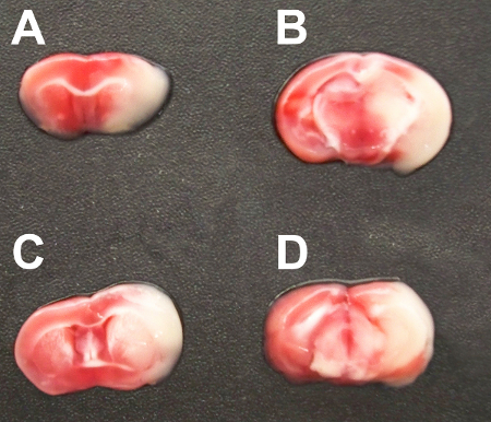
Figure 1. TTC-stained serial coronal brain sections (2 mm) from mice subjected to MCA occlusion.
