The myofibroblast is an influential stromal cell of the gastrointestinal tract that regulates important physiologic processes in both normal and disease states. We describe a technique that allows for the isolation of primary myofibroblasts from both mouse and human colon tissue, which can be utilized for in vitro experimentation.
The myofibroblast is a stromal cell of the gastrointestinal (GI) tract that has been gaining considerable attention for its critical role in many GI functions. While several myofibroblast cell lines are commercially available to study these cells in vitro, research results from a cell line exposed to experimental cell culture conditions have inherent limitations due to the overly reductionist nature of the work. Use of primary myofibroblasts offers a great advantage in terms of confirming experimental findings identified in a cell line. Isolation of primary myofibroblasts from an animal model allows for the study of myofibroblasts under conditions that more closely mimic the disease state being studied. Isolation of primary myofibroblasts from human colon tissue provides arguably the most relevant experimental data, since the cells come directly from patients with the underlying disease. We describe a well-established technique that can be utilized to isolate primary myofibroblasts from both mouse and human colon tissue. These isolated cells have been characterized to be alpha-smooth muscle actin and vimentin-positive, and desmin-negative, consistent with subepithelial intestinal myofibroblasts. Primary myofibroblast cells can be grown in cell culture and used for experimental purposes over a limited number of passages.
The regulation of gastrointestinal (GI) tract function involves complex, dynamic interactions between the mucosal layer and its underlying mesenchyme. These interactions normally serve to maintain homeostasis but may also lead to the development of disease states under pathologic conditions1. Myofibroblasts are a subpopulation of stromal cells located subjacent to the epithelial layer that communicate in a paracrine fashion with surrounding cell subpopulations to regulate a number of critical GI tract processes that include mucosal regeneration, repair, and fibrosis2.
The 18Co cell line is an example of a human colonic myofibroblast cell line that has been widely used to study myofibroblast function because it shares many of the characteristics of primary in situ myofibroblasts. These include the protein expression of a-smooth muscle actin (α-SMA) and vimentin, as well as the expression of a number of cell surface receptors such as the epidermal growth factor receptor, or receptors for lysophosphatidic acid3,4. Because of these shared ultrastructural characteristics, as well as similarities in biologic function5, the 18Co cell line has been used extensively to study myofibroblast function in the context of many disease states such as inflammatory bowel disease or colorectal cancer3,6. However, there are inherent limitations and concerns when using a cell line. These include genotypic instability over time that differentiates cell lines from primary cells, altering the phenotype and biologic function of the cell, including growth rates and interactions with other cell populations. Cell lines also lack the normal components of the GI microenvironment (epithelial, stromal, and vascular components), which is a major limitation to its use. Therefore, the conclusions drawn from experimental cell line research requires further validation to reduce the risk of misinterpretation.
Primary myofibroblasts can be obtained from human and mouse colon tissue to confirm experimental findings identified in a cell line7. The method was originally described by Mahida, et al.8, and our protocol is one of many well-described techniques that can be used to isolate primary myofibroblasts from mouse and human colon9. For any mouse model of colonic disease, this technique can be used to isolate primary myofibroblasts to study how they interact with neighboring cells or contribute to both normal or pathologic GI processes10,11. This technique can be used to study how myofibroblasts contribute to mucosal healing, fibrosis, and the development of colorectal adenomas and carcinomas. Primary myofibroblasts can also be obtained from human colon tissue that has been resected at the time of operation for benign (inflammatory bowel disease, strictures, diverticulitis) or malignant conditions. Isolated primary cells from human and mouse colon tissue can be grown in cell culture and utilized over a limited number of passages.
1. Obtain Colon Tissue
Mouse
A protocol to perform animal experiments, detailing the rationale and objectives of the research, was submitted and approved by our institutional Office of Animal Research Oversight.
Human
A protocol to obtain human tissue from surgical patients, detailing the importance of obtaining this tissue as well as an assessment of potential risks and benefits, was submitted and approved by our institutional Office of the Human Research Protection Program.
2. Denude Epithelial Cells
3. Myofibroblast Isolation and Culture
Abbreviations
HBSS: Hank’s balanced salt solution; RPMI-5: Roswell Park Memorial Institute media; ACK: Ammonium-Chloride-Potassium; FCS: fetal calf serum; TC-treated: tissue culture treated
Solutions
ACK lysis buffer – (4.15 g NH4Cl, 0.5 g KHCO3, 18.6 mg Na2EDTA, 400 ml H2O, adjust pH to 7.2-7.4 with 1 N HCl). Filter sterilize through a 0.2 µm filter and store at 4 °C. RPMI-5 – (to make 500 ml: 454.5 ml RPMI, 25 ml FCS, 5 ml 200 mM L-glutamine, 5 ml 1 M HEPES, pH 7.4, 5 ml 100 mM sodium pyruvate, 5 ml 100x Pen-Strep, 500 ml 50 mM B-ME in PBS).
Once isolated, primary human myofibroblasts can be grown in cell culture and used over a limited number of passages (up to passage 4). These cells are characterized as being a-smooth muscle actin and vimentin positive, and desmin-negative (Figure 1A), consistent with intestinal subepithelial myofibroblasts5,7. They also have a characteristic stellate morphology (Figure 1B). Primary myofibroblasts can then be used to confirm experimental findings identified in a cell line. This approach was utilized to demonstrate that the pro-inflammatory cytokine TNF-α (10 ng/ml) induced the upregulation of EGF receptor expression and signaling in these cells7. Upregulated EGF receptor expression and signaling was initially found utilizing the previously mentioned human colonic myofibroblast cell line 18Co (Figure 2). In Figure 2A, we demonstrate that exposure of 18Co cells to TNF-α led to a time-dependent increase in EGF receptor expression. This correlated with enhanced EGF-induced COX-2 expression and p42/44 MAPK phosphorylation in these cells (Figure 2B). To validate these experimental findings, experiments were repeated using primary human myofibroblasts isolated from surgically resected colon of patients with colorectal cancer. As shown in Figure 2C and 2D, primary human myofibroblasts closely mimic the experimental findings initially identified in the 18Co cell line7.
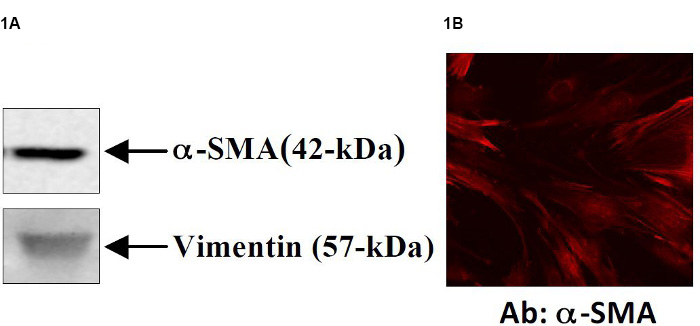
Figure 1. Primary myofibroblasts can be isolated from human colon tissue7. Isolated primary cells demonstrate a myofibroblast-like phenotype that are consistently a-SMA and vimentin positive (Figure 1A) and demonstrate a stellate morphology (Figure 1B).
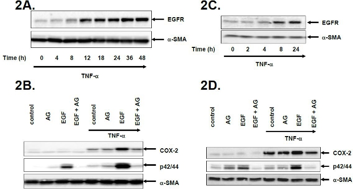
Figure 2. Experimental findings in a cell line are substantiated using primary human myofibroblast cells7. The human colonic myofibroblast cell line 18Co was used to demonstrate that TNF-α induces the upregulation of EGF receptor expression and signaling in these cells (Figure 2A). This upregulation of EGF receptor expression was associated with enhanced EGF receptor signaling, shown in Figure 2B, where subsequent exposure to EGF led to enhanced p42/44 MAPK phosphorylation and COX-2 expression. These effects were completely inhibited with the EGF receptor inhibitor AG1478. Primary human myofibroblasts isolated from surgically resected colon of patients with colorectal cancer confirm these experimental findings (Figures 2C and 2D). Tumor necrosis factor-alpha=TNF-α, EGFR=EGF receptor, AG=AG1478, COX-2=cyclo-oxygenase-2. Click here to view larger figure.
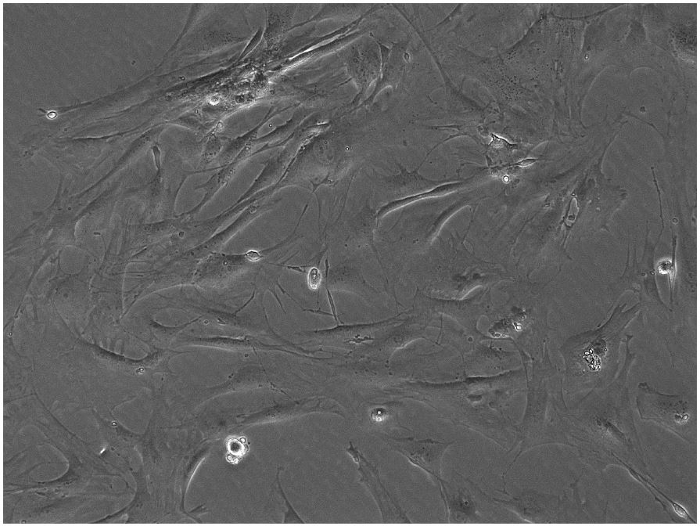
Figure 3. Primary myofibroblasts can be isolated from mouse colon tissue. A 10X view of primary mouse myofibroblast cells. Click here to view larger figure.
The general technique is similar when using either mouse or human colon. This technique can also be used to isolate myofibroblasts from the small intestine. Specific details for the isolation of myofibroblasts from 1. mouse and 2. human colon are discussed below.
1. Mouse Colon
Irrigation of the mouse colon is facilitated by attaching a 4 in, 16 G Popper blunt-end needle to one end of the colon. This also helps when cutting the colon lengthwise. You can accordion the colon tissue onto the needle, then place one tip of the scissor with the sharp end up and slide the colon tissue over the scissor edge while cutting to open it lengthwise.
In step 2.1, it is important to understand that nuclear material from dead cells can lead to cell clumping. The addition of DNase (50 mg/ml) may be used to address this problem.
The most critical step is step 2.4. Keep a careful watch of the colon tissue. When the colon begins to look stringy, remove the colon from the enzymes. You do not need to incubate the colon for the entire 60 min. If the colon tissue is exposed to the dispase and collagenase D for too long, the cells will die. This is a common mistake that will result in a low cell yield.
Contamination of the cells is another common problem. Careful technique, appropriate washing and filtering will minimize the risk of contamination, along with standard sterile techniques used for cell culture work.
2. Human Colon
The length of time for incubation with the enzymes depends on the size of the specimen that is being digested. We typically receive a ½ inch full thickness strip of human colon wall. There is a modification in step 2.3 when isolating myofibroblasts from human colon tissue. For step 2.3, for human colon you must double the amount of dispase (20 U) and collagenase D (4000 U) used. An even larger segment of colon will require a longer incubation time. Also, keep in mind that the specific activity of the enzymes may vary with lots, so you may have to adjust the incubation time accordingly.
For both primary mouse and human myofibroblasts, 0nce grown in culture, the isolated cells should be evaluated to assess for purity of the cells and to confirm that the cells are myofibroblast-like. This can be done with a variety of technique including immunohistochemical staining (antibodies to myofibroblast markers include a-smooth muscle actin and vimentin; monoclonal antibody to CD68 is specific to macrophages, CD31 is specific to endothelial cells, and CD45 is specific to immune cells; antibodies to e-cadherin and various cytokeratins are specific for epithelial cells. mRNA expression can also be assessed by RT-PCR12. Flow cytometry can also be used for cell analysis13.
Primary myofibroblasts can be studied, frozen in liquid nitrogen for subsequent use at a later date, or used for in vitro and co-culture experimentation. Primary cells can also be implanted into the subcutaneous tissue of mice to study cell-cell interactions14. Future applications may involve re-implantation of primary myofibroblasts into the colon wall of a syngeneic mouse utilizing mouse colonoscopy following genetic manipulation.
A number of alternative methods to isolate primary myofibroblasts from human and mouse colon have also been described. Two are listed below:
The authors have nothing to disclose.
This work was supported by National Institutes of Health Grant 5KO8DK085136-02 to JY.
Table of specific reagents and equipment:
Name of the reagent | Company | Catalogue number | Comments (optional) |
---|---|---|---|
Dispase I | Invitrogen/GIBCO | #17105-041 | |
Collagenase D | Roche | #1 088 858 | |
4 in, 16 G Popper blunt-end needle | Fisher | #14-825-16K |