This protocol highlights a method to rapidly assess the biocompatibility of a crystalline nanocellulose (CNC)/agarose composite hydrogel biomaterial ink with mouse bone marrow-derived mast cells in terms of cell viability and phenotypic expression of the cell surface receptors, Kit (CD117) and high-affinity IgE receptor (FcεRI).
Three-dimensional (3D) bioprinting utilizes hydrogel-based composites (or biomaterial inks) that are deposited in a pattern, forming a substrate onto which cells are deposited. Because many biomaterial inks can be potentially cytotoxic to primary cells, it is necessary to determine the biocompatibility of these hydrogel composites prior to their utilization in costly 3D tissue engineering processes. Some 3D culture methods, including bioprinting, require that cells be embedded into a 3D matrix, making it difficult to extract and analyze the cells for changes in viability and biomarker expression without eliciting mechanical damage. This protocol describes as proof of concept, a method to assess the biocompatibility of a crystalline nanocellulose (CNC) embedded agarose composite, fabricated into a 24-well culture system, with mouse bone marrow-derived mast cells (BMMCs) using flow cytometric assays for cell viability and biomarker expression.
After 18 h of exposure to the CNC/agarose/D-mannitol matrix, BMMC viability was unaltered as measured by propidium iodide (PI) permeability. However, BMMCs cultured on the CNC/agarose/D-mannitol substrate appeared to slightly increase their expression of the high-affinity IgE receptor (FcεRI) and the stem cell factor receptor (Kit; CD117), although this does not appear to be dependent on the amount of CNC in the bioink composite. The viability of BMMCs was also assessed following a time course exposure to hydrogel scaffolds that were fabricated from a commercial biomaterial ink composed of fibrillar nanocellulose (FNC) and sodium alginate using a 3D extrusion bioprinter. Over a period of 6-48 h, the FNC/alginate substrates did not adversely affect the viability of the BMMCs as determined by flow cytometry and microtiter assays (XTT and lactate dehydrogenase). This protocol describes an efficient method to rapidly screen the biochemical compatibility of candidate biomaterial inks for their utility as 3D scaffolds for post-print seeding with mast cells.
The recent interest in 3D culture systems and 3D bioprinting has focused attention on hydrogels and hydrogel composites. These composites serve as viscous yet porous biomimetics and can be composed of up to 99% water content by weight, which is comparable to biological tissues1,2,3. These features of hydrogel composites thereby permit the growth of cells without affecting their viability and function. One such composite is crystalline nanocellulose (CNC), which has been used as a reinforcing material in hydrogel composites, cell scaffolds in the development of biomaterial implants, and in two-dimensional (2D) and 3D in vitro cell culture4,5. For the most part, matrices composed of CNC are not overtly cytotoxic to human corneal epithelial cells6, intestinal epithelial cells7, human bone marrow-derived mesenchymal stem cells8, or neuron-like cells9. However, metabolic activity and proliferation of human bone marrow-derived mesenchymal stem cells decreases in correlation with the increased viscosity of wood-based nanocellulose composites, suggesting that the composition of the matrix must be carefully tested for its deleterious effects on cell functions8.
Similarly, CNC can induce inflammatory responses in macrophages upon internalization, which could have serious consequences in 3D immune cell culture systems10,11. In fact, there is very little data available on how CNC may influence other immune cell responses, particularly allergic inflammatory responses that are initiated by mast cells. Mast cells are granulated leukocytes that express the high-affinity IgE receptor, FceRI, responsible for activating inflammatory responses to allergens. Their proliferation and differentiation are dependent-upon stem cell factor (SCF), which binds the tyrosine receptor, Kit. Mast cells are derived from bone marrow progenitor cells that enter the circulation and subsequently migrate peripherally to disperse ubiquitously in all human tissues12. As mast cells function in a 3D tissue environment, they are an ideal immune cell candidate for studying immunological processes in in vitro 3D tissue models. However, to date, there is no viable in vitro 3D tissue model containing mast cells.
Due to the highly sensitive nature of mast cells and their propensity to elicit pro-inflammatory responses to external stimuli, careful consideration of the 3D matrix constituents and the bioprinting method of introducing mast cells into the 3D scaffold is required, as discussed further. Tissue constructs can be biofabricated from two broad categories of biomaterials, i.e., bioinks and biomaterial inks. The distinction lies in the fact that bioinks are cell-laden hydrogel composites, whereas biomaterial inks are hydrogel composites that are devoid of cells, as defined by Groll et al.13,14. Hence, 3D constructs printed with bioinks contain cells pre-embedded within the hydrogel matrix, whereas 3D constructs printed with biomaterial inks need to be seeded with cells post-printing. The biofabrication of culture scaffolds from hydrogel-based bioinks/biomaterial inks is most commonly performed using extrusion 3D bioprinters, which extrude the bioink/biomaterial ink through a microscale nozzle under pressure via either a pneumatically or mechanically driven piston14. Extrusion bioprinters fabricate 3D scaffolds by depositing the bioink in 2D cross-sectional patterns that are sequentially stacked upon each other in a 'bottom-up' approach.
To be compatible with extrusion bioprinting, the hydrogel-based bioink/biomaterial ink must possess thixotropic (shear-thinning) properties, whereby the constituent hydrogel polymers of the bioink/biomaterial ink flow like a fluid through a microchannel nozzle when subjected to shear stress, but revert to a viscous, gel-like state upon removal of the shear stress15. Due to their high water content, the polymers of hydrogel-based bioinks/biomaterial inks must be crosslinked, either physically or covalently, to maintain the architecture and structural integrity of the 3D bioprinted structure. In the case of cell-laden bioinks, the cells are directly subjected to chemical stresses during the crosslinking process. The process of extruding cells encapsulated within the bioink hydrogel matrix also subjects the cells to shear stress, which can lead to reduced viability and/or cell death. Once the 3D tissue model has been bioprinted, it is difficult to discriminate between the levels of cytotoxicity elicited by the hydrogel matrix itself and the extrusion and crosslinking processes, respectively. This is particularly challenging in the context of 3D scaffolds where the cells are pre-embedded within the hydrogel matrix, thus making it difficult to remove the cells for subsequent analyses, which would be detrimental to the viability of mast cells.
A gentler approach to generating 3D tissue constructs containing mast cells involves seeding the cells into pre-printed, porous biomaterial ink 3D scaffolds from a cell culture suspension, which leverages the innate ability of mast cells to migrate from the circulation into peripheral tissues. The benefits of this cell seeding approach are two-fold: (i) the mast cells are not subjected to shear and chemical stresses from the extrusion and crosslinking processes, respectively, and (ii) the cells can be easily removed from the 3D scaffold after exposure by gentle washing for analysis without adversely affecting their viability. The additional benefit of seeding and analyzing the cell viability of mast cells on 3D bioprinted, porous hydrogel scaffolds as opposed to 2D hydrogel discs is that the 3D bioprinted hydrogel scaffolds recapitulate microscale topographical features of in vivo tissues, which are not present in bulk, 2D planar hydrogel discs. This approach is a suitable, rapid, and cost-effective approach to determine the potentially catastrophic cytotoxic effects of candidate bioink hydrogel matrices on mast cells, as well as other immunological cells, prior to investment in costly 3D tissue engineering experiments.
NOTE: This protocol is composed of five sections: (1) isolation of mouse bone marrow and differentiation of mouse bone marrow-derived mast cells (BMMCs), (2) fabrication of CNC/agarose/D-mannitol hydrogel substrates in a 24-well system and culture of BMMCs on the substrates, (3) removal of BMMCs from the CNC/agarose/D-mannitol hydrogel substrates and analysis of viability and biomarker expression using flow cytometry, (4) 3D bioprinting of hydrogel scaffolds from a commercially available fibrillar nanocellulose (FNC)/sodium alginate composite biomaterial ink, and (5) culture of BMMCs on FNC/sodium alginate hydrogel scaffolds and analysis of viability using flow cytometry, XTT, and lactate dehydrogenase (LDH) microtiter assays.
1. Generation of the BMMC culture
NOTE: Mice were euthanized by CO2 asphyxiation following isoflurane anesthesia. The tibia and femur were isolated, and whole bone marrow was harvested. All animal studies were conducted in accordance with the Canadian Council on Animal Care Guidelines and Policies with approval from the Health Science Animal Care and Use Committee for the University of Alberta.
2. Fabrication of the CNC/agarose/D-mannitol hydrogel substrates and BMMC culture
3. Flow cytometric analyses
4. 3D Bioprinting of FNC/sodium alginate hydrogel substrates
NOTE: The 3D bioprinter used in this study is a pneumatic-extrusion system equipped with two independent, temperature-controlled printheads. The biomaterial ink used to 3D bioprint the hydrogel scaffolds is formulated of (a) highly hydrated fibrillar nanocellulose (FNC), which is morphologically similar to collagen, (b) sodium alginate, and (c) D-mannitol. It is supplied as a sterile hydrogel suspension in 3 mL cartridges to which sterile luer-lock conical bioprinting nozzles (22, 25, or 27 G) can be attached.
5. Incubation of BMMCs on 3D bioprinted rectilinear scaffolds and viability testing
One of the most crucial characteristics of a successful biomaterial ink or culture substrate is that of biocompatibility. Primarily, the substrate must not induce cellular death. There are several microtiter-based and flow cytometric methods of quantifying cell viability and necrosis; however, these methods are not amenable to analyzing cells embedded within a hydrogel matrix. In this protocol, the above mentioned limitation is circumvented by seeding the BMMCs onto the hydrogel substrate or bioprinted scaffold. After a specific incubation period (6-48 h in this study), the BMMCs are easily harvested by micropipetting without the need for mechanical disruption or hydrolysis of the hydrogel substrate or bioprinted scaffold, which would otherwise cause physical damage to the cells. The viability of the BMMCs is then rapidly analyzed using the PI permeability method via flow cytometry. PI is a membrane-impermeant dye that is excluded from viable cells with intact cell membranes and fluoresces only upon intercalating between the bases pairs of DNA16.
As such, only cells with compromised cell membranes are permeable to PI and therefore, will fluoresce. PI permeability analysis demonstrated no changes in BMMC viability when they are cultured on the CNC/agarose/D-mannitol substrate. In fact, none of the CNC concentrations tested (up to 12.5% w/v) elicited any adverse effects on BMMC viability as compared to the BMMCs cultured in the absence of the CNC/agarose/D-mannitol hydrogel substrates (Figure 3B,C). It is also vital that the bioink or culture substrate does not alter the morphology of the cells. Based on the flow cytometric FSC vs. SSC plots, the hydrogel substrate with the highest CNC concentration (12.5% w/v) did not alter the native size (FSC) or granularity (SSC) of the BMMCs as compared to the BMMCs cultured in the absence of the CNC/agarose/D-mannitol hydrogel substrates (Figure 3A(i), (ii)).
Another important characteristic of a biomaterial ink or culture substrate is that it must possess the ability to maintain the differentiation and phenotype of the cells it supports. Two of the most quintessential biomarkers of mast cells are the high-affinity IgE receptor, FcεRI, which facilitates BMMC responses to antigens and the stem cell factor receptor, Kit (CD117), which is required for mast cell survival and differentiation. Mature mast cells are defined by the expression of these two surface receptors, and for a bioink substrate to maintain them in culture, it must not significantly modify the expression of these two receptors. The CNC/agarose/D-mannitol substrate appeared to increase FcεRI and Kit expression at CNC concentrations ≥ 2.5% (Figure 4A(iii),B(iii)). Interestingly, the elevated FcεRI and Kit expression levels remained relatively consistent between 2.5% and 12.5% CNC, which is indicative of a plateau effect and suggests an effect that is not dependent upon the concentration of CNC, but on some other parameter of the hydrogel composite.
The 3D bioprinted hydrogel biomaterial ink scaffolds generated in this study consist of fibrillar nanocellulose (FNC), instead of crystalline nanocellulose, and sodium alginate as the gelator, instead of agarose. Fibrillar nanocellulose exhibits distinct nanoscale topographical features as compared to CNC17 which, in addition to the microscale architecture of the 3D bioprinted FNC hydrogel bioink substrates, could potentially affect the viability of the BMMCs. Following the 3D bioprinting and ionic crosslinking of FNC/alginate/D-mannitol bioink scaffolds (Figure 6), BMMCs were cultured either alone or on the 3D bioprinted hydrogel scaffolds for 6, 18, 24, and 48 h, respectively, in order to assess the dynamic changes in the viability of the BMMCs in response to the FNC/alginate/D-mannitol scaffolds, if any, over extended periods of time. The XTT assay indicated that the metabolic activity of the BMMCs cultured on the 3D bioprinted hydrogel scaffolds remained relatively consistent at ~100% across all tested time points when compared to BMMCs cultured alone (Figure 7A). The lysis of cells results in the release of LDH into the cell culture medium, which the LDH assay detects as a function of its oxido-reductive enzymatic activity.
The BMMCs cultured on the 3D bioprinted hydrogel scaffolds exhibited a gradual time-dependent increase in LDH release when compared to BMMCs cultured alone; however, this trend had a non-significant standard deviation of less than 9% (Figure 7B). PI staining of the BMMCs cultured on the 3D bioprinted hydrogel scaffolds revealed no significant changes in viability compared to BMMCs cultured alone at each time point (Figure 7C). Notably, the MFI of PI-stained BMMCs cultured on the 3D bioprinted hydrogel scaffolds remained relatively consistent (PI-MFI of 7000) across all time points and was similar to the BMMCs cultured on the CNC/agarose/D-mannitol substrates, which exhibited a consistent PI-MFI of 7000 across all CNC concentrations (2.5-12.5%). Collectively, these data demonstrate that the FNC/alginate/D-mannitol hydrogel scaffolds do not adversely affect the viability of the BMMCs.
Figure 1: Anatomy of mouse leg, depicting the tibia and femur from which bone marrow is isolated. Please click here to view a larger version of this figure.
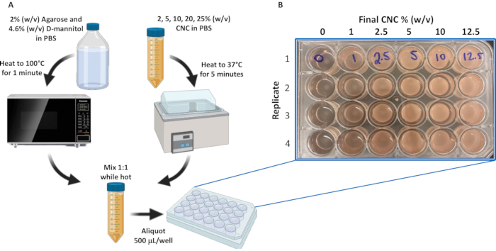
Figure 2: Preparation of the CNC/agarose/D-mannitol hydrogel substrate. (A) Schematic of CNC/agarose/D-mannitol hydrogel substrate preparation protocol. (B) CNC/agarose/D-mannitol preparations (0, 1, 2.5, 5, 10, and 12.5% (w/v) CNC in PBS/agarose/D-mannitol) were loaded onto a 24-well plate in quadruplicate as illustrated. Abbreviations: PBS = phosphate-buffered saline; CNC = crystalline nanocellulose. Please click here to view a larger version of this figure.
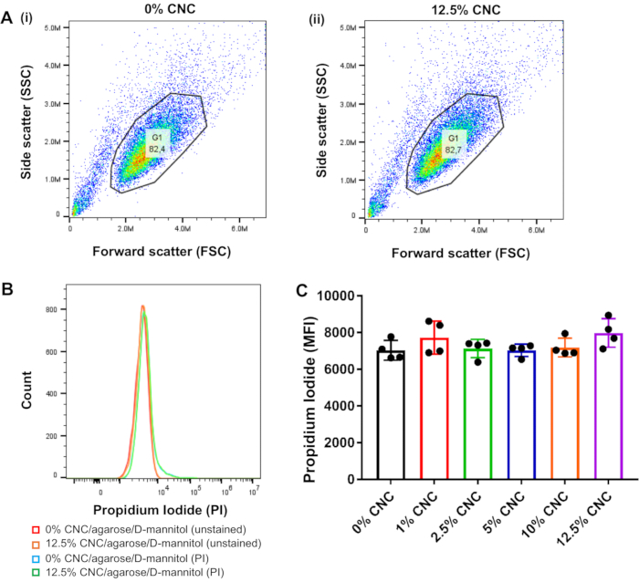
Figure 3: Flow cytometric analysis of BMMC viability via propidium Iodide exclusion following incubation on CNC/agarose/D-mannitol substrates. BMMCs were removed from the CNC/agarose/D-mannitol hydrogel substrates, washed twice, resuspended in PBS-0.5% w/v BSA, stained with PI for 1 h at 4 °C, and analyzed by flow cytometry (n=4). A total of 20,000 cells per sample were acquired including PI fluorescence emission detection in the PE channel. Data analysis was performed using flow cytometry analysis software. (A) Forward scatter (x-axis) versus side scatter (y-axis) dot plot analysis of total cell population from (i) untreated control BMMC (0% CNC/agarose/D-mannitol) and (ii) BMMCs cultured on 12.5% CNC/agarose/D-mannitol, depicting the gated cell population used for data analysis. (B) Histogram overlay profile of gated cells (unstained or stained with PI), from untreated control BMMCs (0% CNC/agarose/D-mannitol) and BMMCs cultured on 12.5% CNC/agarose/D-mannitol. (C) BMMCs were cultured on different CNC/agarose/D-mannitol substrates (1-12.5% (w/v) CNC) for 18 h, and cell viability was determined by flow cytometric analysis of PI-stained cells. Graphical representation of PI MFIs for BMMCs incubated on different CNC/agarose/D-mannitol substrates (1-12.5% (w/v) CNC) relative to BMMCs that were untreated and stained with PI. Abbreviations: BMMCs = bone marrow-derived mast cells; CNC = crystalline nanocellulose; PBS = phosphate-buffered saline; BSA = bovine serum albumin; PI = propidium iodide; PE = phycoerythrin. Please click here to view a larger version of this figure.
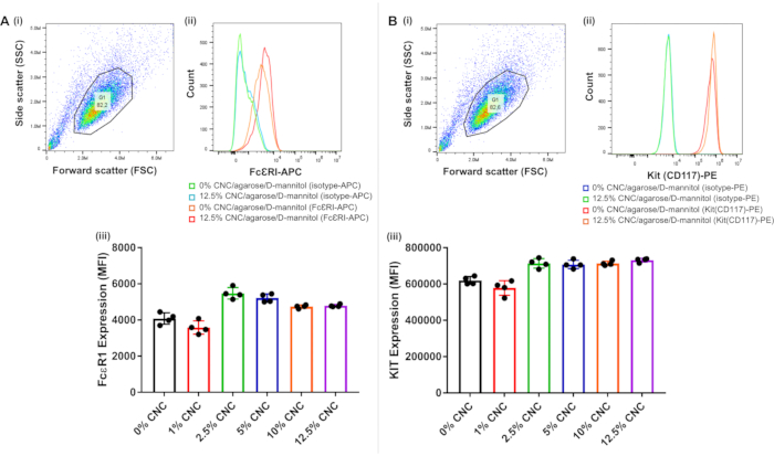
Figure 4: Flow cytometric analysis of FcεRI and Kit (CD117) cell surface receptor expression. BMMCs were cultured either in the absence or presence of CNC/agarose/D-mannitol substrates for 18 h, removed, and analyzed for receptor expression. Data is representative of 4 replicates. Forward scatter (x-axis) versus side scatter (y-axis) dot plot of the total cell population in the untreated BMMC sample (0% CNC/agarose/D-mannitol), stained with (A)(i) the isotype control antibody-APC or (B)(i) the isotype control antibody-PE, and the gated cell population used for data analysis. Histogram overlay profiles of gated BMMCs (stained with isotype control antibody or (A)(ii) anti-FcεRI-APC antibody or (B)(ii) anti-Kit-PE antibody), incubated either alone (0% CNC/agarose/D-mannitol) or on 12.5% CNC/agarose/D-mannitol substrates. BMMCs were cultured for 18 h on different CNC/agarose/D-mannitol substrates (1-12.5% (w/v) CNC) followed by FcεRI and Kit surface receptor expression analysis, respectively, via flow cytometry (n=4). Graphical representation of MFIs of BMMCs stained with (A)(iii) anti-FcεRI-APC or (B)(iii) anti-Kit-PE antibodies, respectively, following culture on different CNC/agarose/D-mannitol substrates (1-12.5% (w/v) CNC) relative to cells that were untreated (0% CNC) and stained similarly. Abbreviations: CNC = crystalline nanocellulose; APC = allophycocyanin; PE = phycoerythrin; BMMCs = bone marrow-derived mast cells; MFI = mean fluorescence intensities. Please click here to view a larger version of this figure.
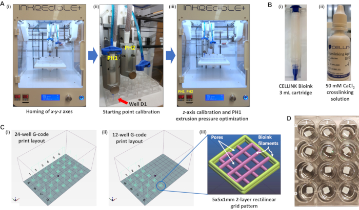
Figure 5: 3D bioprinter equipment and consumables required to bioprint 5 x 5 x 1 mm 2-layer rectilinear bioink hydrogel scaffolds in a 24-well plate format. (A)(i) A pneumatic-extrusion 3D bioprinter with the printhead assembly position at its resting position upon completing the homing of its x-y-z axes. (A)(ii) Starting point calibration of printhead 1 with a bioink cartridge installed and placement of the print nozzle directly over the middle of well D1 in the x-y-z dimensions. (A)(iii) Position of PH1 following z-axis calibration and the extrusion pressure of PH1 set to 12 kPa. (B)(i) Bioink cartridge (3 mL) containing NFC/Alginate/D-mannitol biomaterial ink formulation. (B)(ii) Droplet dispenser of 50 mM CaCl2 crosslinking solution. (C)(i) Schematic representation of a print layout from a G-code file encoding the printing of 5 x 5 x 1 mm 2-layer rectilinear bioink hydrogel scaffolds in all wells of a 24-well plate. (C)(ii) Schematic representation of a print layout from a G-code file encoding the printing of 5 x 5 x 1 mm 2-layer rectilinear bioink hydrogel scaffolds only in wells A1-3, B1-3, C1-3, and D1-3 of a 24-well plate. (C)(iii) Expanded view of the 5 x 5 x 1 mm 2-layer rectilinear bioink hydrogel scaffold pattern in the slicing program, Slic3r. (D)Topview of actual 3D bioprinted 5 x 5 x 1 mm 2-layer rectilinear bioink hydrogel scaffolds in a 24-well plate format immersed in PBS. Abbreviations: 3D = three-dimensional; PH1 = printhead 1; NFC = nanofibrillar cellulose; G-code = geometric code; PBS = phosphate-buffered saline. Please click here to view a larger version of this figure.
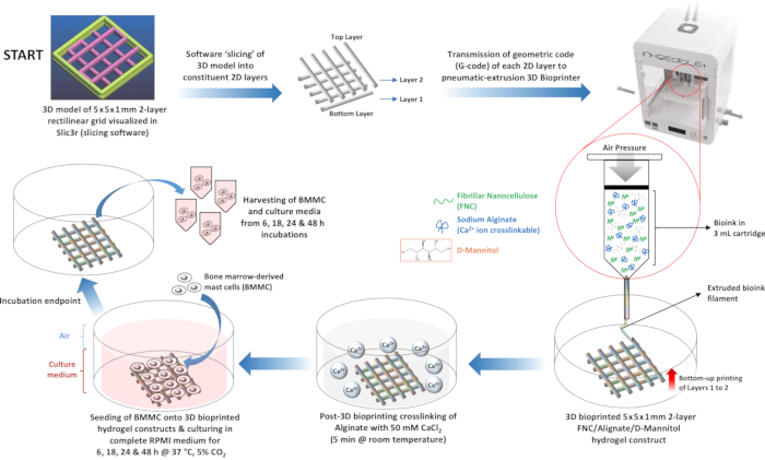
Figure 6: Schematic diagram depicting workflow of 3D bioprinting and BMMC culture on FNC/alginate/D-mannitol hydrogel scaffolds. 3D bioprinting of hydrogel scaffolds with FNC/Alginate/D-mannitol bioink from a G-code file encoding a 5 x 5 x 1 mm 2-layer grid pattern, crosslinking of the hydrogel scaffolds with CaCl2, and culturing of BMMCs on the crosslinked FNC/Alginate/D-mannitol hydrogel constructs. Abbreviations: 3D = three-dimensional; 2D = two-dimensional; G-code = geometric code; FNC = fibrillar nanocellulose; BMMCs = bone-marrow-derived mast cells. Please click here to view a larger version of this figure.
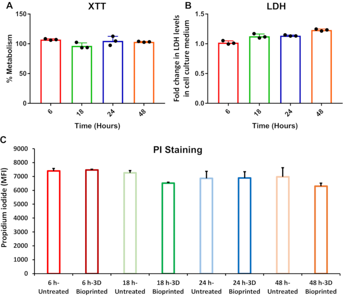
Figure 7: Flow cytometric (PI) and microtiter (XTT and LDH) assays of BMMC viability following incubation on FNC/alginate/D-mannitol scaffolds. (A) Cell proliferation (XTT) metabolic assay data for BMMCs cultured on FNC/Alginate/D-mannitol bioink substrates for 6, 18, 24, and 48 h, respectively. Values are presented as a percentage of the XTT metabolic data for BMMCs cultured alone for 6, 18, 24, and 48 h, respectively. Error bars indicate standard deviation (n=3). (B) LDH enzyme release assay data for BMMCs cultured on FNC/Alginate/D-mannitol bioink scaffolds for 6, 18, 24, and 48 h, respectively. Values are presented as fold-changes relative to the LDH enzyme released by BMMCs cultured alone for 6, 18, 24, and 48 h, respectively. Error bars indicate standard deviation (n=3). (C) Flow cytometric data of PI-stained BMMCs that were cultured either alone or on FNC/Alginate/D-mannitol bioink scaffolds for 6, 18, 24, and 48 h, respectively. Error bars indicate standard deviation (n=3). Abbreviations: LDH = lactate dehydrogenase; BMMCs = bone marrow-derived mast cells; FNC = fibrillar nanocellulose; PI = propidium iodide; MFI = mean fluorescence intensity. Please click here to view a larger version of this figure.
1.1.1. 500 mL bottle of RPMI (HyClone, GE Healthcare, USA) |
1.1.2. 4 mM L-glutamine (Gibco, Waltham Massachusetts, USA) |
1.1.3. 50 mM β-Mercaptoethanol (Fisher Scientific, Hampton, New Hampshire, USA) |
1.1.4. 1 mM Sodium Pyruvate (Gibco) |
1.1.5. 100 U/mL penicillin (Gibco) |
1.1.6. 100 µg/mL streptomycin (Gibco) |
1.1.7. 0.1 mM MEM non-essential amino acids (Gibco) |
1.1.8. 25 mM HEPES (Fisher) |
1.1.9. 10% heat inactivated FBS (Gibco) |
1.1.10. 30 ng/mL mouse recombinant IL-3 (Peprotech, Rocky Hill, New Jersey, USA) |
Table 1: Complete RPMI-1640 media supplements.
Supplemental File 1. Please click here to download this File.
Supplemental File 2. Please click here to download this File.
Supplemental File 3. Please click here to download this File.
The fabrication of 3D biomimetic tissues requires the successful amalgamation of the bioink, which mimics components of the extracellular matrix, with the cellular component(s) to create physiological analogs of in vivo tissues. This necessitates the use of primary cells, and not transformed cells, when fabricating physiological biomimetic tissues. Primary immunological cells, such as mast cells, however, are particularly susceptible to cytotoxic effects and phenotypic changes that may be elicited by the bioink matrix itself, which is undesirable. Therefore, the ability to rapidly assess the effects of the candidate biomaterial ink on the viability and phenotype (surface receptor expression) of the mast cells is highly advantageous, especially prior to 3D bioprinting of complex tissues containing different cell types embedded within the bioink matrix, which is costly and time-consuming.
The approach of this protocol involves culturing BMMCs, an example of primary mast cells, on the surface of preformed candidate hydrogel bioink substrates (1-12.5% CNC embedded in agarose), which permits the easy retrieval of the BMMCs for subsequent analysis of cell viability and surface receptor expression by flow cytometry. The isolation of bone marrow from mouse femur and tibia requires precision and attention to detail due to the fragility and small size of these bones. After successful isolation and deposition of the bone marrow into cell culture medium, it is essential to maintain the cell culture with 30 ng/mL mouse recombinant IL-3 continuously for 4 weeks, which ensures sustained stimulation to differentiate the hematopoietic progenitor cells into mast cells that are double-positive for the Kit (CD117) and FcεRI cell surface receptors.
Flow cytometric analysis of mast cells for the surface expression of Kit (CD117) and FcεRI receptors should employ isotype antibody controls to assist with differentiating non-specific background signal from the specific signals of the antibodies used to target these receptors, as demonstrated in Figure 4A(ii),B(ii). It is also essential to gate on a well-defined mast cell population to exclude cell debris as shown in Figure 4A(i) and 4B(i), which can also non-specifically bind antibodies. Gentle micropipetting should be used when retrieving the BMMCs from the surface of the bioink hydrogel substrates to reduce the shear forces exerted on the cells, which could damage the cell membrane and result in artificially elevated staining with PI. The PI-stained BMMCs should be analyzed by flow cytometry immediately after the staining incubation period ends as the PI staining medium, which is based on PBS-0.5% w/v BSA, is not intended to sustain cells for extended periods of time outside of cell culture medium. Because PI only permeates cells with compromised cell membranes, it is able to stain necrotic cells with high selectivity and sensitivity. However, PI is unable to detect apoptotic cells, which maintain intact cell membranes.
The PI-exclusion flow cytometric assay described in this protocol may be augmented with Annexin-V-fluorescein isothiocyanate (FITC), which specifically binds to the phospholipid, phosphatidylserine, that is translocated to the outer leaflet of the cell membrane of apoptotic cells. However, compensation is necessary to account for the fluorescence emission bleed-through of FITC into the detector channel used to acquire fluorescence emission from PI and vice versa. In preparing the bioprinter for 3D bioprinting from the provided 24-well plate G-code, it is essential that the starting point calibration be performed accurately, whereby the x– and y-coordinates of the center of well D1 are recorded, and the G-code file is updated with these x– and y-coordinates on line 1. If these initial calibration steps are not performed accurately, the printheads will not move to the correct starting point at the commencement of printing. Correct calibration of the z-axis is equally important as failure to do so can result in the nozzle of the printhead colliding with the printbed or the 24-well plate at the commencement of printing.
The schematic diagram of the 5 x 5 x 1 mm 2-layer rectilinear grid construct (Figure 5C(iii)), illustrates the presence of pores between the extruded bioink filaments that form the substrate. The size of the pores and diameter of the filaments are dependent on three parameters: (i) the travel speed of the print nozzle, (ii) the extrusion pressure applied to the bioink within the cartridge, and (iii) the print nozzle diameter. Bioink substrates with large filament diameters and small pores can be printed using a slower print nozzle speed, higher extrusion pressure (>12 kPa), and larger diameter print nozzle (22 G). Conversely, a faster print nozzle speed, lower extrusion pressure (<12 kPa), and smaller diameter print nozzle (27 G) will result in substrates with finer filaments and larger pores. However, an insufficiently high extrusion pressure will result in inconsistent clumps of bioink being extruded instead of continuous filaments, which will adversely affect the quality and structural integrity of the 3D bioprinted substrate.
The CNC/agarose/D-mannitol candidate bioink presented in this protocol undergoes thermal gelation when agarose cools down to room temperature. In contrast, the commercial bioink used for 3D bioprinting in this protocol undergoes ionic crosslinking with CaCl2 due to the inclusion of sodium alginate in the bioink formulation. It is, therefore, necessary to immerse the bioprinted FNC/alginate/D-mannitol scaffolds in 50 mM CaCl2 solution after bioprinting to facilitate crosslinking (gelation) of the substrate. Omission of the ionic crosslinking step with CaCl2 will result in the dissolution of the FNC/alginate/D-mannitol scaffolds when they are immersed in cell culture medium. The foremost limitation of the CNC/agarose/D-mannitol bioink formulation in its application in actual 3D bioprinting is the exceptionally high melting temperature of agarose (90-95 °C). Even though the printheads of the 3D bioprinter used in this study can reach a maximum temperature of 130 °C, the extruded filaments of CNC/agarose/D-mannitol will likely disperse rapidly as successive layers are printed to construct the substrate. This limitation of the CNC/agarose/D-mannitol bioink formulation can be circumvented by either printing the CNC/agarose/D-mannitol bioink directly onto a cooled printbed to accelerate gelation, or substituting agarose with sodium alginate, which can be extruded at room temperature and undergoes rapid ionic crosslinking with 50 mM CaCl2 within 5 min.
This protocol offers a reliable approach to rapidly screen and exclude bioink formulations that exhibit poor biochemical compatibility with sensitive immunological cells, such as mast cells, that should not be subjected to pneumatic-extrusion 3D bioprinting. Furthermore, this protocol is cost-effective as it requires relatively low quantities of cells to perform a multiplexed screening assay. In contrast, pre-formulated bioink-cell mixtures need to be bioprinted at very high cell densities (>107 cells/mL), which can prove to be costly and time-consuming to perform a biocompatibility screening assay especially when culturing primary cells that are dependent on expensive recombinant growth factors.
The authors have nothing to disclose.
We thank Alberta Innovates for providing the CNC and Ken Harris and Jae-Young Cho for their technical advice when preparing the CNC/agarose/D-mannitol matrix. We also thank Ben Hoffman, Heather Winchell and Nicole Diamantides for their technical advice and support with the setup and calibration of the INKREDIBLE+ 3D bioprinter.
A | |||
Acetic Acid (glacial) | Sigma Aldrich | AX0074-6 | |
Agarose (OmniPur) | EMD Millipore Corporation | 2125-500GM | |
Armenian Hamster IgG Isotype Control, APC (Clone: eBio299Arm) | Thermo Fisher Scientific | 17-4888-82 | |
B | |||
b-Mercaptoethanol | Fisher Scientific | O3446I-100 | |
b-Nicotinamide adenine dinucleotide sodium salt (NAD) | Sigma Aldrich | N0632-5G | |
BD 5 mL Syringe (Luer-Lok Tip) | BD | 309646 | |
BD PrecisionGlide Needle 26G x 1/2 in | BD | 305111 | |
BioLite 24 Well Multidish | Thermo Fisher Scientific | 930-186 | |
BioLite 96 Well Multidish | Thermo Fisher Scientific | 130-188 | |
BioLite 175 cm2 Flask Vented | Thermo Fisher Scientific | 130-191 | |
Biosafety Cabinet Class II | Microzone Corp., Canada | BK-2-6-B3 | |
BSA, Fraction V (OmniPur) | EMD Millipore Corporation | 2930-100GM | |
C | |||
C57BL/6 mice | The Jackson Laboratory | 000664 | |
CD117 (c-Kit) Monoclonal Antibody, PE (Clone: 2B8) | Thermo Fisher Scientific | 12-1171-82 | |
CELLINK BIOINK (3 x 3 mL Cartridge) | CELLINK LLC | IK1020000303 | |
CELLINK CaCl2 Crosslinking Agent – Sterile Bottle 1 x 60 mL | CELLINK LLC | CL1010006001 | |
CELLINK Empty Cartridges 3cc with End and Tip Caps | CELLINK LLC | CSC0103000102 | |
CELLINK HeartWare for PC | CELLINK LLC | Version 2.4.1 | |
CELLINK INKREDIBLE+ 3D BIOPRINTER | CELLINK LLC | S-10003-001 | |
CELLINK Sterile Standard Conical Bioprinting Nozzles 22G | CELLINK LLC | NZ4220005001 | |
CELLINK Sterile Standard Conical Bioprinting Nozzles 25G | CELLINK LLC | NZ4250005001 | |
CELLINK Sterile Standard Conical Bioprinting Nozzles 27G | CELLINK LLC | NZ4270005001 | |
Cell Proliferation Kit II (XTT) (Roche) | Sigma Aldrich | 11465015001 | |
Centrifuge (Benchtop) | Eppendorf | 5804R | |
Corning Costar 96 Well Clear Flat-Bottom Non-Treated PS Microplate | Sigma Aldrich | CLS3370 | |
CO2 Incubator | Binder GmbH, Germany | 9040-0113 | |
CytoFLEX Flow Cytometer | Beckman Coulter | A00-1-1102 | |
D | |||
D-mannitol (MilliporeSigma Calbiochem) | Fisher Scientific | 44-390-7100GM | |
F | |||
Falcon 15 mL Polystyrene Conical Tubes, Sterile | Corning | 352095 | |
Falcon 50 mL Polystyrene Conical Tubes, Sterile | Corning | 352070 | |
FceR1 alpha Monoclonal Antibody, APC (Clone: MAR-1) | Thermo Fisher Scientific | 17-5898-82 | |
Fetal Bovine Serum (FBS), qualified, heat inactivated | Thermo Fisher Scientific | 12484028 | |
FlowJo Software | Becton Dickinson & Co. USA | Version 10.6.2 | |
G | |||
GraphPad Prism | GraphPad Software, LLC | Version 8.4.3 | |
H | |||
Hemacytometer (Improved Neubauer 0.1 mmm deep levy) | VWR | 15170-208 | |
HEPES Sodium Salt | Fisher Scientific | BP410-500 | |
I | |||
Iodonitrotetrazolium chloride (INT) | Sigma Aldrich | I10406-5G | |
L | |||
L-Glutamine 200 mM (Gibco) | Thermo Fisher Scientific | 25030-081 | |
Lithium L-lactate | Sigma Aldrich | L2250-100G | |
M | |||
MEM Non-Essential Amino Acids 100 mL 100x (Gibco) | Thermo Fisher Scientific | 11140-050 | |
1-Methoxy-5-methylphenazinium methyl sulfate (MPMS) | Sigma Aldrich | M8640 | |
Microtubes (1.7 mL clear) | Axygen | MCT-175-C | |
Microtubes (2.0 mL clear) | Axygen | MCT-200-C | |
MilliQ Academic (for producing MilliQ ultrapure water) | Millipore | ZMQS60001 | |
N | |||
Nalgene Rapid-Flow 90 mm Filter Unit (0.2 mm Pore size, 500 mL) | Thermo Fisher Scientific | 566-0020 | |
Nalgene Syringe filter (0.2 mm PES, 25 mm) | Thermo Fisher Scientific | 725-2520 | |
P | |||
Penicillin Streptomycin 100 mL (Gibco) | Thermo Fisher Scientific | 15140-122 | |
PBS pH 7.4, No Calcium/Magnesium, 500 mL (Gibco) | Thermo Fisher Scientific | 10010-023 | |
Propidium iodide, 1.0 mg/mL (Invitrogen) | Thermo Fisher Scientific | P3566 | |
R | |||
Rat IgG2b kappa Isotype Control, PE (Clone: eB149/10H5) | Thermo Fisher Scientific | 12-4031-82 | |
Recombinant Murine IL-3 | PeproTech, Inc. | 213-13 | |
RPMI-1640 Medium 1X + 2.05 mM L-Glutamine (HyClone) | GE Healthcare | SH30027.01 | |
S | |||
Sarstedt 96 well round base PS transparent micro test plate (82.1582.001) | Fisher Scientific | NC9913213 | |
Sodium Azide, 500 g | Fisher Scientific | BP922I-500 | |
Sodium Pyruvate (100 mM) 100X (Gibco) | Thermo Fisher Scientific | 11360-070 | |
T | |||
Tris Base (2-amino-2(hydroxymethyl)-1,3-propanediol) | Sigma Aldrich | 252859 | |
Trypan Blue solution (0.4%, for microscopy) | Sigma Aldrich | 93595 | |
V | |||
VARIOSKAN LUX Microplate Spectrophotometer (Type: 3020) | Thermo Fisher Scientific | VLBL00D0 |