Single-Cell Suspension Preparation from Nile Tilapia Intestine for Single-Cell Sequencing
Summary
Here, we demonstrate the preparation of a high-quality single-cell suspension of tilapia intestine for single-cell sequencing.
Abstract
Nile tilapia is one of the most commonly cultured freshwater fish species worldwide and is a widely used research model for aquaculture fish studies. The preparation of high-quality single-cell suspensions is essential for single-cell level studies such as single-cell RNA or genome sequencing. However, there is no ready-to-use protocol for aquaculture fish species, particularly for the intestine of tilapia. The effective dissociation enzymes vary depending on the tissue type. Therefore, optimizing the tissue dissociation protocol by selecting the appropriate enzyme or enzyme combination to obtain enough viable cells with minimum damage is essential. This study illustrates an optimized protocol to prepare a high-quality single-cell suspension from Nile tilapia intestine with an enzyme combination of collagenase/dispase. This combination is highly effective for dissociation with the utilization of bovine serum albumin and DNase to reduce cell aggregation after digestion. The cell output satisfies the requirements for single-cell sequencing, with 90% cell viability and a high cell concentration. This protocol can also be modified to prepare a single-cell suspension from the intestines of other fish species. This research provides an efficient reference protocol and reduces the need for additional trials in the preparation of single-cell suspensions for aquaculture fish species.
Introduction
Cells are the fundamental units of organisms. Compared to bulk-tissue studies, single-cell level studies can reflect cell heterogeneity and provide higher-resolution information1. In recent years, researchers have applied single-cell sequencing technologies for genome, transcriptome, epigenome, or multi-omic studies at the single-cell level in mammals, zebrafish, and other model organisms and reported great breakthroughs2,3,4,5,6,7. While most studies have focused on model organisms, there are few reference protocols or commercial dissociation kits for single-cell sequencing in economic fish species, which limits the application of single-cell sequencing in aquaculture research. Therefore, developing tissue dissociation protocols that produce high-quality single-cell suspensions with high cell viability and nucleic acid integrity is crucial.
Optimizing the tissue dissociation protocol with the appropriate enzyme or enzyme combination to obtain enough viable cells with minimum damage is essential. The most effective enzyme for tissue dissociation varies depending on the tissue type. In mammals, several enzymes have been used to prepare single-cell suspensions for mammalian solid tissues, including collagenase, dispase, trypsin, papain, elastase, hyaluronidase, liberase, accutase, and trypLE8,9. Trypsin digestion combined with mechanical disruption has commonly been used to dissociate tissues for cell culture in fish10,11,12,13,14. Trypsin has also been used or added into the digestion cocktail for tissue dissociation in the rat intestine15 and zebrafish gill tissue16. However, for several reasons, trypsin is not the best option for single-cell sequencing. Trypsin alone is usually ineffective for tissue dissociation. Additionally, trypsin induces DNA strand breaks17,18 and RNA degradation19.
Papain degrades the proteins that make up the tight junctions between cells. In mammalian nerve and smooth muscle cells, papain is more efficient and less destructive than other proteases20,21. However, like trypsin, papain results in the free DNA-induced aggregation of cells due to the cell lysis that occurs during enzymatic digestion9. Elastase breaks down elastin, which is typically found in the skin, lungs, ligaments, tendons, and vascular tissues22. It is often used in combination with collagenase, dispase, or trypsin for dissociating lung tissue8. Hyaluronidase cleaves the glycosidic bonds of hyaluronan, contributing to the digestion of the extracellular matrix in various connective tissues and skin9,23.
In general, collagenase and dispase are good options for extracellular matrix breakdown. They have been used in the dissociation of human, mouse, and zebrafish intestines24,25,26,27. Collagenase destroys the peptide bond in collagen, promotes the digestion of the extracellular matrix, and releases the cells into suspension, and, thus, collagenase is often used for human and mouse solid tissue dissociation, including for the liver28,29, spleen30, pancreas31, and intestine25. Dispase is a protease that hydrolyzes the N-terminal peptide bonds of nonpolar amino acid residues and is milder than collagenase. It cleaves the extracellular matrix components, such as fibronectin, type IV collagen, and to a lesser extent, type I collagen, without affecting cell-cell junctions. Dispase is used separately or with other enzymes for tissue dissociation, such as for intestine25,32, brain33, liver34, etc. In addition, commercially available digestion cocktails, including liberase, accutase, and trypLE, are also good alternatives for solid tissue dissociation, especially for the skin, liver, and kidney8,9.
Nile tilapia (Oreochromis niloticus) belongs to the family Cichlidae of the order Perciformes. It is one of the most cultured freshwater fish species in tropical and subtropical areas, with an annual production of 4.5 million tons in 202235. It is one of the best-studied aquaculture fish species with a well-annotated genome. Nile tilapia is an ideal research model for aquaculture fish species due to its short generation time, ease of rearing, and adaptability to a wide range of rearing environments. The intestine is of great research interest as it is the organ of nutrition digestion and absorption, metabolism, and mucosal immunity. The intestine is the habitat of microbial populations and is an essential immune tissue36. It is immunologically active due to the presence of numerous immune cell types, including macrophages, B cells, granulocytes, and T cells.
In the current study, we developed a protocol for preparing a high-quality single-cell suspension from the Nile tilapia intestine to facilitate single-cell level studies in aquaculture fish species. According to the characteristics of these tissue-specific enzymes and preliminary work, collagenase/dispase is appropriate for dissociating tilapia intestine tissue. The final enzyme type to consider in preparing single-cell suspensions is DNase-I, which prevents cell aggregation by degrading free DNA released through dead cell lysis during enzymatic digestion without initiating apoptotic pathways9 and increases the live cell yield36. Additionally, bovine serum albumin (BSA) is added to the washing buffer to reduce cell clumping and improve cell viability. Several reagent companies describe BSA as an enzyme stabilizer. The addition of 0.04%-1% BSA to PBS (phosphate-buffered saline) has been used to develop a washing solution for preparing single-cell sequencing suspensions without adverse effects38. The addition of a low ratio of BSA could help maintain cell viability and avoid the free DNA-induced aggregation of cells due to cell lysis. This protocol can also provide a valuable reference for developing cell dissociation protocols from the intestines of other aquaculture fish species.
Protocol
All animal protocols during this study were approved by the Hainan University Institutional Animal Care and Use Committee (Protocol number: HNUAUCC-2022-00063; Approval date: 2022-03-03). A list of the equipment and supplies used in this experiment can be found in the Table of Materials. A summary of the current protocol is illustrated in Figure 1.
1. Fish preparation
- Obtain 6 month old Nile tilapia with a mean body weight of 100 g from a reliable source. Select fish that are free of any signs of disease.
- Stop feeding the fish 24 h before proceeding to the next step.
NOTE: Fish fasting reduces the volume of intestinal contents and facilitates the preparation of intestinal segments. - Rinse the fish in well-aerated sterile water for 15 min to wash off loosely bound bacteria on the body surface.
- Euthanize the fish with 300 mg/L tricaine methanesulfonate (MS-222) for about 10 min until all movement stops.
2. Reagent preparation
- Prepare 0.08% BSA-Dulbecco's phosphate-buffered saline (BSA-DPBS, for cell rinsing and resuspension). Dissolve 0.08 g of BSA (0.8% w/v) in 100 mL of 1x DPBS. Store at 4 °C, and pre-cool on ice when used.
NOTE: The addition of BSA is beneficial for minimizing cell clumping. The BSA ratio can be increased from 0.04% to 1% according to the cell clumping conditions. - Prepare the enzymatic cell dissociation reagent.
- Dilute the collagenase/dispase with 1x PBS to a final concentration of 1 mg/mL (0.1 U/mL collagenase and 0.8 U/mL dispase). Ensure that PBS and not DPBS is used.
NOTE: PBS provides the calcium ions needed for the enzyme to function properly. DPBS should not be used in this step because it is Ca2+/Mg2+ free. Additionally, collagenase type I is used in the current protocol. - Filter the dilution through a sterile 0.22 µm membrane filter.
- Prepare a cell dissociation reagent that is composed of a 95% volume of collagenase/dispase solution and a 5% volume of fetal bovine serum (FBS).
NOTE: The dissociation reagent should be made fresh every time it is used. Using 5% FBS helps maintain the cell viability during digestion.
- Dilute the collagenase/dispase with 1x PBS to a final concentration of 1 mg/mL (0.1 U/mL collagenase and 0.8 U/mL dispase). Ensure that PBS and not DPBS is used.
- Prepare trypan blue solution.
- Take the trypan blue solution from the 4 °C refrigerator, and keep it at 15-20 °C for a few minutes before use to prevent precipitation.
- Filter the solution through a sterile 0.22 µm membrane.
3. Equipment preparation
- Pre-cool the refrigerated centrifuge to 4 °C before use.
- Obtain sterile RNase-free 1.5 mL centrifuge tubes, 50 mL conical tubes, wide-bore glass pipettes and tips, membrane filters, cell strainers, and all the low retention tubes and tips.
- Autoclave glass pipettes and dissection tools, including forceps, scissors, and scalpels, and pre-treat them with the RNase removal solution to prevent the adverse effects of RNase for single-cell RNA-seq.
4. Tissue dissection and cell dissociation
- Put the euthanized fish on ice for dissection. Aseptically, collect 3-4 cm of the mid-section of the intestine, and excise as much fat and mesentery as possible. Carefully remove the fat attached to the intestinal surface and the layer of elastic and malleable clear mucosa with forceps.
NOTE: The fat and mesentery are attached to the external surface of the intestine. - Remove as much fat as possible to prevent its adverse effects on the dissociation protocol. Using a syringe, gently rinse the fragments with sterile ice-cold PBS several times to wash off the intestinal contents and mucus. Avoid rough handling.
- Dissect the segment of intestine into small pieces, and transfer them to a 1.5 mL tube filled with 1 mL of ice-cold PBS.
- Centrifuge at 300 × g for 5 min at 4 °C. Remove the supernatant gently, and replace with 1 mL of ice-cold 0.08% BSA-DPBS. Resuspend the tissue fragments by gentle aspiration using a glass pipette. Repeat this step twice.
- Remove the supernatant, and digest the tissue fragments using 1 mL of the enzymatic dissociation reagent (950 µL of collagenase/dispase solution and 50 µL of FBS) in a 37 °C water bath for 30-60 min.
- Additionally, add 5 µL of RNase inhibitor (40 U/µL in the stock solution) to the dissociation reagent if the samples are prepared for single-cell RNA sequencing.
NOTE: The final concentration of the collagenase/dispase mix is 1 mg/mL, including collagenase at 0.1 U/mL and dispase at 0.8 U/mL.
- Additionally, add 5 µL of RNase inhibitor (40 U/µL in the stock solution) to the dissociation reagent if the samples are prepared for single-cell RNA sequencing.
- Manually invert the tube at 5 min intervals during the 30-60 min water bath.
NOTE: The exact incubation time varies according to the tissues used. Check the progress of dissociation under a microscope until no bulk tissue is seen. Place 10 µL of the cell suspension on a glass slide with a pipette, and examine it under the microscope. Increase the digestion time if the cells are not completely dissociated. - Spin down the tubes at 300 × g for 5 min at 4 °C, and gently remove the enzymatic cell dissociation reagent using wide-bore tips. Add 1 mL of ice-cold 0.08% BSA-DPBS, and gently pipette the cells up and down using a wide-bore glass pipette for 1 min.
- Repeat this step twice.
NOTE: Pipetting up and down should be performed with care using wide-bore and low-retention tips to prevent cell disruption.
- Repeat this step twice.
- Pre-wet a 40 µm cell strainer with 0.08% BSA-DPBS, and place it into a 50 mL conical tube. Pass the cell suspension through the strainer. Tap and rinse with an additional 200 µL DPBS, and collect the suspension at the bottom of the tube.
- Transfer the suspension into a 1.5 mL tube. Add 5 µL of DNase I (1 U/µL), and incubate for 15 min at 15-20 °C. Centrifuge at 300 × g for 5 min at 4 °C.
- Remove the supernatant, and resuspend the cell pellet in 400 µL of ice-cold DPBS (Ca2+/Mg2+ free). Gently pipette up and down with wide-bore tips to mix the cells. Repeat this step twice.
5. Staining and microscopic examination
- For staining, mix the cell suspension with 0.4% trypan blue solution at a 1:1 ratio. Incubate for 3 min at room temperature.
- Add 5-10 µL of the mixture onto a hemocytometer, and examine under the microscope.
NOTE: Viable cells will have unstained cytoplasm, while dead cells will have a blue cytoplasm (Figure 2). - Count the viable and dead cells in three different fields under the microscope. Calculate the cell viability percentage by dividing the number of viable cells by the total number of cells in a field.
NOTE: The cell viability should be more than 85%, and cell concentration should not be less than 1 x 10⁵-1 x 10⁷/mL. The cell suspension background should be clean, without clumps, fragments, or impurities.
Representative Results
This protocol describes the preparation of a high-quality single-cell suspension of Nile tilapia intestine for single-cell sequencing (Figure 1). This research shows that the collagenase/dispase mix has a good dissociation effect and is mild for intestine tissue. The selection of the optimal digestion enzyme is essential for preparing a high-quality single-cell suspension. In the preliminary work, the dissociation efficiencies of several commonly used enzymes were compared, and the results are listed in Table 1. The collagenase/dispase mix was verified to have a much better dissociation effect than collagenase, dispase, or trypsin alone and several other enzyme mixtures, including liberase, trypsin/elastase, collagenase/elastase, and collagenase/trypsin. Moreover, the collagenase/dispase also yielded the most viable cells at the end of the experiment.
During the incubation time, the progress of dissociation was checked under a microscope. After 30 min, the bulk tissue was almost gone, and generally, no bulk tissue was seen after 45 min. After digestion, the cell suspension filtered through the strainer easily. Following this protocol, the trypan blue staining and microscopic examination showed that the intestinal cells had high cell viability (more than 90%; Figure 2, and that the cell concentration could be up to 1 x 10⁷/mL. The cells were dispersed as single cells. Most cells were bright and white (viable cells), whereas only a few were dark or blue (dead cells). Dead cells are stained dark blue because of the permeabilized cell membrane. The cell viability and cell concentration satisfied the requirements of single-cell sequencing.
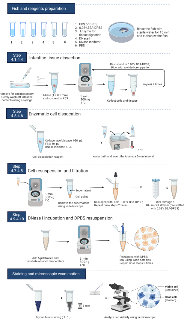
Figure 1: Summary flow diagram of the Nile tilapia intestine dissociation and single-cell suspension preparation protocols. The reagents are prepared (sections 1-2) before the excision of the intestine. The intestines are minced and centrifugally washed in PBS (steps 4.1-4.4). The tissue fragments are digested using collagenase/dispase solution in a 37 °C water bath to isolate the cells (steps 4.5-4.6). The digested cell suspension is rinsed with 0.08% BAS-DPBS and filtered through a 40 µm cell strainer (steps 4.7-4.8). After DNase I incubation, the cell pellet is resuspended in DPBS (steps 4.9-4.10). Trypan blue staining determines the cell concentration and viability (section 5). Please click here to view a larger version of this figure.
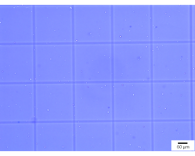
Figure 2: Microscopic examination of a single-cell suspension prepared from Nile tilapia intestinal tissue and stained with trypan blue. Viable cells are white and bright, whereas dead cells are dark and blue. Please click here to view a larger version of this figure.
| Enzyme | Best working concentration (mg/mL) | Dissociation effect |
| Collagenase II | 1 | +++ |
| Dispase | 0.25 | ++ |
| Trypsin | 2.5 | ++ |
| Liberase | 1 | +++ |
| *Collagenase/dispase | 1 | +++++ |
| Collagenase II/elastase | 1/1 | +++ |
| Trypsin/elastase | 2.5/0.5 | +++ |
| Trypsin/collagenase II | 1/1 | +++ |
Table 1: Comparison results of the dissociation effects of commonly used digestion enzymes for Nile tilapia intestine. More "+" symbols represent a higher degree of dissociation and higher cell viability. The enzyme labeled with "*" is a commercial product, and the 1 mg/mL best working solution includes 0.1 U/mL collagenase I and 0.8 U/mL dispase. The other mixtures of enzymes were mixed when they were used by the researchers.
Discussion
This protocol describes the preparation of a high-quality single-cell suspension of Nile tilapia intestine. Before dissociation, the removal of fat and mesentery from the intestine is necessary, particularly for carnivorous fish intestines with much fat. Using a syringe instead of hard scraping to wash off the intestinal contents reduces the mechanical damage to the cells. To ensure cell viability, it is also essential to maintain the temperature at 20 °C or below for the tissue dissection and rinsing steps. The washing solutions are cooled on ice, and the centrifuge temperature is adjusted to 4 °C in advance. Most importantly, mild and efficient cell dissociation enzymes for the studied tissue should be selected. Based on a previous study, the collagenase/dispase mix is optimal for the Nile tilapia intestine. This enzyme combination is milder and more effective on tilapia intestine than trypsin, dispase, or collagenase alone. Additionally, 5% FBS was included in the dissociation reagent to maintain the cell viability during digestion39. The exact incubation time varies according to the tissues used. The digestion time needs to be increased if the tissue is not completely dissociated. In addition, collagenase/dispase requires Ca2+ to function. Therefore, PBS is used to dilute the enzyme rather than DPBS, which is free from Ca2+/Mg2+. For the washing solutions, DPBS is used in case these ions influence the downstream sequencing steps.
Most dissociation enzymes, including collagenase/dispase, work with maximum speed and efficiency at their optimum temperature, which is commonly 37 °C. At this temperature, genes are transcribed, and their expression levels change in response to handling40,41. This phenomenon has been reported in several studies in mammals and zebrafish. In the mouse kidney, apoptosis and stress-related genes including JUN-B, FOS, and Hsp90 are differentially expressed under different enzymatic dissociation conditions40. The differential expression of immediate early genes and HSP genes is altered in zebrafish fin cells at different dissociation conditions41. It has been shown that early response genes, including multiple members of the FOS and JUN families, are significantly up-regulated after only a few minutes of separation at 37 °C in cell suspensions prepared by the enzymatic dissociation of mammalian tissues6. Therefore, the expression changes in apoptosis and stress-related genes should be considered carefully for the ScRNA-seq data. This disadvantage can be overcome by using appropriate controls for the ScRNA-seq experiments. One of the controls should be treated under the same dissociation conditions so that the artifact changes in gene expression can be detected or avoided.
In this protocol, BSA is added to DPBS primarily to minimize cell losses and cell clumping. Ratios of BSA from 0.04% to 1% can be used to prepare single-cell sequencing suspensions without adverse effects38. Using the correct BSA proportion would reduce cell aggregation. The appropriate ratio should be optimized for different tissues and cell clumping conditions. In this protocol, 0.08% BSA prevented Nile tilapia intestine cell attachment. However, the final cell resuspension should not contain BSA to avoid the influence of Ca2+/Mg2+ on the downstream applications.
Another measure to reduce cell aggregation is the addition of DNase I. Ruptured or dying cells release "sticky" DNA molecules, which are among the principal causes of cell clumping. DNase degrades free DNA and, thus, minimizes cell clumping. Therefore, it is commonly used in cell suspension preparation for single-cell sequencing42,43. Additionally, gentle pipetting using wide-bore glass pipettes or tips helps reduce cell damage. Especially for single-cell RNA-seq, tools such as scissors, tubes, glass pipettes, and cell strainers must be RNase-free.
Above all, the whole process of the preparation of a high-quality single-cell suspension should be done with the appropriate dissociation enzyme, should be performed gently and quickly at a low temperature, and should include measures to prevent cells from aggregating in order to achieve high cell viability and concentration and nucleic acid integrity. This protocol can also be used for the preparation of intestine single-cell suspensions for other fish species with minor modifications. In addition, this protocol can also provide a valuable reference for developing cell dissociation protocols for other fish tissues for single-cell sequencing and single-cell-level studies, such as cell culture and flow cytometry.
Disclosures
The authors have nothing to disclose.
Acknowledgements
The authors wish to acknowledge support from the Hainan Provincial Natural Science Foundation of China (NO. 320QN211) and the Research Fund Program of Guangdong Provincial Key Laboratory of Aquatic Animal Disease Control and Healthy Culture of China (NO. PBEA2021ZD01).
Materials
| 0.22-μm Sterile Filter | Solarbio Life Sciences | SLGV033RB | It is used to filter and sterilize the enzyme solution. |
| 40-μm Cell Strainer | Solarbio Life Sciences | F8200 | Cell Strainer is applied to eliminate undigested tissue pieces. |
| Bovine serum albumin (BSA) | Sigma-Aldrich | SRE0098 | Powder; dilute 0.04 g BSA with 100 mL 1× DPBS to prepare 0.04% BSA-DPBS washing bffer. Store at 2 – 8 °C. |
| Collagenase II | Sangon Biotech | A004202 | Dilute with PBS to a final concentration of 1 mg/mL. |
| Collagenase/dispase | Roche | 10269638-001 | Dilute with PBS to a final concentration of 1 mg/mL. |
| Dispase | Sigma-Aldrich | D4818 | Dilute with PBS to a final concentration of 1 mg/mL. |
| DNase I | Sigma-Aldrich | AMPD1 | DNase I helps reduce cell clumping. |
| Dulbecco's phosphate-buffered saline (DPBS), Ca2+/Mg2+-free | Solarbio Life Sciences | E607009-0500 | Store at room temperature. |
| Elastase | Sangon Biotech | A600438 | Dilute with PBS to a final concentration of 0.5 mg/mL. |
| Fetal bovine serum (FBS) | Gibco | 16000-044 | Serum, used at volume of 5% in digetstion solution. |
| Inverted Microscope | Leica | qTOWER3G | It is used to examine cell viability. |
| Liberase | Roche | 5401119001 | Dilute with PBS to a final concentration of 0.25 mg/mL. |
| Nile tilpia (Oreochromis niloticus) | ProGift Aquaculture Technology Co. Ltd. | NA | Healthy fish with no disease signs (Mean body weight: 100 g). |
| Phosphate-buffered saline (PBS) | Solarbio Life Sciences | P1020 | Store at room temperature. |
| Refrigerated Centrifuge | Eppendorf | 5424 | It is used to spin down the tissue and cell petet. |
| RNase inhibitor | NEB | M0314L | Inhibit RNase activity |
| Solid-phase RNase-Be-Gone Reagent | Sangon Biotech | B644201-0050 | It is used to remove the RNase from tools such as dissecting scissors and glass pipettes. Store at room temperature. |
| Tricaine methanesulfonate (MS-222) | Sigma-Aldrich | E10521 | For fish euthanasia. |
| Trypan Blue | Invitrogen | C0040 | It is used for staining dead cells. |
| Trypsin | Sangon Biotech | E607001 | Dilute with PBS to a final concentration of 1 mg/mL. |
References
- Tang, X., Huang, Y., Lei, J., Luo, H., Zhu, X. The single-cell sequencing: new developments and medical applications. Cell and Bioscience. 9 (1), (2019).
- He, H., et al. Single-cell transcriptome analysis of human skin identifies novel fibroblast subpopulation and enrichment of immune subsets in atopic dermatitis. The Journal of Allergy and Clinical Immunology. 145 (6), 1615-1628 (2020).
- Carmona, S. J., et al. Single-cell transcriptome analysis of fish immune cells provides insight into the evolution of vertebrate immune cell types. Genome Research. 27 (3), 451-461 (2017).
- Wen, L., Tang, F. C. Single cell epigenome sequencing technologies. Molecular Aspects of Medicine. 59, 62-69 (2018).
- Xu, R., et al. Single cell sequencing coupled with bioinformatics reveals PHYH as a potential biomarker in kidney ischemia reperfusion injury. Biochemical and Biophysical Research Communications. 602, 156-162 (2022).
- Potter, S. S. Single-cell RNA sequencing for the study of development, physiology and disease. Nature Reviews Nephrology. 14 (8), 479-492 (2018).
- Andrews, T. S., Hemberg, M. Identifying cell populations with scRNASeq. Molecular Aspects of Medicine. 59, 114-122 (2018).
- Lafzi, A., Moutinho, C., Picelli, S., Heyn, H. Tutorial: Guidelines for the experimental design of single-cell RNA sequencing studies. Nature Protocols. 13 (12), 2742-2757 (2018).
- Reichard, A., Asosingh, K. Best practices for preparing a single cell suspension from solid tissues for flow cytometry. Cytometry Part A. 95 (2), 219-226 (2019).
- Sathiyanarayanan, A., Goswami, M., Nagpure, N., Babu, P. G., Das, D. K. Development and characterization of a new gill cell line from the striped catfish, Pangasianodon hypophthalmus (Sauvage, 1878). Fish Physiology and Biochemistry. 48 (2), 367-380 (2022).
- Ager-Wick, E., et al. Preparation of a high-quality primary cell culture from fish pituitaries. Journal of Visualized Experiments. (138), e58159 (2018).
- Kumar, R., et al. Establishment and characterization of a caudal fin-derived cell line, AOF, from the Oscar, Astronotus ocellatus. FishPhysiology and Biochemistry. 45 (1), 123-131 (2019).
- Schnell, S., et al. Procedures for the reconstruction, primary culture and experimental use of rainbow trout gill epithelia. Nature Protocols. 11 (3), 490-498 (2016).
- Xu, S. H., Cooke, I. M. Voltage-gated currents of tilapia prolactin cells. General and Comparative Endocrinology. 150 (2), 219-232 (2007).
- Ayyaz, A., et al. Single-cell transcriptomes of the regenerating intestine reveal a revival stem cell. Nature. 569 (7754), 121-125 (2019).
- Pan, W., et al. Single-cell transcriptomic analysis of neuroepithelial cells and other cell types of the gills of zebrafish (Danio rerio) exposed to hypoxia. Scientific Reports. 12, 10144 (2022).
- Huang, H. L., et al. Trypsin-induced proteome alteration during cell subculture in mammalian cells. Journal of Biomedical Science. 17 (1), 36 (2010).
- Kapiszewska, M., Reddy, N. M., Lange, C. S. Trypsin-induced changes in cell shape and chromatin structure result in radiosensitization of monolayer Chinese hamster V79 cells. International Journal of Radiation Biology. 60 (4), 635-646 (1991).
- Vrtačnik, P., Kos, &. #. 3. 5. 2. ;., Bustin, S. A., Marc, J., Ostanek, B. Influence of trypsinization and alternative procedures for cell preparation before RNA extraction on RNA integrity. Analytical Biochemistry. 463, 38-44 (2014).
- Huettner, J. E., Baughman, R. W. Primary culture of identified neurons from the visual cortex of postnatal rats. The Journal of Neuroscience. 6 (10), 3044-3060 (1986).
- Kinoshita, K., Sato, K., Hori, M., Ozaki, H., Karaki, H. Decrease in activity of smooth muscle L-type Ca2+ channels and its reversal by NF-kappaB inhibitors in Crohn’s colitis model. American Journal of Physiology. Gastrointestinal and Liver Physiology. 285 (3), 483-493 (2003).
- Chung, M. I., et al. Sequences and domain structures of mammalian, avian, amphibian and teleost tropoelastins: Clues to the evolutionary history of elastins. Matrix Biology. 25 (8), 492-504 (2006).
- Berry, M. N., Friend, D. S. High-yield preparation of isolated rat liver parenchymal cells: A biochemical and fine structural study. Journal of Cell Biology. 43 (3), 506-520 (1969).
- Merlos-Suárez, A., et al. The intestinal stem cell signature identifies colorectal cancer stem cells and predicts disease relapse. Cell Stem Cell. 8 (5), 511-524 (2011).
- Glass, L. L., et al. Single-cell RNA sequencing reveals a distinct population of proglucagon-expressing cells specific to the mouse upper small intestine. Molecular Metabolism. 6 (10), 1296-1303 (2017).
- Herring, C. A., et al. Unsupervised trajectory analysis of single-cell RNA-seq and imaging data reveals alternative tuft cell origins in the gut. Cell Systems. 6 (1), 37-51 (2018).
- Gu, W., et al. Single-cell RNA sequencing reveals size-dependent effects of polystyrene microplastics on immune and secretory cell populations from zebrafish intestines. Environmental Science & Technology. 54 (6), 3417-3427 (2020).
- Yang, W., et al. Single-cell transcriptomic analysis reveals a hepatic stellate cell-activation roadmap and myofibroblast origin during liver fibrosis in mice. Hepatology. 74 (5), 2774-2790 (2021).
- Howard, R. B., et al. The enzymatic preparation of isolated intact parenchymal cells from rat liver. Journal of Cell Biology. 35 (3), 675-684 (1967).
- Pezoldt, J., et al. Single-cell transcriptional profiling of splenic fibroblasts reveals subset-specific innate immune signatures in homeostasis and during viral infection. Communications Biology. 4 (1), 1355 (2021).
- Baron, M., et al. A single-cell transcriptomic map of the human and mouse pancreas reveals inter- and intra-cell population structure. Cell Systems. 3 (4), 346-360 (2016).
- Barriga, F. M., et al. Mex3a Marks a slowly dividing subpopulation of Lgr5+ intestinal stem cells. Cell Stem Cell. 20 (6), 801-816 (2017).
- Volovitz, I., et al. A non-aggressive, highly efficient, enzymatic method for dissociation of human brain-tumors and brain-tissues to viable single-cells. BMC Neuroscience. 17 (1), 30 (2016).
- Chen, L., et al. Combined effects of arsenic and 2,2-dichloroacetamide on different cell populations of zebrafish liver. Science of the Total Environment. 821, 152961 (2022).
- FAO. The state of world fisheries and aquaculture 2022. Towards blue transformation. Food and Agriculture Organization of the United Nations (FAO). , (2022).
- Beck, B. H., Peatman, E. . Mucosal Health in Aquaculture. , (2015).
- Leelatian, N., et al. A Single cell analysis of human tissues and solid tumors with mass cytometry. Cytometry. Part B, Clinical Cytometry. 92 (1), 68-78 (2017).
- Lee, H., Engin, F. Preparing highly viable single-cell suspensions from mouse pancreatic islets for single-cell RNA sequencing. STAR Protocols. 1 (3), 100144 (2020).
- Bresciani, E., Broadbridge, E., Liu, P. P. An efficient dissociation protocol for generation of single cell suspension from zebrafish embryos and larvae. MethodsX. 5, 1287-1290 (2018).
- Denisenko, E., et al. Systematic assessment of tissue dissociation and storage biases in single-cell and single-nucleus RNA-seq workflows. Genome Biology. 21 (1), 130 (2020).
- vanden Brink, S. C., et al. Single-cell sequencing reveals dissociation-induced gene expression in tissue subpopulations. Nature Methods. 14 (10), 935-936 (2017).
- Avey, D., et al. Single-cell RNA-seq uncovers a robust transcriptional response to morphine by glia. Cell Reports. 24 (13), 3619-3629 (2018).
- Herring, C. A., et al. Unsupervised trajectory analysis of single-cell RNA-seq and imaging data reveals alternative tuft cell origins in the gut. Cell Systems. 6 (1), 37-51 (2018).
