Skin tattooing is a potent and safe way to delivery DNA vaccine intradermally. Here, a DNA plasmid encoding EGFP is delivered by tattooing to the skin of a laboratory mouse, and the expression of EGFP in the skin cells is then inspected by confocal microscopy.
Nucleic acid-based vaccination is a topic of growing interest, especially plasmid DNA (pDNA) encoding immunologically important antigens. After the engineered pDNA is administered to the vaccines, it is transcribed and translated into immunogen proteins that can elicit responses from the immune system. Many ways of delivering DNA vaccines have been investigated; however each delivery route has its own advantages and pitfalls. Skin tattooing is a novel technique that is safe, cost-effective, and convenient. In addition, the punctures inflicted by the needle could also serve as a potent adjuvant. Here, we a) demonstrate the intradermal delivery of plasmid DNA encoding enhanced green fluorescent protein (pCX-EGFP) in a mouse model using a tattooing device and b) confirm the effective expression of EGFP in the skin cells using confocal microscopy.
1. Plasmid DNA Purification
2. Tattoo System Preparation
3. Animal Shaving
4. Delivery of Plasmid DNA by Tattooing
5. Confirmation of Antigen Expression
The expression of EGFP with an excitation peak at 488 nm and emission peak at 509 nm can be observed in mouse skin cells. From a 1.875 μg dose of DNA, containing approximately 3×10¹⁷copies of the plasmid, we typically observed 10-20 EGFP signals in the 1 cm2 tattooed area. This relatively low number of transfected cells is consistent with the results of a previous study3. The EGFP expression (Figure 1) provides the evidence that EGFP plasmid was delivered into the animal’s skin cells using the DNA tattooing technique.
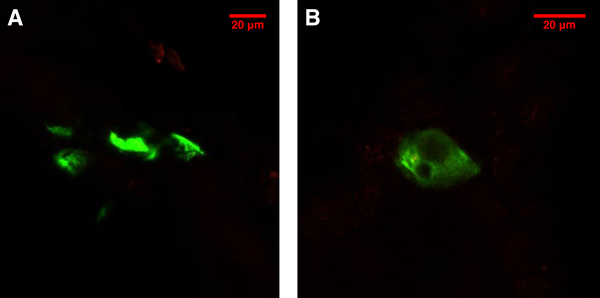
Figure 1. EGFP expression in the skin cells 48 hr after the tattooing treatment on the hindleg of a balb/c mouse viewed with a confocal microscope. A) A projection of EGFP signals from multiple focal planes. B) EGFP expression in a single cell.
Troubleshooting
1. No antigen expression is detected (e.g. no EGFP positive control signals).
2. Severe bleeding or damage to the skin occurs at the tattooing site.
3. The background signal is too high when imaging EGFP.
DNA vaccination is considered safer than traditional vaccination strategies as it does not require manipulation of, or expose the vaccines to, live or attenuated pathogens4. However, the result of DNA vaccination depends heavily on the delivery route. Skin is abundant in antigen-presenting cells, such as Langerhans Cells and dendritic cells1, and thus an ideal site for immunization in terms of immunogenicity and ease of access5,6. As a result, intradermal vaccination strategy is one of the most popular choices for DNA vaccines. As shown in this video, DNA tattooing is a simple yet promising way to administer a DNA vaccine intradermally. Interestingly, the inflammatory responses caused by the tattooing process could also serve as a natural and potent adjuvant1,2. It has been reported that the DNA tattooing can elicit up to a 100-fold increase in T-cell responses in monkeys, as compared to T-cell responses in animals immunized via the intramuscular route7,8. Compared to other dermal delivery methods, such as the gene gun, DNA tattooing holds several advantages. First, DNA tattooing does not require expensive equipment and carriers, i.e. gold particles. This would be a huge advantage in terms of vaccine distribution, especially for developing countries. Secondly, it is known that high air pressure can cause pDNA damage due to shear force, which could decrease the antigen expression level. A study has shown that DNA tattooing damages less than 3% of total pDNA9. Finally, DNA tattooing can cover a large area of skin, which could potentially elicit a stronger immune response. Tattooing devices can also be used to deliver peptide/protein vaccines, and have been proven to induce both humoral and cell-mediated immune responses10. We now use skin tattooing routinely in our animal experiments in a DNA prime-protein boost vaccination protocol (three DNA primes followed by two protein boosts over a period of 16 weeks), and we have successfully induced in mice strong immune responses against HIV-1 gp120.
The authors have nothing to disclose.
We would like to thank all members of the Kong Lab and Dr. Yan Deng at Microscopy Core, Office of Collaborative Science, NYUMC for their assistance and technical support. This work was supported by a pilot grant from the New York University Center for AIDS Research (CFAR, NIH grant AI027742).
Name of Reagent/Material | Company | Catalogue Number | Comments |
pCX-EGFP plasmid DNA | Clontech | ||
STEALTH Rotary Tattoo System | Worldwide Tattoo Supply | STEALTH-L | |
Tattoo needles | Worldwide Tattoo Supply | 1207RSB | |
EndoFree Plasmid Maxi Kit | Qiagen | 12362 | |
0.22 μm PVDF sterile filter | Millipore | SLGV013SL | |
electrical hair trimmer | Commercially available | ||
disposable safety razors | Commercially available | ||
Silver Sulfadiazine Cream | Watson | NDC 0591-0810-55 | |
Ketamine HCL | NDC 0856-2012-01 | ||
Zylazine Sterile Solution | NADA 139-236 |