The following protocol describes the procedure to assemble sandwich-like cultures to be used as an intermediate stage between bi-dimensional (2D) and three-dimensional (3D) cellular environments. The engineered systems can have applications in microscopy, biomechanics, biochemistry and cell biology assays.
Cell culture has been traditionally carried out on bi-dimensional (2D) substrates where cells adhere using ventral receptors to the biomaterial surface. However in vivo, most of the cells are completely surrounded by the extracellular matrix (ECM), resulting in a three-dimensional (3D) distribution of receptors. This may trigger differences in the outside-in signaling pathways and thus in cell behavior.
This article shows that stimulating the dorsal receptors of cells already adhered to a 2D substrate by overlaying a film of a new material (a sandwich-like culture) triggers important changes with respect to standard 2D cultures. Furthermore, the simultaneous excitation of ventral and dorsal receptors shifts cell behavior closer to that found in 3D environments. Additionally, due to the nature of the system, a sandwich-like culture is a versatile tool that allows the study of different parameters in cell/material interactions, e.g., topography, stiffness and different protein coatings at both the ventral and dorsal sides. Finally, since sandwich-like cultures are based on 2D substrates, several analysis procedures already developed for standard 2D cultures can be used normally, overcoming more complex procedures needed for 3D systems.
Traditionally, cell culture has been carried out on bi-dimensional (2D) substrates, though most of the in vivo cellular microenvironments have a three-dimensional (3D) nature. This unnatural 2D environment triggers changes in cell behavior as a way of self-adaptation to a flat world, which directly impacts cell fate1,2. Hence, results obtained on 2D cell cultures are not always reproducible in vivo. This has encouraged the development of new relevant culture systems seeking to provide more physiological-like conditions to get further insights into any dimension-dependent biological mechanism3,4.
One of the main differences between 2D culture and the 3D in vivo environment is the distribution of cell receptors anchored to the extracellular matrix (ECM): whereas on 2D substrates cells adhere ventrally, the majority of cells in vivo are completely surrounded by the ECM and thus cell adhesion occurs through a 3D distribution of receptors. This triggers different cell adhesion signaling pathways thereby modulating important processes such as cell growth, cell differentiation and gene expression. During the last decades, many different 3D culture systems have been established5-8, though their variability and complexity hinder their standardization in common cell culture procedures. Moreover 3D systems are usually not easy to handle and current experimental procedures on 2D substrates cannot be easily established for 3D cultures. In addition, literature rarely compares 3D cultures with the equivalent 2D condition or other 3D systems, hindering the proper understanding of cell behavior in these models.
Once having the cells adhered on a 2D substrate, the excitation of the dorsal receptors — by overlaying a film of a new material (sandwich-like culture) — can trigger cell responses alike 3D environments. The reason behind this is the simultaneous activation of both dorsal and ventral receptors to adhere and spread within the sandwich environment (Figure 1)9,10. As a consequence, cells undergo important changes with respect to 2D cultures11,12. Thus, cell fate is determined during assembly because of the sandwich culture, since the dorsal stimulation triggers changes in key cellular pathways. Therefore, the cell fate is highly determined by the time when the sandwich-like culture is assembled11.
Due to the nature of the system, a sandwich-like culture is a simple and versatile tool that allows the study of different parameters in cell/material interactions such as chemistry, topography, stiffness and protein coatings at both the ventral and dorsal sides. This offers a higher degree of versatility compared to other 3D systems (Figure 2) due to the independent dorsal and ventral combination of a wide variety of surface conditions. Additionally, different cell lines and different times to assemble the sandwich-like culture can be studied, increasing the wide spectra of possibilities.
A standard protocol of the sandwich-like culture is detailed below using either poly-L-lactic acid (PLLA) electrospun fibers or films as dorsal substrates, glass coverslip as ventral substrate and fibronectin as protein coating. Sandwich-like cultures were assembled just after cell seeding or after 3 hr of 2D culture. However, note that other material systems and proteins could be used; likewise the sandwich-like culture can be assembled at different time points.
1. Production of Dorsal Substrates
2. Sandwich Culture
3. Analysis
Note: Sandwich-like cultures are based on 2D substrates, and so can be commonly analyzed by procedures already developed for standard 2D cultures. For example, since PLLA is transparent and cells are constrained to move within the x-y plane, microscopy is done as on 2D substrates. Cell migration can be therefore analyzed as for 2D cultures, without the need of tracking cells in the z-axis as for 3D cultures, which simplifies the experiment and image analysis. To study the wound healing assay by a scratch assay follow this protocol:
Note: Protein and nucleic acid extraction is performed similarly as on 2D substrates. There is only one extra step that consists in disassembling the sandwich-like culture to add the lysis buffer directly on the cells in order to increase the extraction efficiency. For instance, for mRNA extraction:
Note: Immunodetection of proteins can be also performed as on 2D substrates. Since sandwich-like cultures could hinder the correct diffusion of the antibodies and buffers, incubation times should be increased. Also, the sandwich can be disassembled before starting the staining protocol but in this latter case some cells will remain attached to the dorsal substrate and some to the ventral substrate.
The stimulation of dorsal receptors within the sandwich-like culture triggers changes in cell morphology, cell adhesion and intracellular signaling pathways (e.g. focal adhesion kinase, FAK)10-12. As an example, fibroblasts cultured within the sandwich-like system overexpressed the α5 integrin subunit compared to the 2D, as observed for other 3D cultures15,16.
Cell fate is highly dependent on the time when the dorsal receptors are stimulated and by the properties of the dorsal interaction, similarly as happens in other 3D systems such as hydrogels. For example, hydrogels where proteins are tightly bound usually show smaller and rounded cells with undeveloped actin cytoskeleton and diffuse focal adhesions. This can be mimicked in a sandwich-like culture using substrates that adsorb proteins tightly, so that cells are not able to mechanically reorganize this layer of proteins and cell spreading is hindered. Similarly, hydrogels where cells can remodel the ECM can be mimicked with the sandwich system by using substrates that adsorb proteins more loosely10.
The stimulation of the dorsal receptors has been shown to modulate C2C12 cell fate. Dorsal electrospun PLLA fibers direct cell alignment when coated with fibronectin but not when coated with bovine serum albumin (a non-adhesive protein). This result points out cells do biologically sense and react to the dorsal inputs (Figure 4)9. Additionally, sandwich-like cultures with plane dorsal PLLA triggered an increase in the level of myogenesis. This depends also on the dorsal biological stimuli since the interaction with different dorsal proteins results in distinct differentiation rates (Figure 5)9.
Cell migration within the sandwich-like culture is also altered when compared to the 2D culture. It has been shown that in a wound-healing assay, cells within the sandwich culture adopt a highly elongated morphology and migrate shorter distances than on 2D substrates (Movies 1 and 2). Cell migration rates are furthermore related to the nature of the ventral and dorsal stimulation12.
Similarly as within 3D fibronectin and collagen gels17,18, sandwich-like culture increases cell-mediated ECM reorganization (i.e. the ventral fibronectin) in respect to the 2D condition (Figure 6)12. This process relies on the dorsal mechanical stimuli and cytoskeleton stability since the use of contractility inhibitors (blebbistatin, Y27632) hindered the process. Interestingly, the ventral fibronectin was also reorganized when using different dorsal protein coatings (i.e. vitronectin and bovine serum albumin) and even if left uncoated12.
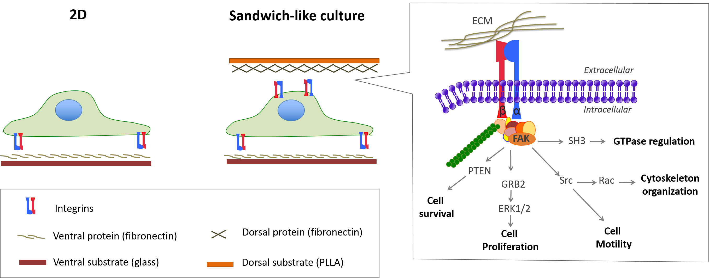
Figure 1: Sketch of standard (2D) and sandwich-like cultures. Stimulation of the dorsal receptors within the sandwich-like culture triggers additional cell adhesion signaling that modulates important cellular processes. Please click here to view a larger version of this figure.
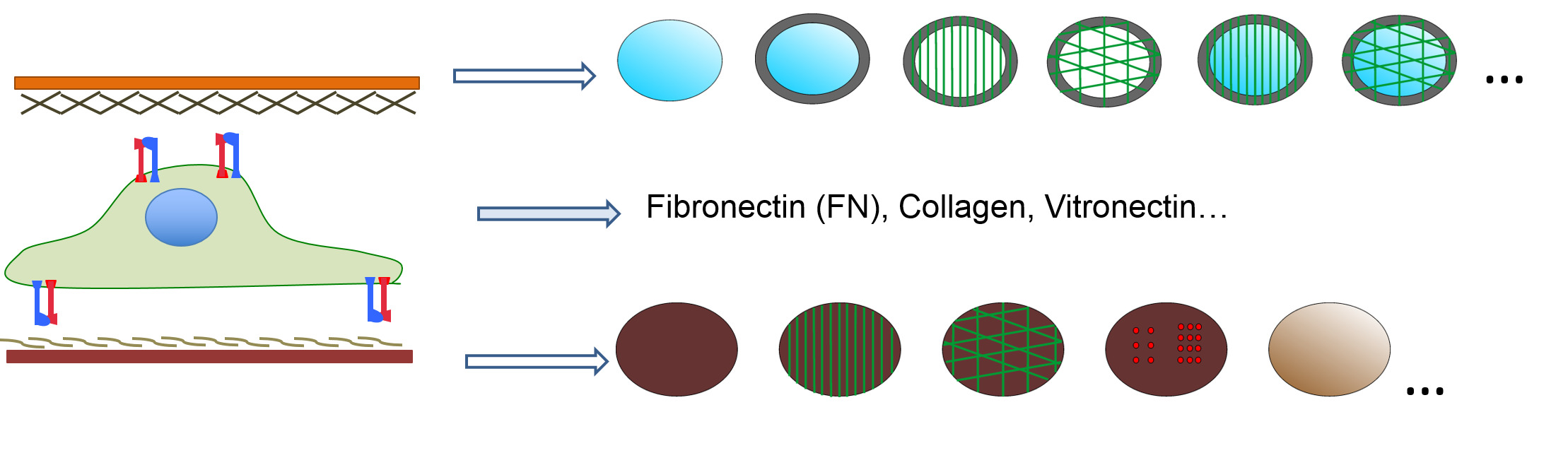
Figure 2: Sandwich-like culture is a versatile system that allows the study of different well-controlled parameters (topographical inputs, stiffness, gradients…) on both the ventral and dorsal substrates.
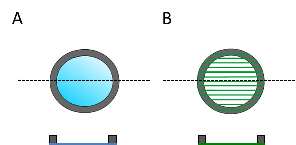
Figure 3: Dorsal substrates are sketched for both the top and cross-section view in order to show that have a designated top and bottom side. (A) Flat PLLA film and (B) electrospun fibers.
Figure 4: C2C12 morphology under different culture conditions including plane (p) and aligned fibers (a) of PLLA that were used as ventral (subscript) or dorsal (superscript) substrates. Dotted lines represent fibers orientation where necessary. Cells cultured on the plane substrate and overlaid with aligned fibers of PLLA (SWpa) sense the dorsal stimuli. Particularly, cells sense the dorsal fibers when coated with fibronectin but not when coated with bovine serum albumin (a non-adhesive protein). Consequently cells adhere to the fibers coated with fibronectin and align in the same direction. Image adapted from reference9. Please click here to view a larger version of this figure.
Figure 5: Cell differentiation in sandwich-like cultures after 4 days in differentiation media. Different culture conditions were analyzed including plane (p) and aligned fibers (a) of PLLA that were used as ventral (subscript) or dorsal (superscript) substrates. Samples were coated with fibronectin in all cases. (A) Fluorescence staining showing sarcomeric myosin positive cells (green) and cell nuclei (red). (B) Differentiated cells orientation as calculated by Fast Fourier Transform. (C) Myogenesis as determined by the percentage of sarcomeric myosin-positive cells. Data is normalized to the gold standard control. Statistically significant differences are indicated with *** P <0.001. Image adapted from reference9. Please click here to view a larger version of this figure.
Figure 6: Reorganization of ventral fibronectin. Sandwich culture triggers ventral FN reorganization by forming new fibronectin fibrils (brush-like labeling; pointed out with white arrows). Ventral fibronectin is reorganized within sandwich-like cultures with different dorsal protein coating (fibronectin, vitronectin and bovine serum albumin) or even when the dorsal substrate is left uncoated. Actin cytoskeleton (green), nuclei (blue) and FN (red) are shown. Please click here to view a larger version of this figure.
Movie 1: Cell migration on 2D. L929 fibroblast migrating on a fibronectin coated glass coverslip in a wound healing assay. Images were acquired for 16 hr (with a frame taken every 20 min). Please click here to view this video.
Movie 2: Cell migration within the sandwich-like culture. L929 fibroblast migrating in a wound healing assay within a sandwich-like culture were the ventral substrate was a fibronectin coated glass coverslip and the dorsal substrate a fibronectin coated PLLA film. Images were acquired for 16 hr (with a frame taken every 20 min). Please click here to view this video.
Nowadays, 3D culture is an important topic for the pharmaceutical and biotechnological industry as well as research in cell biology, including cancer and stem cells. As a consequence several 3D culture systems have been developed. Unfortunately, differences between the 3D systems usually result in different cell behavior, hindering the understanding of cell fate. Besides, experimental procedures are usually not as straightforward as for 2D culture systems. Hence developing new culture systems seeking to overcome some of these drawbacks is highly important.
Sandwich-like culture has been shown to strongly influence key cellular processes such as cell differentiation, cell morphology, cell signaling and cell migration. Furthermore cells share similarities with cells cultured in 3D systems, supporting the statement that sandwich-like culture links 2D with 3D culture systems. Physiological tissues have pores in the range of 3 to 14 µm that constraint cells and therefore influence cellular processes such as migration. This is somehow recapitulated using the sandwich-like system as dorsal and ventral stimuli represent per se a constrained environment that limits cell morphology and will necessarily influence cell migration regardless of the protein coatings.
Due to the simultaneous stimulation of dorsal and ventral receptors, sandwich-like culture is a robust technology to investigate the role of dimensionality in cell behavior. Moreover, as it is based on 2D substrates, this culture system is highly versatile to study different material properties and ECM inputs allowing the study of cell behavior under different microenvironments. Besides, the influence of the time at which dorsal receptors are stimulated on cell fate can be investigated by overlaying the dorsal substrate at different time points. Hence, unlike other 3D systems, the sandwich-like system provides a wide spectrum of well-controlled cellular microenvironments. Consequently the sandwich-like system is an interesting cell culture platform to mimic different physiological environments in order to study cell biology and test cell fate under different culture conditions.
As mentioned before, one advantage of this system is that different ventral and dorsal substrates can be studied using different protein coatings. Therefore, important parameters for cell fate such as ligand density can be tuned by controlling the ligand density of each one of the 2D substrates used (e.g. ventral and dorsal substrates). However, note that different substrates may need changes in the protocol. For example, using bigger dorsal substrates may result in the need to set the proper incubation time to peel the samples off the Petri dish. Likewise, bigger ventral substrates could result in hypoxic areas at the center of the sample because of the limited oxygen permeability of the ventral substrate (due to the glass coverslip). Additionally, analysis procedures will depend on the substrate properties. For instance, using opaque dorsal substrates will hinder standard microscopy protocols though it will still allow protein/nucleic acids extraction. Another key factor is the permeability of the dorsal substrate since cells should be allowed to get nutrients from the medium and discard waste.
To sum up, a sandwich-like culture is a simple system that offers the possibility to mimic different 3D-like microenvironments to investigate cell fate.
The authors have nothing to disclose.
The support from ERC through HealInSynergy (306990) and the FPU program AP2009-3626 are acknowledged.
Ploy(lactic acid) | NatureWorks | 4042D | Reagent |
Cover glasses (12 mmØ) | Marienfeld | 631-0666 | Equipment |
Chloroform | Scharlab | CL0200 | Reagent |
1,1,1,3,3,3-Hexafluoro-2-propanol (HFIP) | Sigma | 105228 | Reagent |
Syringe (1mL) | Henke Sass Wolf | 4010-200V0 | Equipment |
Syringe pump | New Era Pump Systems | NE1000 | Equipment |
High Voltage DC Power Supply | Glassman High Voltage | Series FC | Equipment |
Incubator | Hucoa-Herlös | 3111 | Equipment |
Laminar flow hood | Telstar | AV30/70 | Equipment |
Human Fibronectin | Sigma | F2006 | Reagent |
RNeasy Micro Kit | Qiagen | 74004 | Reagent |
Inverted microscope | Leica Microsystems | DMI 6000 | Equipment |
Triton X-100 | Sigma-Aldrich | T8787 | Reagent |
Albumin | Sigma-Aldrich | A7409 | Reagent |
Tween 20 | Sigma-Aldrich | P2287 | Reagent |