Summary

正常および病的脳機能を研究するために脳深部刺激でSaccadometryを使用します

Published: July 14, 2016
doi:

Summary

This paper describes the use of quantitative measurement of eye movements in conjunction with stimulation of focal areas of the deep brain in order to study physiology, pathophysiology, and the mechanisms of deep brain stimulation.

Abstract

The oculomotor system involves a large number of brain areas including parts of the basal ganglia, and various neurodegenerative diseases including Parkinson’s and Huntington’s can disrupt it. People with Parkinson’s disease, for example, tend to have increased saccadic latencies. Consequently, the quantitative measurement of saccadic eye movements has received considerable attention as a potential biomarker for neurodegenerative conditions. A lot more can be learned about the brain in both health and disease by observing what happens to eye movements when the function of specific brain areas is perturbed. Deep brain stimulation is a surgical intervention used for the management of a range of neurological conditions including Parkinson’s disease, in which stimulating electrodes are placed in specific brain areas including several sites in the basal ganglia. Eye movement measurements can then be made with the stimulator systems both off and on and the results compared. With suitable experimental design, this approach can be used to study the pathophysiology of the disease being treated, the mechanism by which DBS exerts it beneficial effects, and even aspects of normal neurophysiology.

Introduction

近年1を作る神経決定の高レベルのメカニズムに関する情報を得るの定量的および非侵襲的な方法として、反応時間の測定の使用に関心が高まっています。広く研究されてきた反応時間の一つのタイプは、サッケード待ち時間として知られている、視覚刺激の提示にサッカードを開始するのに要する時間です。サッカードは、我々は急速にある場所から別の場所に私たちの視線を移すときに発生する高速な眼球運動です。彼らは典型的には、2つまたは毎秒3の頻度で発生した、私たちが作る眼球運動の最も一般的なタイプです。各サッカードが有効で、別の2のではなく、視覚的な世界では1のキューを見て決定です。

目の動きを制御する神経経路は広く研究されており、かなりよく3を文書化されています。敏感な電子機器を使用して、眼球運動機能の側面を正確かつobjectivelすることができyは定量化。これは、眼球運動そのものの詳細な研究を促進するだけでなく、神経生理学や病態生理の他の領域を調査するためのツールと​​して使用するためにそれらを可能にします。

眼球運動の測定は、疾患状態についての有用な情報を提供することができます。眼球運動は、最近、例えば、ハンチントン4,5およびパーキンソン病6,7を含む神経変性疾患の潜在的なバイオマーカーとして注目されており、十分にサッケード反応時間は、これらの条件下で通常よりも遅くなる傾向があることが確立されています。サッカード測定の潜在的な用途は、診断や病気の追跡への補助があります。サッカードのタスクは、単純なprosaccade(左または右に突然現れた視覚刺激に向かって可能な限り迅速に探している)から、このような抗サッケード(視覚刺激とは反対側に、可能な限り迅速に探して)、またはメモリ – など、より複雑な作業の範囲導かれたサッカード(探し)はもはや存在しないターゲットの思い出した場所に向かって。

脳深部刺激は、いくつかの神経疾患に対する有効な治療法です。最も一般的振戦、硬直、運動緩慢、およびジスキネジアを含むパーキンソン病の運動症状を治療するために使用されます。また、そのような強迫性障害などのあまり一般的ではないが、神経因性疼痛、てんかん、および精神医学的状態のためのジストニアと本態性振戦、およびを含む他の運動障害のために使用されます。これは、科学者は、生体内で人間の脳の深部構造への直接の電気的アクセスを持っているので、実験的な神経学のための貴重な機会を提供する唯一の設定です。種々の標的は、動眼神経経路に関与しているそれらの多くは大脳基底核内のいくつかの場所、を含む、治療される状態に応じて刺激されます。これは、研究の広い範囲は、刺激を送達するためにDBSシステムを用いて行うことができることを意味します与えられた脳位置およびアイトラッキング装置に記録し、その効果を分析します。実験パラダイムによって、このような研究は、疾患、またはDBSは、その特定の設定で作動される機構の効果を刺激される領域の生理機能についての情報を得ることができます。この記事では、脳深部刺激患者における衝動性眼球運動試験の一般的なアプローチについて説明します。

アイトラッキング装置のいくつかの異なる種類があります。このプロトコールに記載された研究のためにポータブルsaccadometer水平衝動性眼球運動を記録するために使用されました。ポータブルsaccadometersはセッションが特に厳しいジスキネジアで苦しむ人々のために、パーキンソン病の患者にとってより快適であることを意味する( 図1参照 )ヘッドレストを必要としないという利点を有します。ここで使用saccadometerは、軽量で約5センチ幅10センチです。 saccadometerのmeasu直接赤外線眼球運動記録法の使用による解像度の眼球運動:内側眼角の使用光の前方に位置する赤外線源とセンサは、ミリ秒間隔で眼球の回転位置を確立するために、角膜からの反射しました。分析のための良質のデータを取得するためにsaccadometerは、少なくとも12ビットの分解能を有する少なくとも1 kHzの速度でサンプリングする必要があります。ここで使用saccadometerでは視覚刺激は、によって生成された光の3赤13のcdメートル-2スポットがあった低出力レーザーに建てられ、1正中線のスポットと±10度で、他の2といくつかの0.1度を従属各スポット( すなわち、)右と左に。

図1
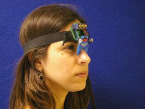
図1. Saccadometer。ヘッドsaccadometer弾性バンドに取り付けられ、鼻の橋の上に載って取り付けられました。四つのミニチュアレーザープロジェクト視標マット面上への、そして参加者の目の動きは、各眼の鼻側の差動赤外線反射率変換器によって測定されます。レーザーターゲットが頭を動かすと、ヘッドレストは必要ありません。 この図の拡大版をご覧になるにはこちらをクリックしてください。

Protocol

地元の倫理委員会は、セクション1で以下に詳述するように、参加者から得た同意、この研究を承認したと伝えました。 1.参加者の同意テストセッションが含まれるかを詳細に説明して情報シートを持つ参加者を提供します。 参加者は同意書上の各点を説明し、それらに任意の質問をする機会を与え、彼らと同意書を通過し、読み、研究では、それらの撮?…

Representative Results

図3は、移植視床下核DBSシステムとパーキンソン病患者から、衝動性眼球運動の軌跡の一例を示す図です。刺激システムがオフ(上のグラフ)と(下のグラフ)に切り替えて二つのグラフは、患者のprosaccadesをプロットします。グラフ上の各トレースは度で目の位置が離れて正中線(y軸)からの時間(x軸)の関数として変化する、すなわち、どの?…

Discussion

良質衝動性データを得るための最も重要な要因は、参加者に与えられた指示は明確かつ正確であることを保証することです。 antisaccadic作業の指示が完全に明らかにされていない場合たとえば、参加者が代わりにprosaccadesを実行する可能性があります。参加者が明らかに刺激またはsaccadometerが正確に目の位置を測定することはできません見ることができない場合は録画も台無しにすることがで?…

Disclosures

The authors have nothing to disclose.

Acknowledgements

Dr. Antoniades was supported by the National Institute of Health Research (NIHR) and by the Dementias and Neurodegenerative Diseases Research Network (DENDRON) and by the Wellcome Trust. Dr FitzGerald was supported by the National Institute for Health Research (NIHR) Oxford Biomedical Research Centre.

Materials

Saccadometer device ( Ober Consulting Poland)
Computer with Windows environment
Software, Latency Meter for downloading the raw data from the saccadometer.

References

  1. Leigh, R. J., & Kennard, C. Using saccades as a research tool in the clinical neurosciences. Brain. 127, 460-477 (2004).
  2. Carpenter, R. H. The neural control of looking. Curr Biol. 10, R291-293 (2000).
  3. Leigh, R. J., & Zee, D. S. The Neurology of Eye Movements. New York: Oxford University Press, (2006).
  4. Antoniades, C. A., Xu, Z., Mason, S. L., Carpenter, R. H., & Barker, R. A. Huntington's disease: changes in saccades and hand-tapping over 3 years. Journal of Neurology. 257, 1890-1898 (2010).
  5. Blekher, T.M., Yee, RD., Kirkwood, SC., Hake, AM., Stout, JC., Weaver, MR., Foroud, TM. Oculomotor control in asymptomatic and recently diagnosed individuals with the genetic marker for Huntington's disease. in Vision Research. Vol. 44 2729-2736 (2004).
  6. Chan, F., Armstrong, I. T., Pari, G., Riopelle, R. J., & Munoz, D. P. Deficits in saccadic eye-movement control in Parkinson's disease. Neuropsychologia. 43, 784-796 (2005).
  7. Antoniades, C. A., Demeyere, N., Kennard, C., Humphreys, G. W., & Hu, M. T. Antisaccades and executive dysfunction in early drug-naive Parkinson's disease: The discovery study. Mov Disord. (2015).
  8. Antoniades, C. et al. An internationally standardised antisaccade protocol. Vision Res. 84, 1-5 (2013).
  9. Ober, J. K. et al. Hand-Held system for ambulatory measurement of saccadic durations of neurological patients. . In: Modelling and Measurement in Medicine. (2003).
  10. Temperli, P. et al. How do parkinsonian signs return after discontinuation of subthalamic DBS? Neurology. 60, 78-81 (2003).
  11. Antoniades, C. A. et al. Deep brain stimulation: eye movements reveal anomalous effects of electrode placement and stimulation. PLoS ONE. 7, e32830 (2012).
  12. Yugeta, A. et al. Effects of STN stimulation on the initiation and inhibition of saccade in Parkinson disease. Neurology. 74, 743-748 (2010).
  13. Terao, Y., Fukuda, H., Ugawa, Y., & Hikosaka, O. New perspectives on the pathophysiology of Parkinson's disease as assessed by saccade performance: a clinical review. Clin Neurophysiol. 124, 1491-1506 (2013).
  14. Temel, Y., Visser-Vandewalle, V., & Carpenter, R. H. Saccadic latency during electrical stimulation of the human subthalamic nucleus. Curr Biol. 18, R412-414 (2008).
  15. Antoniades, C. A. et al. Deep brain stimulation abolishes slowing of reactions to unlikely stimuli. J Neurosci. 34, 10844-10852 1065-14.2014(2014).
  16. Rivaud-Pechoux, S. et al. Improvement of memory guided saccades in parkinsonian patients by high frequency subthalamic nucleus stimulation. J Neurol Neurosurg Psychiatry. 68, 381-384 (2000).
  17. Takikawa, Y., Kawagoe, R., Itoh, H., Nakahara, H., & Hikosaka, O. Modulation of saccadic eye movements by predicted reward outcome. Experimental brain research. Experimentelle Hirnforschung. 142, 284-291 (2002).
  18. Dorris, M. C., & Munoz, D. P. A neural correlate for the gap effect on saccadic reaction times in monkey. Journal of Neurophysiology. 73, 2558-2562, (1995).
  19. Hanes, D. P., & Schall, J. D. Countermanding saccades in macaque. Visual Neuroscience. 12, 929-937, (1995).
  20. Opris, I., Barborica, A., & Ferrera, V. P. On the gap effect for saccades evoked by electrical microstimulation of frontal eye fields in monkeys. Experimental brain research. Experimentelle Hirnforschung. 138, 1-7 (2001).
  21. Takagi, M., Frohman, E. M., & Zee, D. S. Gap-overlap effects on latencies of saccades, vergence and combined vergence-saccades in humans. Vision Res. 35, 3373-3388 (1995).
  22. Schall, J. D. Neuronal activity related to visually guided saccades in the frontal eye fields of rhesus monkeys: comparison with supplementary eye fields. Journal of Neurophysiology. 66, 559-579 (1991).
  23. Pare, M., & Hanes, D. P. Controlled movement processing: superior colliculus activity associated with countermanded saccades. J Neurosci. 23, 6480-6489 (2003).
check_url/kr/53640?article_type=t

Play Video

Cite This Article
Antoniades, C. A., FitzGerald, J. J. Using Saccadometry with Deep Brain Stimulation to Study Normal and Pathological Brain Function. J. Vis. Exp. (113), e53640, doi:10.3791/53640 (2016).

View Video