Summary

समस्याग्रस्त प्लांट, Oomycete, और फफूंद नमूने के लिए इलेक्ट्रॉन माइक्रोस्कोपी (SEM) प्रोटोकॉल स्कैन

Published: February 03, 2017
doi:

Summary

Problems in the processing of biological samples for scanning electron microscopy observation include cell collapse, treatment of samples from wet microenvironments and cell destruction. Low-cost and relatively rapid protocols suited for preparing challenging samples such as floral meristems, oomycete cysts, and fungi (Agaricales) are compiled and detailed here.

Abstract

स्कैनिंग इलेक्ट्रॉन माइक्रोस्कोप (SEM) के साथ टिप्पणियों के लिए जैविक नमूने के प्रसंस्करण में आम समस्याओं सेल पतन, गीला microenvironments और सेल विनाश से नमूने के उपचार में शामिल हैं। नाजुक नमूने यहां बताए गए SEM के तहत छवि पर कब्जा के लिए नमूना उपचार में मुख्य चुनौतियों में से कुछ पर काबू पाने की प्रक्रिया करने के लिए युवा पुष्प ऊतकों, oomycete अल्सर, और कवक बीजाणुओं (Agaricales) उदाहरण के रूप में विशिष्ट प्रोटोकॉल का उपयोग करना,।

पुष्प मेरिस्टेमों एफएए के साथ तय (Formalin-एसिटिक-शराब) और महत्वपूर्ण बिंदु ड्रायर (सीपीडी) के साथ कार्रवाई की सेलुलर दीवारों या विकृत अंगों ढह प्रदर्शित नहीं किया था। इन परिणामों के पुष्प विकास के पुनर्निर्माण के लिए महत्वपूर्ण हैं। ऐसे glutaraldehyde तय oomycete अल्सर के रूप में गीला microenvironments, से नमूने का एक समान सीपीडी आधारित उपचार, सु के विभिन्न प्रकारों पर नैदानिक विशेषताओं का अंतर विकास (जैसे, पुटी कांटा) का परीक्षण करने के लिए इष्टतम हैbstrates। नर्स कवक बीजाणुओं से जुड़ी कोशिकाओं के विनाश पुनर्जलीकरण, निर्जलीकरण, और सीपीडी उपचार, इन कोशिकाओं के आगे कार्यात्मक अध्ययन के लिए एक महत्वपूर्ण कदम के बाद बचा हुआ था।

प्रोटोकॉल यहाँ विस्तृत कम लागत और अच्छी गुणवत्ता के चित्र के अधिग्रहण के विकास की प्रक्रिया को फिर से संगठित करने के लिए और नैदानिक ​​विशेषताओं का अध्ययन करने के लिए तेजी के विकल्प का प्रतिनिधित्व करते हैं।

Introduction

जीव विज्ञान में, स्कैनिंग इलेक्ट्रॉन माइक्रोस्कोपी का उपयोग करें (SEM) संरचनात्मक विकास, तुलनात्मक आकृति विज्ञान, अंग विकास, और आबादी या प्रजातियों 1 के लक्षण वर्णन के अध्ययन के लिए बढ़ा दिया गया है। सूक्ष्म संरचनाओं के अपने दो आयामी दृष्टिकोण के साथ, इस तरह के micromorphology और व्यवस्था जैसे क्षेत्रों 20 वीं सदी की दूसरी छमाही के बाद से SEM तकनीक अग्रिमों से फायदा। उदाहरण के लिए, 1970 के दशक में धूम कोटिंग पद्धति की शुरूआत गैर प्रवाहकीय ऊतकों 2, 3 की इमेजिंग बढ़ाने ऐसे शूट apices और फूल के रूप में नाजुक सामग्री के संभावित अवलोकन किया। SEM के एक उच्च निर्वात वातावरण 4 में स्थलाकृति पुन: पेश करने नमूना की सतह से निकली इलेक्ट्रॉनों का उपयोग करता है।

SEM शामिल अध्ययन दोनों संरचनात्मक पात्रों के अनुमान और growt के पुनर्निर्माण में ध्यान केंद्रित कर रहेज प्रक्रियाओं। न्यू संरचनात्मक वर्गीकरण के लिए प्रासंगिक और वर्ण जीवों की एक विस्तृत श्रृंखला की व्यवस्था SEM टिप्पणियों से खोज की गई है। उदाहरण के लिए, संयंत्र लक्षण इस तरह की लकड़ी 5, कलंक विविधता 6, nectary और पुष्प आकृति विज्ञान 7, 8, trichome विवरण 9, और पराग अनाज की पहनाया हुआ गड्ढ़े 10, 11, ठीक से बिना कल्पना नहीं की जा सकती है के रूप में, प्रजातियों निदान या supraspecific वर्गीकरण के लिए इस्तेमाल किया SEM। पारंपरिक SEM के साथ सफल टिप्पणियों को भी लंबे समय formalin तय जीवों 12 के लिए हासिल किया गया है और संयंत्र वनस्पति संग्रहालय 13 नमूनों।

दूसरी ओर, SEM का उपयोग विकास की प्रक्रिया के पुनर्निर्माण के अध्ययन में इस तरह के अंग विकास 14, infe के रूप में विषयों की एक विस्तृत श्रृंखला शामिलबैक्टीरिया 15, पौधे जड़ शरीर क्रिया विज्ञान 16, परजीवी मेजबान लगाव तंत्र 17, 18, परजीवी 19, mycoparasitism और प्रतिजीविता 20, 21, विकास कुरूपता 22, जंगली और उत्परिवर्ती व्यक्तियों 23 के तुलनात्मक विकास, और पूरे जीवन चक्र पर दवा के प्रभाव से प्रेरित ctions 24। पर्यावरण स्कैनिंग इलेक्ट्रॉन माइक्रोस्कोप (ESEM) 25 विकास की प्रक्रिया में गीला जैविक नमूने के अवलोकन के लिए महत्वपूर्ण लाभ हो सकता है, नाजुक सामग्री अभी भी ESEM) के कम निर्वात हालत में समझौता किया जा सकता है, और नुकसान से बचने के लिए पर्याप्त रूप से संसाधित करने की आवश्यकता के मूल्यवान रूपात्मक अवलोकन।

इस पत्र, तीन रचनाकार के SEM अवलोकन के लिए विशिष्ट प्रोटोकॉल की समीक्षा मेंनमूनों की erent प्रकार प्रस्तुत है: पुष्प मेरिस्टेमों, oomycetes (Saprolegnia), और फंगल सामग्री। इन प्रोटोकॉल हमारे पिछले SEM आधारित अध्ययन 26, 27, 28, 29, 30, 31, 32, 33, जहां विशिष्ट कठिनाइयों और वैकल्पिक समाधान पाया गया है के अनुभव संकलन। संयंत्र तुलनात्मक विकास और संरचनात्मक अध्ययन के मामले में, SEM के उपयोग के 1970 के दशक के 34, 35 में शुरू किया था, और तब से, शोधकर्ताओं ने पाया है कि कुछ पुष्प सुविधाओं को और अधिक अस्थिर तुलना में पहले सोचा 36 हैं। पुष्प विकास के पुनर्निर्माण युवा पुष्प मेरिस्टेमों और anthesis के बीच सभी चरणों का कब्जा शामिल है। इस उद्देश्य तक पहुंचने के लिए, यह Esse हैntial कि नमूना स्थलाकृति और कोशिका दीवार अखंडता निर्धारण और बाद में निर्जलीकरण के बाद समझौता नहीं कर रहे हैं। (आंकड़े 1 ए, 1 बी) के युवा पुष्प मेरिस्टेमों विशेष रूप से सेल की दीवार ढहने की चपेट में हैं। इसी तरह, इस तरह के nectaries, पंखुड़ी, कलंक और sporangia के रूप में नाजुक संरचनाओं प्रभावी और undamaging प्रोटोकॉल की आवश्यकता होती है। इस समीक्षा SEM इमेजिंग के लिए बरकरार युवा और नाजुक ऊतकों को रखने के लिए एक इष्टतम प्रोटोकॉल का सार।

Oomycetes (Stramenopiles) परजीवी के सबसे विविध और व्यापक समूहों में से -एक, रोगाणुओं और पौधों से अकशेरुकी और रीढ़ 37 से लेकर मेजबान टीम के साथ के मामले में – वहाँ बीजाणुओं कि आगे बढ़ने और एक गीला वातावरण में विकसित कर रहे हैं। क्योंकि बीजाणुओं एक पर्याप्त सब्सट्रेट मानक SEM प्रोटोकॉल के लिए उपयुक्त नहीं की जरूरत है इस हालत SEM अवलोकन के लिए एक चुनौती का प्रतिनिधित्व करता है। Oomycetes के बीच, Saprolegnia की प्रजातियों में विशेष रुचि के हैं क्योंकि वे सीएn aquacultures, मत्स्य पालन, और उभयचर आबादी 38 में गंभीर कटौती के कारण। इस तरह के अल्सर के आदी कांटा के रूप में Micromorphological विशेषताओं, Saprolegnia, जो संक्रमण नियंत्रण और संभावित उपचार 39 की स्थापना के लिए मौलिक है की प्रजातियों की पहचान करने के लिए उपयोगी होना पाया गया है। इधर, विभिन्न substrates पर अल्सर की रीढ़ की हड्डी विकास के पैटर्न की तुलना करने के लिए और महत्वपूर्ण बिंदु ड्रायर (सीपीडी) तैयार करने और बाद में SEM अवलोकन के लिए नमूना हेरफेर करने के लिए एक प्रयोगात्मक प्रोटोकॉल है।

एक तिहाई मामले में, वहाँ दिलचस्प निष्कर्ष है कि कवक Phellorinia herculanea च के बीजाणुओं की एक निरीक्षण के बाद आया है। stellata च। नोवा (Agaricales) 31। साथ में बीजाणुओं के साथ, अप्रत्याशित नर्सरी कोशिकाओं का एक समूह SEM के तहत पहचान की थी। पिछले पारंपरिक प्रोटोकॉल और इलाज सामग्री के साथ, नर्स कोशिकाओं कहां आयाटी पूरी तरह से (चित्रा -1 सी) ढह गई। बीजाणुओं के लिए जुड़े विशेष ऊतकों के बारे में अधिक अनुमान मानक दृष्टिकोण यहाँ वर्णित (चित्रा -1) करने के लिए आसान है, लेकिन महत्वपूर्ण संशोधनों के साथ बनाया जा सकता है।

इस समीक्षा में, वहाँ विस्तृत SEM प्रोटोकॉल है कि में SEM अवलोकन के साथ जुड़े विभिन्न समस्याओं से निपटने के लिए इस्तेमाल किया जा सकता है वनस्पतियों, oomycetes, और इस तरह के सेल पतन और मेरिस्टेमेटिक ऊतक सिकुड़ते, पुटी कांटा के गैर इष्टतम विकास, और के विनाश के रूप Agaricales, अल्पकालिक ऊतकों, क्रमशः।

आकृति 1
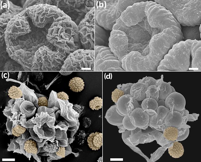
चित्रा 1: बिना (क, ग) और (बी, डी) प्रोटोकॉल एफएए-इथेनॉल सीपीडी इलाज के नमूने की तुलना। (एकख) Anacyclus clavatus, मध्य विकास के फूलों की कलियों। बड आज़मियम tetroxide 46 <के साथ इलाज/ sup> (क) और एफएए-सीपीडी प्रोटोकॉल (ख) के साथ इलाज कली। (गघ) के Phellorinia herculanea च बीजाणुओं के साथ नर्स कोशिकाओं। stellata। बिना किसी उपचार (ग) और प्रोटोकॉल यहाँ Agaricales (घ) के लिए वर्णित के साथ सूखे नमूने हैं। नारंगी में बीजाणुओं। तराजू: (एबी) 100 माइक्रोन, (सीडी) 50 माइक्रोन। Photos वाई रूज़-Leon द्वारा उठाए गए थे। यह आंकड़ा का एक बड़ा संस्करण देखने के लिए यहां क्लिक करें।

Protocol

नोट: इस प्रोटोकॉल छह मुख्य वर्गों, विशेष जीवों (वर्गों 1-3), और तीन सब (4-6) के लिए आम प्रक्रियाओं का वर्णन करने के लिए तीन समर्पित भी शामिल है। तारों (*) प्रयोगकर्ताओं द्वारा संशोधित कदम से संकेत मिलता है। <p class…

Representative Results

पुष्प विकास और विकास के फिक्सेशन और पूरी तरह से बनाई संयंत्र संरचनाएं एफएए-सीपीडी यहाँ वर्णित प्रोटोकॉल का प्रयोग, युवा और परिपक्व संयंत्र के ऊतकों बेहतर तय की और SEM ?…

Discussion

मानक SEM प्रोटोकॉल के लिए सम्मान के साथ, यहाँ प्रस्तुत प्रक्रियाओं अपेक्षाकृत तेजी से पालन करने के लिए आसान है, और कम लागत के तरीके शामिल हैं। नमूने की राशि पर और प्रसंस्करण में आसानी पर निर्भर करता है, यह…

Disclosures

The authors have nothing to disclose.

Acknowledgements

इस परियोजना अनुदान समझौते सं 634429. इस प्रकाशन केवल लेखक के विचारों को प्रतिबिंबित करता है के तहत यूरोपीय संघ के क्षितिज 2020 अनुसंधान और नवाचार कार्यक्रम से धन प्राप्त हुआ है, और यूरोपीय आयोग जो जानकारी का बनाया जा सकता है किसी भी उपयोग के लिए जिम्मेदार नहीं ठहराया जा सकता उसमें निहित। हम यह भी असली Jardin Botanico, CSIC द्वारा की गई वित्तीय योगदान को स्वीकार करते हैं। एसआर Saprolegnia में उसे अनुसंधान के समर्थन के लिए यूरोपीय संघ [ITN-SAPRO-238550] करने के लिए आभारी है। हम भी फ्रांसिस्को Calonge धन्यवाद कृपया नमूने प्रोसेसिंग (चित्रा 5) के लिए Phellorinia herculanea छवियों और बी Pueyo प्रदान करना चाहते हैं। सभी छवियों मैड्रिड में रियल Jardin Botanico-CSIC पर SEM सेवा द्वारा उठाए गए थे।

Materials

Acetic acid No specific supplier Skin irritation, eye irritation
aluminium stubs Ted Pella, Inc. 16221 www.tedpella.com
Centrifuge tubes No specific supplier
Critical Point Dryer Polaron Quatum Technologies CPD7501
D (+) Glucose Merck 1,083,421,000
Double sided sellotape No specific supplier
Ethanol absolute No specific supplier. Flammable
European bacteriological agar Conda 1800.00 www.condalab.com
Filter paper No specific supplier
Forceps No specific supplier
Formalin 4% No specific supplier. Harmful, acute toxicity, skin sensitisation, carcinogenicity. Flammable
Glass cover slips No specific supplier
Glass hermetic container  No specific supplier
Glutaraldehyde 25% DC 253857.1611  (L) Dismadel S.L. 3336 www.dismadel.com
Mycological peptone Conda 1922.00 www.condalab.com
needles No specific supplier
Petri dishes No specific supplier
Plastic containers No specific supplier
Sample holder with lid  for the critical point dryer  Ted Pella, Inc. 4591 www.tedpella.com
scalpels No specific supplier
Scanning Electron Microscope Hitachi S3000N
Software for SEM
Solution A: NaH2PO4
Solution B: Na2HPO4
Specimen holders No specific supplier
Sputter coater Balzers SCD 004
Stereomicroscope No specific supplier
Transmission Electron Microscope (TEM) grids Electron Microscopy Sciences G200 (Square Mesh) www.emsdiassum.com
Tweezers No specific supplier

References

  1. Endress, P. K., Baas, P., Gregory, M. Systematic plant morphology and anatomy: 50 years of progress. Taxon. 49 (3), 401-434 (2000).
  2. Falk, R. H., Gifford, E. M., Cutter, E. G. Scanning electron microscopy of developing plant organs. Science. 168 (3938), 1471-1474 (1970).
  3. Damblon, F. Sputtering, a new method of coating pollen grains in scanning electron microscopy. Grana. 15 (3), 137-144 (1975).
  4. Everhart, T. E., Thornley, R. F. M. Wide-band detector for micro-microampere low-energy electron currents. J. Sci. Instrum. 37 (7), 37246-37248 (1960).
  5. Collins, S. P., et al. Advantages of environmental scanning electron microscopy in studies of microorganisms. Microsc. Res. Techniq. 25 (5-6), 398-405 (1993).
  6. Fannes, W., Vanhove, M. P. M., Huyse, T., Paladini, G. A scanning electron microscope technique for studying the sclerites of Cichlidogyrus. Parasitol. Res. 114 (5), 2031-2034 (2015).
  7. Erbar, C., Leins, P. Portioned pollen release and the syndromes of secondary pollen presentation in the Campanulales-Asterales complex. Flora. 190 (4), 323-338 (1995).
  8. Jansen, S., Smets, E., Baas, P. Vestures in woody plants: a review. IAWA Journal. 19 (4), 347-382 (1998).
  9. Bortolin Costa, M. F., et al. Stigma diversity in tropical legumes with considerations on stigma classification. Bot. Rev. 80 (1), 1-29 (2014).
  10. Almeida, O. J. G., Cota-Sánchez, J. H., Paoli, A. A. S. The systematic significance of floral morphology, nectaries, and nectar concentration in epiphytic cacti of tribes Hylocereeae and Rhipsalideae (Cactaceae). Perspect. Plant Ecol. 15 (5), 255-268 (2013).
  11. Konarska, A. Comparison of the structure of floral nectaries in two Euonymus L. species (Celastraceae). Protoplasma. 252 (3), 901-910 (2015).
  12. Giuliani, C., Maleci Bini, L. Insight into the structure and chemistry of glandular trichomes of Labiatae, with emphasis on subfamily Lamioideae. Plant Syst. Evol. 276 (3-4), 199-208 (2008).
  13. Li, K., Zheng, B., Wang, Y., Zhou, L. L.Breeding system and pollination biology of Paeonia delavayi (Paeoniaceae), an endangered plant in the Southwest of China. Pak. J. Bot. 46 (5), 1631-1642 (2014).
  14. García, L., Rivero, M., Droppelmann, F. Descripción morfológica y viabilidad del polen de Nothofagus nervosa (Nothofagaceae). Bosque. 36 (3), 487-496 (2015).
  15. Prenner, G., Klitgaard, B. B. Towards unlocking the deep nodes of Leguminosae: floral development and morphology of the enigmatic Duparquetia orchidacea (Leguminosae, Caesalpinioideae). Am. J. Bot. 95 (11), 1349-1365 (2008).
  16. Ratnayake, K., Joyce, D. C., Webb, R. I. A convenient sample preparation protocol for scanning electron microscope examination of xylem-occluding bacterial biofilm on cut flowers and foliage. Sci. Hortic-Amsterdam. 140 (1), 12-18 (2012).
  17. Çolak, G., Celalettin Baykul, M., Gürler, R., Çatak, E., Caner, N. Investigation of the effects of aluminium stress on some macro and micro-nutrient contents of the seedlings of Lycopersicon esculentum Mill. by using scanning electron microscope. Pak. J. Bot. 46 (1), 147-160 (2014).
  18. Arafa, S. Z. Scanning electron microscope observations on the monogenean parasite Paraquadriacanthus nasalis from the nasal cavities of the freshwater fish Clarias gariepinus in Egypt with a note on some surface features of its microhabitat. Parasitol. Res. 110 (5), 1687-1693 (2012).
  19. Uppalapatia, S. R., Kerwinb, J. L., Fujitac, Y. Epifluorescence and scanning electron microscopy of host-pathogen interactions between Pythium porphyrae (Peronosporales, Oomycota)and Porphyra yezoensis (Bangiales, Rhodophyta). Bot. Mar. 44 (2), 139-145 (2001).
  20. Meaney, M., Haughey, S., Brennan, G. P., Fairweather, I. A scanning electron microscope study on the route of entry of clorsulon into the liver fluke, Fasciola hepatica. Parasitol. Res. 95 (2), 117-128 (2005).
  21. Sundarasekar, J., Sahgal, G., Subramaniam, S. Anti-candida activity by Hymenocallis littoralis extracts for opportunistic oral and genital infection Candida albicans. Bangladesh J. Pharmacol. 7 (3), 211-216 (2012).
  22. Benhamou, N., Rey, P., Picard, K., Tirilly, Y. Ultrastructural and cytochemical aspects of the interaction between the mycoparasite Pythium oligandrum and soilborne plant pathogens. Phytopathology. 89 (6), 506-517 (1999).
  23. Singh, A., et al. First evidence of putrescine involvement in mitigating the floral malformation in mangoes: A scanning electron microscope study. Protoplasma. 251 (5), 1255-1261 (2014).
  24. Xiang, C., et al. Fine mapping of a palea defective 1 (pd1), a locus associated with palea and stamen development in rice. Plant Cell Rep. 34 (12), 2151-2159 (2015).
  25. Mendoza, L., Hernandez, F., Ajello, L. Life cycle of the human and animal oomycete pathogen Pythium insidiosum. J. Clin. Microbiol. 31 (11), 2967-2973 (1993).
  26. Bello, M. A., Rudall, P. J., González, F., Fernández, J. L. Floral morphology and development in Aragoa (Plantaginaceae) andrelated members of the order Lamiales. Int. J. Plant Sci. 165 (5), 723-738 (2004).
  27. Bello, M. A., Hawkins, J. A., Rudall, P. J. Floral morphology and development in Quillajaceae and Surianaceae (Fabales), the species-poor relatives of Leguminosae and Polygalaceae. Ann. Bot. 100 (4), 1491-1505 (2007).
  28. Bello, M. A., Hawkins, J. A., Rudall, P. J. Floral ontogeny in Polygalaceae and its bearing on the homologies of keeled flowers in Fabales. Int. J. Plant Sci. 171 (5), 482-498 (2010).
  29. Bello, M. A., Alvarez, I., Torices, R., Fuertes-Aguilar, J. Floral development and evolution of capitulum structure in Anacyclus (Anthemideae, Asteraceae). Ann. Bot. 112 (8), 1597-1612 (2013).
  30. Bello, M. A., Martínez-Asperilla, A., Fuertes-Aguilar, J. Floral development of Lavatera trimestris and Malva hispanica reveals the nature of the epicalyx in the Malva generic alliance. Bot. J. Linn. Soc. 181 (1), 84-98 (2016).
  31. Calonge, F. D., Martínez, A. J., Falcó, I., Samper, L. E. Phellorinia herculanea f. stellata f. nova encontrada en España. Bol. Soc. Micol.Madrid. 35 (1), 65-70 (2011).
  32. Liu, Y., et al. Deciphering microbial landscapes of fish eggs to mitigate emerging diseases. ISME J. 8 (10), 2002-2014 (2014).
  33. Sandoval-Sierra, J. V., Diéguez-Uribeondo, J. A comprehensive protocol for improving the description of Saprolegniales (Oomycota): two practical examples (Saprolegnia aenigmatica sp. nov. and Saprolegnia racemosa sp. nov.). PLOS one. , (2015).
  34. Endress, P. K. Zur vergleichenden Entwicklungsmorphologie, Embryologie und Systematik bei Laurales. Bot. Jahrb. Syst. 92 (2), 331-428 (1972).
  35. Tucker, S. Floral development in Saururus cernuus (Saururaceae):1. Floral initiation and stamen development. Am. J. Bot. 62 (3), 993-1005 (1975).
  36. Endress, P. K., Matthews, M. L. Progress and problems in the assessment of flower morphology in higher-level systematics. Plant Syst. Evol. 298 (2), 257-276 (2012).
  37. Beakes, G. W., Glockling, S. L., Sekimoto, S. The evolutionary phylogeny of the oomycete "fungi&#34. Protoplasma. 249 (1), 3-19 (2012).
  38. Romansic, J. M., et al. Effects of the pathogenic water mold Saprolegnia ferax on survival of amphibian larvae. Dis. Aquat. Organ. 83 (3), 187-193 (2009).
  39. van West, P. Saprolegnia parasitica, an oomycete pathogen with a fishy appetite: new challengues for an old problem. Mycologist. 20 (3), 99-104 (2006).
  40. Johansen, D. A. . Plant microtechnique. , (1940).
  41. Unestam, T. Studies on the crayfish plague fungus Aphanomyces astaci. Some factors affecting growth in vitro. Physiol. Plantarum. 18 (2), 483-505 (1965).
  42. Cerenius, L., Söderhäll, K. Repeated zoospore emergence from isolated spore cysts of Aphanomyces astaci. Exp. Mycol. 8 (4), 370-377 (1984).
  43. Diéguez-Uribeondo, J., Cerenius, L., Söderhäll, K. Repeated zoospore emergence in Saprolegnia parasitica. Mycol. Res. 98 (7), 810-815 (1994).
  44. Söderhäll, K., Svensson, E., Unestam, T. Chitinase and protease activities in germinating zoospore cysts of a parasitic fungus, Aphanomyces astaci, Oomycetes. Mycopathologia. 64 (1), 9-11 (1978).
  45. Echlin, P. . Handbook of sample preparation for scanning electron microscopy and X-Ray Microanalysis. , (2009).
  46. Osumi, M., et al. Preparation for observation of fine structure of biological specimens by high-resolution SEM. Microscopy. 32 (4), 321-330 (1983).
  47. Rezinciuc, S. . The Saprolegniales morpho-molecular puzzle: an insight into markers identifying specific and subspecific levels in main parasites. , (2013).
check_url/kr/55031?article_type=t

Play Video

Cite This Article
Bello, M. A., Ruiz-León, Y., Sandoval-Sierra, J. V., Rezinciuc, S., Diéguez-Uribeondo, J. Scanning Electron Microscopy (SEM) Protocols for Problematic Plant, Oomycete, and Fungal Samples. J. Vis. Exp. (120), e55031, doi:10.3791/55031 (2017).

View Video