Summary

문제있는 공장, 난균 강 및 곰팡이 샘플에 대한 전자 현미경 (SEM) 프로토콜을 스캔

Published: February 03, 2017
doi:

Summary

Problems in the processing of biological samples for scanning electron microscopy observation include cell collapse, treatment of samples from wet microenvironments and cell destruction. Low-cost and relatively rapid protocols suited for preparing challenging samples such as floral meristems, oomycete cysts, and fungi (Agaricales) are compiled and detailed here.

Abstract

주사 전자 현미경 (SEM)으로 관찰 용 생물학적 시료를 처리 일반적인 문제는 세포 붕괴 습식 미세 환경과 세포 파괴로부터의 샘플의 처리를 포함한다. 주사 전자 현미경에서 이미지 캡처를위한 샘플 처리의 주요 과제 중 일부를 극복 여기에 설명되어 섬세한 샘플을 처리하기 위해 특정 프로토콜을 젊은 꽃 조직, 난균 강 낭종 및 예제 등의 곰팡이 포자 (주름 버섯 목) 사용.

FAA 고정 꽃 분열 조직은 (포르말린 – 기타 – 아세트산)과 중요 포인트 건조기 (CPD)으로 처리가 세포 벽이나 왜곡 기관을 축소 표시되지 않았습니다. 이러한 결과는 꽃 개발의 재건을 위해 중요하다. 예를 들면 글루 타르 알데하이드로 고정 난균 강 낭종 등의 습식 미세 환경에서 샘플 유사한 CPD 기반 치료, 스와의 다른 유형에 대한 진단 특성의 차이 성장 (예를 들어, 낭종 쪽)을 테스트하는 최적bstrates. 곰팡이 포자 부착 간호사 세포의 파괴는 재수, 탈수 및 CPD 처리, 이러한 세포의 추가 기능 연구를위한 중요한 단계 후 피해야한다.

여기에 설명 된 프로토콜은 저비용과 성장 과정을 재구성 및 진단 특성을 연구하기 위해 좋은 품질의 이미지의 취득을위한 급속 대안을 나타낸다.

Introduction

생물학에서, 주사 전자 현미경을 사용 (SEM)의 구조의 진화, 비교 형태, 장기 개발, 집단 또는 종 (1)의 특성에 관한 연구로 확장되었다. 미세 구조의 두 개의 차원보기와 같은 micromorphology 및 계통 등의 분야는 20 세기 후반 이후 SEM 기술의 발전에서 이익을. 예를 들어, 1970 년대에 스퍼터 코팅 방법의 도입 비 – 전도성 조직 (2) (3)의 영상을 향상 같은 촬영 정점 꽃과 같은 민감한 물질 가능 관찰했다. SEM은 고진공 환경 4 지형 재현 시험편의 표면으로부터 방출 된 전자를 사용한다.

SEM과 관련된 연구는 구조적인 문자의 추론과 growt의 재건에 모두 초점을 맞추고있다시간 처리합니다. 새로운 구조적 분류에 관련된 문자 나 유기체의 광범위한 계통은 SEM 관찰 결과로부터 발견되었다. 예를 들어, 식물의 특성은 10, 11, 제대로없이 시각화 할 수없는 나무 5, 오명 다양성 (6), 꿀샘과 꽃 형태 7, 8, trichome 정보 9, 꽃가루 입자의 옷을 입은 피트로, 종의 진단 또는 supraspecific 분류에 사용 SEM. 기존의 SEM 성공적인 관측도 오랜 포르말린 고정 생물 (12)에 대해 달성 된 식물 식물 표본 13 표본.

한편, SEM을 사용하여 성장 과정 재구성 연구는 장기 개발 14 infe 같은 주제의 다양한 참여박테리아 (15), 식물 뿌리의 생리 (16), 기생충 – 호스트 연결 메커니즘 (17), (18), 기생충 (19), mycoparasitism 및 항균 20, 21, 성장 기형 (22), 야생 및 돌연변이 개인 (23)의 비교 개발 및 전체 수명주기에 약물 효과에 의해 유도 ctions 24. 환경 주사 전자 현미경 (ESEM) (25)의 성장 공정에서 습식 생물학적 시료의 관찰 중요한 장점을 가질 수 있지만 섬세한 재료는 아직까지도 ESEM)의 저 진공 상태에서 손상 및 손실을 방지하기 위해 적절하게 처리 할 수있다 의 가치 형태 학적 관찰.

본 논문에서는 세 DIFF의 SEM 관찰을위한 특정 프로토콜의 리뷰샘플 erent 유형이 표시됩니다 : 꽃 분열 조직, oomycetes (Saprolegnia가 번식), 및 곰팡이 소재. 이러한 프로토콜은 특정한 어려움을 대체 해법이 발견 된 이전 SEM 기반 연구 26, 27, 28, 29, 30, 31, 32, 33, 경험을 컴파일한다. 비교 식물 발달 및 구조 연구의 경우, SEM의 사용은 1970 (34), (35)에 시작하고, 이후, 연구자들은 특정 꽃의 기능은 이전에 36 생각보다 불안정한 것을 발견했다. 꽃 개발의 재건 젊은 꽃 분열 조직과 개화 사이의 모든 단계의 캡처를 포함한다. 이 목적을 달성하기 위해, ESSE 인샘플 지형 세포벽 무결성 정착 이후의 탈수 후 손상되지 않도록 ntial. 젊은 꽃 분열 조직 세포 벽 붕괴에 특히 취약하다 (도 1A, 1B). 마찬가지로, nectaries에, 꽃잎, 암술과 sporangia와 같은 섬세한 구조는 효과적이고 undamaging 프로토콜을 필요로한다. 이 평가는 SEM 이미징 그대로 젊고 섬세한 조직을 유지하는 최적의 프로토콜을 요약 한 것입니다.

미생물과 식물에서 무척추 동물과 척추 동물 (37)에 이르기까지 호스트와 oomycetes (Stramenopiles) 기생충의 가장 다양하고 광범위한 그룹의 온의 경우, – 성장하고 젖은 환경에서 개발 포자가있다. 포자 표준 SEM 프로토콜에 적합하지 않은 적절한 기판을 필요로하기 때문에이 조건은 SEM 관찰에 대한 도전을 나타냅니다. oomycetes 중 Saprolegnia가 번식 종은 특히 관심있는 그들은 캘리포니아 때문에N aquacultures, 어업 및 수륙 양용 인구 38 심각한 감소를 야기한다. 예컨대 낭종 후크 등뼈 같은 Micromorphological 특성, 감염 제어 및 잠재적 치료 39 설정할 근본적인 Saprolegnia가 번식의 종류를 식별하는 것이 유용한 것으로 밝혀졌다. 여기서, 다른 기판에 낭종 척추 성장의 패턴과 비교하고 임계점 건조기 (CPD) 제조 이후의 SEM 관찰 용 시료를 조작하는 실험 프로토콜이있다.

세 번째 경우에, 버섯 Phellorinia의 herculanea (F)의 포자의 검사 후에 와서 흥미로운 연구 결과가있다. stellata 바. 노바 (주름 버섯 목) 31. 함께 포자로, 예상치 못한 보육 셀 그룹은 SEM에서 확인되었다. 이전의 전통적인 프로토콜 및 치료 재료, 간호사 세포 ou는왔다t 완전히 (그림 1C)을 축소. 포자에 관련된 특정 조직에 대한 자세한 추론은 여기서 설명 (그림 1D) 표준 방식으로 간단하지만 중요한 수정을 할 수있다.

이 리뷰에서의 SEM 관찰과 관련된 다양한 문제를 해결하는 데 사용할 수있는 상세한 SEM 프로토콜이있다 피자 식물, 휴대 붕괴와 분열 조직 축소, 낭종 쪽의 비 최적의 성장, 그리고 파괴 등 oomycetes 및 주름 버섯 목, 각각 임시 조직.

그림 1
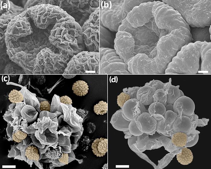
그림 1 : (A, C)과과 (B, D) 프로토콜 FAA-에탄올 CPD없이 처리 된 샘플의 비교. (AB) Anacyclus의 clavatus, 중앙 개발의 꽃 봉오리. 버드 사 산화 오스뮴 46 <처리/ SUP> (a) 및 FAA-CPD 프로토콜 (b)로 처리 새싹. (CD) Phellorinia의 herculanea (F)의 포자와 간호사 세포. stellata. 임의의 처리 (c)없이 여기 주름 버섯 목 (d)에 기술 된 프로토콜로 샘플을 건조. 오렌지 포자. 저울 : (AB) 100 μm의 (CD) 50 μm의. 사진은 Y. 루이즈 – 레온에 의해 촬영되었다. 이 그림의 더 큰 버전을 보려면 여기를 클릭하십시오.

Protocol

참고 :이 프로토콜은 모든 (4-6)에 공통의 절차를 설명하는 특정 생물 (섹션 1-3)과 3-3 헌신 6 가지 주요 섹션이 포함되어 있습니다. 별표 (*)는 실험자에 의해 수정 단계를 나타냅니다. 1. 개발의 연구 및 완전 성형 공장 구조 수집 및 고정 식물 물질은 흄 후드없이 액세스 할 수있는 장소에 수집되면, 도입 원심 분리기 튜브에서 70 % 에탄올에 담가 재료…

Representative Results

꽃 개발 및 개발의 고정 및 완전 성형 공장 구조 여기에 설명 된 FAA-CPD 프로토콜을 사용하여, 젊고 성숙한 식물 조직은 최적의 고정 및 SEM 이미징 탈수 있습니다. 새싹의 지형과 모양이 축소 세포 (도 1B, 1D,도 4a-F)에 의해 왜곡되지 않기 때문에 이러한 꽃 개발과 같은 프로세스는 복원 할 수 있습니다. 복잡한 형상을 …

Discussion

표준 SEM 프로토콜에 대해, 여기에 제시된 절차는 비교적 빠른 따라하기 쉽고, 저렴한 방법 등이 있습니다. 샘플의 양 및 처리의 용이성에 따라, 양호한 품질의 이미지를 획득 4-5 일 걸린다. CPD 및 SEM 작업에 대한 적절한 안전주의 사항을 포함하는 절차는 취급이 용이하다. 특히주의 (1.1.3 및 프로토콜의 2.1.5에 단계 1.1.1 참조) 포르말린과 글루 타르 알데히드에주의해야한다. 샘플 손상 또는 이전 단?…

Disclosures

The authors have nothing to disclose.

Acknowledgements

이 프로젝트는 보조금 협정 제이 책은 저자의 의견을 반영 634429.에서 유럽 연합 (EU)의 호라이즌 (2020)의 연구 및 혁신 프로그램에서 자금을 받았고, 유럽위원회 (European Commission)가 정보로 만들어 질 수있는 사용에 대한 책임을지지 않습니다 거기에 포함. 우리는 또한 리얼 자르 딘 보타니 코, CSIC에 의한 재정 기여를 인정합니다. SR은 Saprolegnia가 번식 그녀의 연구 지원을위한 유럽 연합 (EU) [ITN-SAPRO-238550]에 감사합니다. 우리는 또한 친절에 대한 시스코 칼론 감사 샘플을 (그림 5)을 처리하기위한 Phellorinia의 herculanea 이미지와 B의 Pueyo을 제공하고자합니다. 모든 이미지는 마드리드에서 리얼 자르 딘 보타니-CSIC의 SEM 서비스에 의해 촬영되었다.

Materials

Acetic acid No specific supplier Skin irritation, eye irritation
aluminium stubs Ted Pella, Inc. 16221 www.tedpella.com
Centrifuge tubes No specific supplier
Critical Point Dryer Polaron Quatum Technologies CPD7501
D (+) Glucose Merck 1,083,421,000
Double sided sellotape No specific supplier
Ethanol absolute No specific supplier. Flammable
European bacteriological agar Conda 1800.00 www.condalab.com
Filter paper No specific supplier
Forceps No specific supplier
Formalin 4% No specific supplier. Harmful, acute toxicity, skin sensitisation, carcinogenicity. Flammable
Glass cover slips No specific supplier
Glass hermetic container  No specific supplier
Glutaraldehyde 25% DC 253857.1611  (L) Dismadel S.L. 3336 www.dismadel.com
Mycological peptone Conda 1922.00 www.condalab.com
needles No specific supplier
Petri dishes No specific supplier
Plastic containers No specific supplier
Sample holder with lid  for the critical point dryer  Ted Pella, Inc. 4591 www.tedpella.com
scalpels No specific supplier
Scanning Electron Microscope Hitachi S3000N
Software for SEM
Solution A: NaH2PO4
Solution B: Na2HPO4
Specimen holders No specific supplier
Sputter coater Balzers SCD 004
Stereomicroscope No specific supplier
Transmission Electron Microscope (TEM) grids Electron Microscopy Sciences G200 (Square Mesh) www.emsdiassum.com
Tweezers No specific supplier

References

  1. Endress, P. K., Baas, P., Gregory, M. Systematic plant morphology and anatomy: 50 years of progress. Taxon. 49 (3), 401-434 (2000).
  2. Falk, R. H., Gifford, E. M., Cutter, E. G. Scanning electron microscopy of developing plant organs. Science. 168 (3938), 1471-1474 (1970).
  3. Damblon, F. Sputtering, a new method of coating pollen grains in scanning electron microscopy. Grana. 15 (3), 137-144 (1975).
  4. Everhart, T. E., Thornley, R. F. M. Wide-band detector for micro-microampere low-energy electron currents. J. Sci. Instrum. 37 (7), 37246-37248 (1960).
  5. Collins, S. P., et al. Advantages of environmental scanning electron microscopy in studies of microorganisms. Microsc. Res. Techniq. 25 (5-6), 398-405 (1993).
  6. Fannes, W., Vanhove, M. P. M., Huyse, T., Paladini, G. A scanning electron microscope technique for studying the sclerites of Cichlidogyrus. Parasitol. Res. 114 (5), 2031-2034 (2015).
  7. Erbar, C., Leins, P. Portioned pollen release and the syndromes of secondary pollen presentation in the Campanulales-Asterales complex. Flora. 190 (4), 323-338 (1995).
  8. Jansen, S., Smets, E., Baas, P. Vestures in woody plants: a review. IAWA Journal. 19 (4), 347-382 (1998).
  9. Bortolin Costa, M. F., et al. Stigma diversity in tropical legumes with considerations on stigma classification. Bot. Rev. 80 (1), 1-29 (2014).
  10. Almeida, O. J. G., Cota-Sánchez, J. H., Paoli, A. A. S. The systematic significance of floral morphology, nectaries, and nectar concentration in epiphytic cacti of tribes Hylocereeae and Rhipsalideae (Cactaceae). Perspect. Plant Ecol. 15 (5), 255-268 (2013).
  11. Konarska, A. Comparison of the structure of floral nectaries in two Euonymus L. species (Celastraceae). Protoplasma. 252 (3), 901-910 (2015).
  12. Giuliani, C., Maleci Bini, L. Insight into the structure and chemistry of glandular trichomes of Labiatae, with emphasis on subfamily Lamioideae. Plant Syst. Evol. 276 (3-4), 199-208 (2008).
  13. Li, K., Zheng, B., Wang, Y., Zhou, L. L.Breeding system and pollination biology of Paeonia delavayi (Paeoniaceae), an endangered plant in the Southwest of China. Pak. J. Bot. 46 (5), 1631-1642 (2014).
  14. García, L., Rivero, M., Droppelmann, F. Descripción morfológica y viabilidad del polen de Nothofagus nervosa (Nothofagaceae). Bosque. 36 (3), 487-496 (2015).
  15. Prenner, G., Klitgaard, B. B. Towards unlocking the deep nodes of Leguminosae: floral development and morphology of the enigmatic Duparquetia orchidacea (Leguminosae, Caesalpinioideae). Am. J. Bot. 95 (11), 1349-1365 (2008).
  16. Ratnayake, K., Joyce, D. C., Webb, R. I. A convenient sample preparation protocol for scanning electron microscope examination of xylem-occluding bacterial biofilm on cut flowers and foliage. Sci. Hortic-Amsterdam. 140 (1), 12-18 (2012).
  17. Çolak, G., Celalettin Baykul, M., Gürler, R., Çatak, E., Caner, N. Investigation of the effects of aluminium stress on some macro and micro-nutrient contents of the seedlings of Lycopersicon esculentum Mill. by using scanning electron microscope. Pak. J. Bot. 46 (1), 147-160 (2014).
  18. Arafa, S. Z. Scanning electron microscope observations on the monogenean parasite Paraquadriacanthus nasalis from the nasal cavities of the freshwater fish Clarias gariepinus in Egypt with a note on some surface features of its microhabitat. Parasitol. Res. 110 (5), 1687-1693 (2012).
  19. Uppalapatia, S. R., Kerwinb, J. L., Fujitac, Y. Epifluorescence and scanning electron microscopy of host-pathogen interactions between Pythium porphyrae (Peronosporales, Oomycota)and Porphyra yezoensis (Bangiales, Rhodophyta). Bot. Mar. 44 (2), 139-145 (2001).
  20. Meaney, M., Haughey, S., Brennan, G. P., Fairweather, I. A scanning electron microscope study on the route of entry of clorsulon into the liver fluke, Fasciola hepatica. Parasitol. Res. 95 (2), 117-128 (2005).
  21. Sundarasekar, J., Sahgal, G., Subramaniam, S. Anti-candida activity by Hymenocallis littoralis extracts for opportunistic oral and genital infection Candida albicans. Bangladesh J. Pharmacol. 7 (3), 211-216 (2012).
  22. Benhamou, N., Rey, P., Picard, K., Tirilly, Y. Ultrastructural and cytochemical aspects of the interaction between the mycoparasite Pythium oligandrum and soilborne plant pathogens. Phytopathology. 89 (6), 506-517 (1999).
  23. Singh, A., et al. First evidence of putrescine involvement in mitigating the floral malformation in mangoes: A scanning electron microscope study. Protoplasma. 251 (5), 1255-1261 (2014).
  24. Xiang, C., et al. Fine mapping of a palea defective 1 (pd1), a locus associated with palea and stamen development in rice. Plant Cell Rep. 34 (12), 2151-2159 (2015).
  25. Mendoza, L., Hernandez, F., Ajello, L. Life cycle of the human and animal oomycete pathogen Pythium insidiosum. J. Clin. Microbiol. 31 (11), 2967-2973 (1993).
  26. Bello, M. A., Rudall, P. J., González, F., Fernández, J. L. Floral morphology and development in Aragoa (Plantaginaceae) andrelated members of the order Lamiales. Int. J. Plant Sci. 165 (5), 723-738 (2004).
  27. Bello, M. A., Hawkins, J. A., Rudall, P. J. Floral morphology and development in Quillajaceae and Surianaceae (Fabales), the species-poor relatives of Leguminosae and Polygalaceae. Ann. Bot. 100 (4), 1491-1505 (2007).
  28. Bello, M. A., Hawkins, J. A., Rudall, P. J. Floral ontogeny in Polygalaceae and its bearing on the homologies of keeled flowers in Fabales. Int. J. Plant Sci. 171 (5), 482-498 (2010).
  29. Bello, M. A., Alvarez, I., Torices, R., Fuertes-Aguilar, J. Floral development and evolution of capitulum structure in Anacyclus (Anthemideae, Asteraceae). Ann. Bot. 112 (8), 1597-1612 (2013).
  30. Bello, M. A., Martínez-Asperilla, A., Fuertes-Aguilar, J. Floral development of Lavatera trimestris and Malva hispanica reveals the nature of the epicalyx in the Malva generic alliance. Bot. J. Linn. Soc. 181 (1), 84-98 (2016).
  31. Calonge, F. D., Martínez, A. J., Falcó, I., Samper, L. E. Phellorinia herculanea f. stellata f. nova encontrada en España. Bol. Soc. Micol.Madrid. 35 (1), 65-70 (2011).
  32. Liu, Y., et al. Deciphering microbial landscapes of fish eggs to mitigate emerging diseases. ISME J. 8 (10), 2002-2014 (2014).
  33. Sandoval-Sierra, J. V., Diéguez-Uribeondo, J. A comprehensive protocol for improving the description of Saprolegniales (Oomycota): two practical examples (Saprolegnia aenigmatica sp. nov. and Saprolegnia racemosa sp. nov.). PLOS one. , (2015).
  34. Endress, P. K. Zur vergleichenden Entwicklungsmorphologie, Embryologie und Systematik bei Laurales. Bot. Jahrb. Syst. 92 (2), 331-428 (1972).
  35. Tucker, S. Floral development in Saururus cernuus (Saururaceae):1. Floral initiation and stamen development. Am. J. Bot. 62 (3), 993-1005 (1975).
  36. Endress, P. K., Matthews, M. L. Progress and problems in the assessment of flower morphology in higher-level systematics. Plant Syst. Evol. 298 (2), 257-276 (2012).
  37. Beakes, G. W., Glockling, S. L., Sekimoto, S. The evolutionary phylogeny of the oomycete "fungi&#34. Protoplasma. 249 (1), 3-19 (2012).
  38. Romansic, J. M., et al. Effects of the pathogenic water mold Saprolegnia ferax on survival of amphibian larvae. Dis. Aquat. Organ. 83 (3), 187-193 (2009).
  39. van West, P. Saprolegnia parasitica, an oomycete pathogen with a fishy appetite: new challengues for an old problem. Mycologist. 20 (3), 99-104 (2006).
  40. Johansen, D. A. . Plant microtechnique. , (1940).
  41. Unestam, T. Studies on the crayfish plague fungus Aphanomyces astaci. Some factors affecting growth in vitro. Physiol. Plantarum. 18 (2), 483-505 (1965).
  42. Cerenius, L., Söderhäll, K. Repeated zoospore emergence from isolated spore cysts of Aphanomyces astaci. Exp. Mycol. 8 (4), 370-377 (1984).
  43. Diéguez-Uribeondo, J., Cerenius, L., Söderhäll, K. Repeated zoospore emergence in Saprolegnia parasitica. Mycol. Res. 98 (7), 810-815 (1994).
  44. Söderhäll, K., Svensson, E., Unestam, T. Chitinase and protease activities in germinating zoospore cysts of a parasitic fungus, Aphanomyces astaci, Oomycetes. Mycopathologia. 64 (1), 9-11 (1978).
  45. Echlin, P. . Handbook of sample preparation for scanning electron microscopy and X-Ray Microanalysis. , (2009).
  46. Osumi, M., et al. Preparation for observation of fine structure of biological specimens by high-resolution SEM. Microscopy. 32 (4), 321-330 (1983).
  47. Rezinciuc, S. . The Saprolegniales morpho-molecular puzzle: an insight into markers identifying specific and subspecific levels in main parasites. , (2013).
check_url/kr/55031?article_type=t

Play Video

Cite This Article
Bello, M. A., Ruiz-León, Y., Sandoval-Sierra, J. V., Rezinciuc, S., Diéguez-Uribeondo, J. Scanning Electron Microscopy (SEM) Protocols for Problematic Plant, Oomycete, and Fungal Samples. J. Vis. Exp. (120), e55031, doi:10.3791/55031 (2017).

View Video