Summary

في الوقت الحقيقي الرحلان الشاردي مع تيتراميثيلامونيوم لكمية حجم الكسر و تورتوسيتي من الدماغ خارج الخلية الفضاء

Published: July 24, 2017
doi:

Summary

يصف هذا البروتوكول الرحلان الشاردي في الوقت الحقيقي، وهي الطريقة التي يقيس المعلمات المادية من الفضاء خارج الخلية (إكس) من أدمغة المعيشة. يتم استخدام انتشار جزيء خامل صدر في إكس لحساب كسر حجم إكس و تورتوسيتي. وهو مثالي لدراسة التغيرات العكسية الحادة ل إكس الدماغ.

Abstract

يصف هذا الاستعراض المفاهيم الأساسية والبروتوكول لأداء طريقة الرحلان الشاردي في الوقت الحقيقي (رتي)، ومعيار الذهب لاستكشاف وتحديد مساحة خارج الخلية (إكس) من الدماغ الحية. ويحيط إكس جميع خلايا الدماغ ويحتوي على كلا السائل الخلالي ومصفوفة خارج الخلية. نقل العديد من المواد اللازمة لنشاط الدماغ، بما في ذلك الناقلات العصبية والهرمونات، والمواد الغذائية، يحدث عن طريق نشر من خلال إكس. تحدث تغييرات في حجم وهندسة هذا الفضاء أثناء عمليات الدماغ العادية، مثل النوم، والظروف المرضية، مثل نقص التروية. ومع ذلك، فإن هيكل وتنظيم المخ الدماغ، وخاصة في الدول المريضة، لا يزال غير مستكشفة إلى حد كبير. طريقة رتي يقيس اثنين من المعلمات الفيزيائية للدماغ الحية: حجم الكسر و تورتوسيتي. حجم جزء هو نسبة حجم الأنسجة التي يشغلها إكس. يعد التعفن مقياسا للعوائق النسبية التي تواجهها المادة عندما تنتشر عن طريق إعادة الدماغجيون بالمقارنة مع وسيلة مع عدم وجود عوائق. في رتي، يتم نبض جزيء خامل من ميكرولكترود مصدر في إكس الدماغ. كما تنتشر جزيئات بعيدا عن هذا المصدر، يتم قياس تركيز تغيير أيون مع مرور الوقت باستخدام ميكرولكترود أيون انتقائية وضعه ما يقرب من 100 ميكرون بعيدا. من منحنى الانتشار الناتج، يمكن حساب كل من حجم الكسر و تورتوسيتي. وقد استخدمت هذه التقنية في شرائح الدماغ من أنواع متعددة (بما في ذلك البشر) وفي الجسم الحي لدراسة التغيرات الحادة والمزمنة ل إكس. خلافا لغيرها من الأساليب، رتي يمكن استخدامها لدراسة كل من التغييرات عكسها ولا رجعة فيه إلى إكس الدماغ في الوقت الحقيقي.

Introduction

الفضاء خارج الخلية (إكس) هو شبكة من قنوات مترابطة الخارجي لجميع خلايا الدماغ ويحتوي على كلا السائل الخلالي ومصفوفة خارج الخلية ( الشكل 1A والشكل 1B ). توزيع العديد من المواد اللازمة لوظيفة الخلايا الدماغية، بما في ذلك المواد الغذائية والهرمونات والناقلات العصبية، يحدث عن طريق نشر من خلال إكس. التغيرات في المعلمات الفيزيائية لهذا الفضاء، بما في ذلك حجم والهندسة، ومصفوفة خارج الخلية، يمكن أن تؤثر بشكل كبير الانتشار من خلال إكس وتركيزات أيون المحلية الاستحمام خلايا الدماغ، والتي لها تأثير عميق على وظيفة خلايا الدماغ 1 ، 2 .

في الوقت الحقيقي يستخدم الرحلان الشاردي (رتي) لتحديد اثنين من الخصائص الهيكلية للمنطقة الدماغ: حجم الكسر و تورتوسيتي 3 ، 4 ،"كريف"> 5. جزء حجم ( α ) هي نسبة حجم الأنسجة التي يشغلها إكس ( V إكس ) بالنسبة إلى حجم الأنسجة الكلي ( V الأنسجة ) في حجم الابتدائية ممثل؛

معادلة

إن العسر ( λ ) هو العائق النسبي الذي تواجهه المادة عندما تنتشر عن طريق منطقة الدماغ بالمقارنة مع وسط دون عوائق؛

معادلة

حيث D * (سم 2 ق -1 ) هو معامل الانتشار الفعال للمادة في الدماغ و D (سم 2 ق -1 ) هو معامل الانتشار الحر للمادة في وسط الحرة، مثل هلام الاغاروز المخفف.

اليوم، مادة التحقيق الأكثر شيوعا ل Rطريقة تي هي كاتيون تيتراميثيلامونيوم صغيرة (تما). تما لديه الوزن الجزيئي من 74 غرام / مول، ينفصل تماما في الحل، ولها شحنة موجبة واحدة. وقد أظهرت الدراسات رتي مع هذا أيون أن α معادلة 0.2 و λ معادلة 1.6 1 ، 2 . وهذا يعني أن إكس هو ما يقرب من 20٪ من حجم الدماغ الكلي، وأن نشر جزيء صغير، خامل يحدث ما يقرب من 2.5 مرات أبطأ في إكس من في وسط مع عدم وجود عوائق 3 . ومع ذلك، على حد سواء α و λ تختلف مع عمر الدماغ، والمنطقة، والدولة وفي الحالات المرضية 1 . وقد ارتبطت تغيرات هذه المعلمات بتطور المخ، والشيخوخة، والنوم، والصرع، والعديد من العمليات الأساسية الأخرى وأمراض الدماغ 1، 6 . في حين تقيس تقنيات أخرى α و λ ، رتي يمكن قياس سواء في المناطق المحلية من الأنسجة الحية في الوقت الحقيقي. لهذا السبب، أصبح رتي أداة لا غنى عنها للتحقيق في التغييرات في α و λ خلال التحديات الحادة والتي يمكن عكسها.

تم التحقق من صحة نظرية رتي أصلا من قبل نيكولسون وفيلبس، وقد استخدمت هذه التقنية على نطاق واسع منذ ذلك الوقت 4 ، 7 . التجارب التي تستخدم رتي تبدأ مع الافراج عن نبض تما من مصدر ميكرولكترود بواسطة الرحلان الشاردي في هلام الاغاروز مخفف. مرة واحدة طرد، الأيونات تنتشر بحرية بعيدا عن مصدر نقطة، واختيار من عدد لا حصر له من المسارات العشوائية ( الشكل 1D ). يتم قياس تركيز أيون المتغيرة مع مرور الوقت باستخدام ميكرولكترود أيون انتقائي (إيسم) وضعه تقريبا100 ميكرون بعيدا ( الشكل 1C ). التغيرات في تركيز تما هي الرسوم البيانية وتركيبها على منحنى يسمح لحساب كل من D وعدد النقل من ميكرولكترود الرحلان الشاردي (المعلمات التي نوقشت في البروتوكول). مع هذه القيم، يتم تكرار الإجراء في منطقة الدماغ التي تهم الحصول على D * وحساب كل من α و λ . السيطرة على ميكرولكترود الرحلان الشاردي، وجمع البيانات والرسوم البيانية وتركيب منحنى تركيز تما، وحساب المعلمات التجريبية كلها عادة ما يتم من قبل البرامج واندا و والتر، والتي تم تصميمها خصيصا لهذا الغرض (البرنامج وأدلة هم متاحة مجانا من المؤلفين عند الطلب).

يصف القسم بروتوكول من هذا الاستعراض الإجراءات الأساسية اللازمة لتصميم وأداء رتي في شرائح الدماغ القوارض. وقد استخدمت هذه التقنية أيضا في غير قضيبنماذج الأنف والحنجرة، بما في ذلك شرائح الدماغ البشري وفي الأعمال التحضيرية المجراة الدماغ 9. يقدم قسم النتائج ممثل كل من النتائج المثالية وغير مثالية لتسليط الضوء على الفروق الدقيقة في تفسير البيانات. وأخيرا، يغطي قسم المناقشة باختصار تقنيات استكشاف الأخطاء وإصلاحها، وقيود رتي، والتقنيات البديلة المستخدمة لدراسة إكس، والتطبيقات المستقبلية ل رتي.

شكل 1
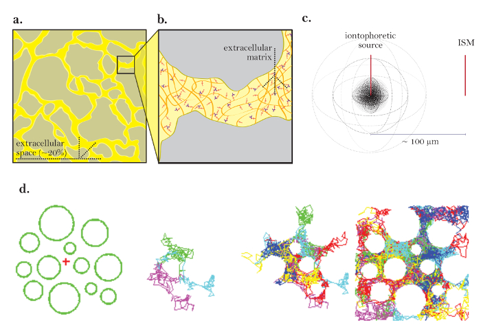
الشكل 1: الرسوم البيانية من الانتشار من خلال إكس. ( أ ) مخطط إكس: يوضح حجم وموقع إكس في قسم الدماغ نموذجي. الأصفر يصادف إكس بين عمليات خلايا الدماغ الرمادي. حجم إكس هو ما يقرب من 20٪ من إجمالي حجم الأنسجة ( أي حجم حجم = 0.2) في ظل الظروف الفسيولوجية. ( ب ) رسم تخطيطي مكبر لل إكس: يسلط الضوء على المعلمات المادية المساهمة في تورتوسيتي، بما في ذلك هندسة خلايا الدماغ (الرمادي) والمصفوفة خارج الخلية (رسم بياني كشبكة من غليكوسامينوغليكانز متعددة الألوان والبروتيوغليكان). ( ج ) رسم بياني ثلاثي الأبعاد للانتشار من مصدر نقطة: يوضح الحركة الصافية للجزيئات الخاملة من مصدر اليونتوفيريت إلى إيسم. وباستثناء حواجز الانتشار والامتصاص الخلوي، تنتشر الجزيئات إلى الخارج في جميع الاتجاهات، وتنتج جبهة تركيز كروية. و إيسم يحد من التركيز المحلي للجزيئات الخاملة صدر من مصدر اليونتوفوريتيك. ( د ) محاكاة الكمبيوتر للانتشار في إكس من الدماغ: [أقصى اليسار] الإعداد لمحاكاة مونت كارلو. وتمثل المجالات الخضراء عمليات الخلايا الدماغية ويمثل الصليب الأحمر مصدر نقطة. هذا نماذج الإعداد المخططة أنسجة المخ في الشكل 1A . [الصور الوسطى] 3 و6 جزيئات تؤدي حركات عشوائية لأنها تنتشر من خلال الفضاء خارج الخلية من الدماغ، مبين في 2 أبعاد. [أقصى اليمين] يمشي عشوائية من العديد من الجزيئات الصادرة من مصدر نقطة. الحركة الصافية لجميع الجزيئات من مصدر نقطة هو الخارج كما هو مبين في الشكل 1C . وتحدد المسارات العشوائية التراكمية المسافات بين الخلايا ( أي إكس؛ انظر المرجع 5 لمزيد من التوضيح). الرجاء انقر هنا لعرض نسخة أكبر من هذا الرقم.

Protocol

وكانت جميع الإجراءات الحيوانية، وتستخدم للحصول على عينات الأنسجة، موافقة لجنة الأخلاقيات الحيوانية في مركز سوني الطبي دونستات. 1. إعداد الحلول والمعدات إعداد 150 ملي كلوريد الصود?…

Representative Results

ويتجلى فائدة تقنية رتي في تجربة مصممة لقياس التغيرات في α وخلال تحدي هايبوسمولار ( الشكل 8 والشكل 9 ). وقد تبين سابقا أن تقليل الأسمولية من إكس عن طريق الغسيل على أسف منخفض التوتر سيؤدي إلى انخفاض في α وزيادة ف…

Discussion

الشكل 10
الشكل 10: البيانات غير المثالية التي تبين القضايا التقنية المشتركة. ( أ ) الرسوم البيانية من القضايا التقنية المشتركة مع ميكرولترودس الرحلان الشاردي: مقارنة الإفراج العادي من تما من ميكرولكترود الش…

Disclosures

The authors have nothing to disclose.

Acknowledgements

وأيد العمل من قبل المعاهد الوطنية للصحة نيندس منحة R01 NS047557.

Materials

A/D and D/A converter National Instruments Corporation NI USB-6221 DAQ The NI USB-6221 is still sold as a 'Legacy' device by NI. They recommend using NI USB-6341 X Series DAQs for new installations, however we have not tested the newer units. We describe the use of the NI USB-6221 with MATLAB and Windows 7 (32-bit). Alternatives: the much older PCI-MIO-16E-4 A/D converter (Used under Windows XP or older OS only) with BNC-2090 BNC connector panel and SH68-68-EP cable. As noted in the Wanda Manual, an experimental MATLAB program to use Axon Binary Files is available.
agarose Lonza NuSieve GTG Agarose #50081 to prepare dilute agarose gel for RTI measurements
amplifier for ISM Dagan Model IX2-700 Dual Intracellular Preamplifier ion and reference voltage amplifier with N=0.1 (for reference barrel) and N=0.001 (for ion barrel) headstages
biological compound miscroscope (with 4x and 10x objective) for chipping the microelectrode tips and inspecting microelectrodes; various suppliers, e.g. AmScope
borosilicate theta capillary glass tubing Harvard Apparatus Warner Instruments model TG200-4; order #64-0811 double-barreled glass tubing for ion-selective microelectrodes and iontophoretic microelectrodes; O.D. 2.0 mm, I.D. 1.4 mm, septum 0.2 mm, length 10 cm
brush Winsor & Newton University Series 233, size 0 round shoft handle brush, available from Amazon
bunsen burner Fisher
camera for visualizing micropipettes Olympus OLY-150 requires monitor, IR filter on substage illuminator is optional
chart recorder to record continuously voltages on ion-selective microelectrode during calibration in tetramethylammonium standards and during RTI experiment; e.g. Kipp & Zonen type BD112 dual-cannel chart recorded, available refurbished
chlorotrimethylsilane, puriss., > 99% Sigma-Aldrich catalog # 92360 for silanization; CAUTION: flammable, acute toxicity (oral, dermal, inhalation), skin corrosion, eye damage, reacts violently with water, see Sigma-Aldrich Safety Information for full description
Commercial Software The MathWorks MATLAB, Data acquisition toolbox for data acquisition and analysis using Wanda and Walter programs. Note that an academic license is available.
eye protective goggles Fisher
fixed-stage compound microscope Olympus BX51WI can use other compound microscopes with fixed stages
forceps Fine Science Tools #11251-10 to chip glass capillary; Dumond #5, preferably used and no longer needed for fine work
fume hood for silanization and filling the tip of ion-selective barrel with liquid ion exchanger; various supliers, e.g. Captair with approriate filter sold by Erlab
glass microscope slide Fisher #12-550A to chip microelectrode tips
heater/stirrer Fisher Corning PC-420D to prepare dilute agarose gel and stir solutions
iontophoretic unit Dagan ION-100 and PS-100 ION-100 is a single channel iontophoresis unit +/- 130 V compliance; PS-100 is an external power supply; alternatives: e.g. Axoprobe-1A made by Axon Instruments (now Molecular Devices), out of production, check for availability of refurbished units (eBay and other sites)
liquid ion exchanger (LIX) for tetramethylammonium World Precision Instruments IE190 Potassium Ion Exchanger Note: this is equivalent to the original Corning potassium exchanger 477317 based on tetraphenlyborate – do not confuse with neutral carrier potassium exchanger originating from the laboartory of Dr. Simon, ETH, Zurich, which does not sense tetramethylammonium, and is sold by Fluka. You can also make liquid ion exchanger for tetramethylammonium yourself: 3% by weight potassium tetrakis = (p-chlorophenyl) borate dissolved in 2,3-dimethylnitrobenzene. Buy chemicals from Fluka (now part of Sigma). See Oehme and Simon (1976) Anal. Chim. Acta 86: 21-25; CAUTION: The toxicological properties of this liquid ion exchanger have not been fully determined. Ingestion or contact with the human body may be harmful. Exercise due care! Liquid ion exchangers should be stored in a cool place out of direct sunlight.
microelectrode holder WPI M3301EH to hold ion-selective microeletrode prefabricate for silanization and filling the tip of ion-selective barrel with liquid ion exchanger; WPI sells two versions of this holder, clear M3301EH and black M3301EH. In our experience, the clear M3301EH appears to be sturdier then the black M3301EH.
micromanipulator Narishige MM-3 to position ion-selective microelectrode prefabricate during silanization and filling the tip of ion-selective barrel with liquid ion exchanger; can be substituted with any three-axis micromanipulator in good working condition
micropipette puller Sutter Instruments Model P-97 to pull double-barreled glass tubing; other pullers can be used as long as they can accommodate large diameter double-barreled glass tubing
microprobe thermometer Physiotemp Model BAT-12R fine probe of this thermometer is placed close to recording site
needle BD Syringes and Needles # 305122 (25 gauge) for silanization; BD PrecisionGlide needles 25 G x 5/8 in (0.5mm x 16mm)
objective 5x dry Olympus MPlan N
objective 10x water immersion Olympus UMPlan FL N 10x objective is water immersion, numerical aperture is 0.3, working distance is 3.3 mm
plastic containers (with lids) Fisher #14-375-148 to store tetramethylammonium standard solutions and microelectrodes
platform and x-y translation stage for fixed-stage microscope EXFO Gibraltar Burleigh platform holds slice chamber, micromanipulators and accesorries, x-y translational stage moves microscope without compromising recording stability
porous minicup for RTI measurements in a dilute agarose gel; homemade
reusable adhesive Bostik Blu-Tack for securing microelectrodes to holding vessel and other uses; various suppliers, available from Amazon
robotic micromanipulator with precise x,y,z positioning Sutter Instruments MP-285 two mircomanipulators are needed to hold separately ion-selective microelectrode and iontophoretic microelectrode. Also possible to glue micropipettes in a spaced array (see text).
signal conditioning unit with low-pass filter Axon Instruments CyberAmp 320 or 380 no longer available from the manufacturer but may be available from E-Bay; alternatives: e.g. FLA-01 Filter/Amplifier from Cygnus Technology. This is a single channel instrument with a minimum cutoff at 10 Hz using a multipole Bessel filter but the company may be willing to modify it for a lower cutoff frequency (2 Hz) if needed.
silver wire A-M Systems #7830 diameter 0.015", bare (no coating)
slice chamber Harvard Apparatus Warner Model RC-27L this is submersion slice chamber; do not use interface slice chamber
stereomicroscope for silanization and filling the tip of ion-selective barrel with liquid ion exchanger; horizontally mounted; various suppliers
syringe, 10 mL BD Syringes and Needles #309604 to backfill microelectrodes and for silanization; BD Luer-Lok tip
syringe filter 0.22µm pore Whatman #6780-1302 to filter backfill solutions; available from Fisher
syringe needle, 28 gauge, 97mm World Precision Instruments MicroFil MF28G-5 to backfill microelectrodes
Teflon (=PTFE) tubing Component Supply STT-28 PTFE tube light wall (28 gauge) for silanization of ion-selective barrel; fits on BD PrecisionGlide needles 25 G x 5/8 in. Note: Teflon is essential, PVC tubing would melt by hot wax.
temperature control system Harvard Apparatus Warner Models TC-344B and SH-27A TC-344B is a dual automatic temperature controller, SH-27A is an in-line heater; controller and heater work with Warner slice chambers
tetramethyammonium (TMA) chloride Sigma-Aldrich T-3411 5 M solution; CAUTION: acute toxicity (oral, dermal, inhalation), carcinogenicity, hazardous to the aquatic environment, see Sigma-Aldrich Safety Information for full description
vibrating blade microtome Leica VT1000S to cut brain slices
xylenes Fisher X5-1 for silanization; CAUTION: flammable, acute toxicity (oral, dermal, inhalation), skin corrosion, eye damage, carcinogenicity, see Fisher Safety Information for full description

References

  1. Sykova, E., Nicholson, C. Diffusion in brain extracellular space. Physiol Rev. 88 (4), 1277-1340 (2008).
  2. Nicholson, C. Diffusion and related transport mechanisms in brain tissue. Rep Prog Phys. 64 (7), 815-884 (2001).
  3. Nicholson, C. Ion-selective microelectrodes and diffusion measurements as tools to explore the brain cell microenvironment. J Neurosci Methods. 48 (3), 199-213 (1993).
  4. Nicholson, C., Phillips, J. M. Ion diffusion modified by tortuosity and volume fraction in the extracellular microenvironment of the rat cerebellum. J Physiol. 321, 225-257 (1981).
  5. Nicholson, C., Sykova, E. Extracellular space structure revealed by diffusion analysis. Trends Neurosci. 21 (5), 207-215 (1998).
  6. Xie, L. L., et al. Sleep drives metabolite clearance from the adult brain. Science. 342 (6156), 373-377 (2013).
  7. Hrabetova, S., Nicholson, C., Michael, A. C., Borland, L. M. Biophysical properties of brain extracellular space explored with ion-selective microelectrodes, integrative optical imaging and related techniques. Electrochemical Methods for Neuroscience Neuroscience. , 167-204 (2007).
  8. Rice, M. E., Okada, Y. C., Nicholson, C. Anisotropic and heterogeneous diffusion in the turtle cerebellum: implications for volume transmission. J Neurophysiol. 70 (5), 2035-2044 (1993).
  9. Vargova, L., et al. Diffusion parameters of the extracellular space in human gliomas. Glia. 42 (1), 77-88 (2003).
  10. Haack, N., Durry, S., Kafitz, K. W., Chesler, M., Rose, C. Double-barreled and concentric microelectrodes for measurement of extracellular ion signals in brain tissue. J Vis Exp. (103), (2015).
  11. Xiao, F., Hrabetova, S. Enlarged extracellular space of aquaporin-4-deficient mice does not enhance diffusion of Alexa Fluor 488 or dextran polymers. 신경과학. 161 (1), 39-45 (2009).
  12. Sherpa, A. D., Pvan de Nes, ., Xiao, F., Weedon, J., Hrabetova, S. Gliotoxin-induced swelling of astrocytes hinders diffusion in brain extracellular space via formation of dead-space microdomains. Glia. 62 (7), 1053-1065 (2014).
  13. Kume-Kick, J., et al. Independence of extracellular tortuosity and volume fraction during osmotic challenge in rat neocortex. J Physiol. 542 (Pt 2), 515-527 (2002).
  14. Saghyan, A., Lewis, D. P., Hrabe, J., Hrabetova, S. Extracellular diffusion in laminar brain structures exemplified by hippocampus. J Neurosci Methods. 205 (1), 110-118 (2012).
  15. Fedirko, N., Svichar, N., Chesler, M. Fabrication and use of high-speed, concentric H+- and Ca2+-selective microelectrodes suitable for in vitro extracellular recording. J Neurophys. 96 (2), 919-924 (2006).
  16. Nicholson, C. Diffusion from an injected volume of a substance in brain tissue with arbitrary volume fraction and tortuosity. Brain Res. 333 (2), 325-329 (1985).
  17. Nicholson, C., Tao, L. Hindered diffusion of high molecular weight compounds in brain extracellular microenvironment measured with integrative optical imaging. Biophys J. 65 (6), 2277-2290 (1993).
  18. Thorne, R. G., Nicholson, C. In vivo diffusion analysis with quantum dots and dextrans predicts the width of brain extracellular space. Proc Natl Acad Sci U S A. 103 (14), 5567-5572 (2006).
  19. Wolak, D. J., Thorne, R. G. Diffusion of macromolecules in the brain: implications for drug delivery. Mol Pharm. 10 (5), 1492-1504 (2013).
check_url/kr/55755?article_type=t

Play Video

Cite This Article
Odackal, J., Colbourn, R., Odackal, N. J., Tao, L., Nicholson, C., Hrabetova, S. Real-time Iontophoresis with Tetramethylammonium to Quantify Volume Fraction and Tortuosity of Brain Extracellular Space. J. Vis. Exp. (125), e55755, doi:10.3791/55755 (2017).

View Video