Summary

生体内でPH、 pO2、酸化還元状態とリン酸濃度および腫瘍微小環境におけるグルタチオンの EPR 評価

Published: March 16, 2018
doi:

Summary

乳がんのマウス ・ モデルにおける腫瘍微小環境における重要な生理学的パラメーターの評価のため水溶性次亜硝酸、トリチル プローブを用いた低磁場 (L バンド、1.2 GHz) 電子常磁性共鳴を示した。

Abstract

このプロトコルは、低磁場電子スピン共鳴 (EPR) の機能を示します-化学がん微小環境 (TME) の定量的な情報を提供するために機能の常磁性プローブとの組み合わせで技術をベースを含むpO2pH、酸化還元状態、間質性無機リン酸 (Pi) および細胞内グルタチオン (GSH) の濃度。特に、最近開発された水溶性多機能トリチル プローブのアプリケーションを提供比類のない機会を生体内での pH pO2 P同時測定Eでxtracellular スペース (希望プローブ)。プローブの分布と測定の時間の独立した相関分析の単一のプローブを使用して 3 つのパラメーターの測定を許可します。

Introduction

がんの進行と治療における TME の重要な役割はますます高く評価1です。固形腫瘍、組織低酸素症2アシドーシス3,4、高還元能力5高濃度の細胞内 GSH67TME の重要な生理学的パラメーターの間で間質性 Pi8はよく記載されています。非侵襲体内 pO2pH、Pi、GSH、レドックス評価は TME、生物学的過程のユニークな洞察を提供し、抗がん剤と TME をターゲットとした治療戦略の前臨床スクリーニングのための事前のツールを助けます。磁気共鳴画像 (MRI) や低磁場 EPR ベース テクニックによる組織の合理的な高周波溶込み深さはこれらの TME パラメーターの非侵襲的評価への最も適切なアプローチとなります。MRI 画像水プロトンに頼ってと解剖学的解像度を提供するために臨床の現場で広く、機能解像度を欠いています。細胞外の Pi 濃度と内因性リン酸からの信号に基づいて pH のリン 31 磁気共鳴 (31P NMR) 測定は TME 特性評価の可能性がある魅力的なが、通常数回によってマスクされ高い細胞内 Pi 濃度9,10。対照をなして、EPR 測定分光法に依存し、常磁性プローブ機能解像度を提供するために設計された特別のイメージングします。外因性の EPR プローブは外因性優位 NMR プローブ EPR の多くの高い固有の感度と内因性背景 EPR 信号の不在のため。11プローブのデュアル機能 pH および酸化還元 nitroxyl の最近の開発と、多機能トリチル プローブ12 TME のいくつかのパラメーターの同時測定体内の比類のない機会を提供します、相関解析プローブ分布と測定の時間に依存しません。私たちの知る限り、体内 pO2pHePi、還元グルタチオンなど、被験者の生活で生理学的に重要な化学 TME パラメーターを同時に評価するために使用できる他の方法がないです。

用プローブ生体内で機能の測定:

常磁性プローブ粒子および溶性プローブ TME のパラメーターにアクセスするために使用の化学構造を図 1に示します。高感度機能、安定性、生体組織と最小限の毒性は、EPR パルスオキシメータ体内の水溶性プローブ優先粒子状のプローブを作るいくつかの利点です。たとえば、粒子プローブでは、数週間にわたって組織pO2の縦断的測定を可能にする可溶性のプローブと比較して組織インプラントのサイトで保持時間を増加しています。その一方で、可溶性プローブ EPR ベース イメージングできるよう複数の機能から併用解析を用いた空間分解測定を提供することにより粒子状のプローブをアウトパ フォームする (pO2pH、Pi、酸化還元とGSH)。

Figure 1
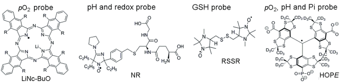
図 1。TME 評価分析を組み立てる常磁性プローブの化学構造。粒子pO2プローブ、LiNc BuO 含まれます (R = O (2CH) の3CH3)、および可溶性プローブ: デュアル機能 pH および酸化還元プローブ、NR;グルタチオン (gsh) 敏感なプローブ、RSSR;多機能pO2, pH, および細胞外微小環境、希望プローブ.の Pi プローブこれらのプローブの合成は、提供された参照11,12に記載されています。この図の拡大版を表示するのにはここをクリックしてください

Protocol

WVU IACUC 承認プロトコルに従ってすべての動物の仕事を行った。 1. プローブの合成と校正 粒子pO2-敏感な LiNc BuO プローブ注:LiNc BuO 微結晶を合成して、参照13で説明されています。彼らは非常に安定して、年室温で保管することができます。LiNc BuO 粒子プローブの EPR 線幅はpO2-機密性の高いパラメータ?…

Representative Results

組織pO2プローブを用いた LiNc BuO 評価: 手順 1.1 で説明されている手順を使用して、作りたて LiNc BuO 微結晶懸濁液の校正を行った。図 2は、LiNc BuO プローブと測定バッファー懸濁液と細胞の注入によって腫瘍開始後成長して?…

Discussion

提示方法すなわちpO2pH、酸化還元状態、および間質性 Pi および細胞内 GSH 濃度化学 TME の重要なパラメーターの生体内非侵襲的評価を可能にします。MRI や低磁場 EPR などの磁気共鳴技術は、非侵襲体内のこれらの TME パラメーターのプロファイルの選択の方法です。MRI は、解剖学的構造を可視化するが、機能の感度を欠いています。MRI とは対照的 EPR 技術は微小環?…

Disclosures

The authors have nothing to disclose.

Acknowledgements

この作品は、NIH によって部分的に支持された CA194013、CA192064、U54GM104942 を付与します。VVK, ab 級, し、TDE の立ち上げのため、WVCTSI を認めた著者は、博士・ m ・ Gencheva、k. ・ シュタインバーガーを例示実験支援ありがちましょう。内容は著者の責任と NIH の公式見解を必ずしも表さない。

Materials

L-band EPR spectrometer Magnettech, Germany L-band (1.2 GHz) electron paramagnetic resonance (EPR) spectrometer for collection in vitro and in vivo spectra of paramagnetic molecules
 Temperature & Gas Controller  Noxygen, Germany Temperature & Gas Controller designed to control and adjust the temperature and gas composition  
Sonicator Fisher Scientific
GSH (L-Glutathione reduced) Sigma-Aldrich G4251
MMTV-PyMT  mice In house
DMEM Thermo Fisher Scientific 11995065
Met-1 murine breast cancer cells In house
C57Bl/6 wild type mice  Jackson Laboratory
Trypsin Thermo Fisher Scientific 25200056
Trypan Blue Exclusion Dye  Thermo Fisher Scientific T10282
Ohmeda Fluotec 3 
Isoflurane (IsoFlo) Abbott Laboratories
Sodium phosphate dibasic Sigma-Aldrich S9763
Sodium phosphate monobasic sigma-Aldrich S07051
Sodium Chloride sigma-Aldrich S7653
Hydrochloric acid sigma-Aldrich 320331
Sodium Hydroxide sigma-Aldrich S8045
Glucose sigma-Aldrich
Glucose oxydase sigma-Aldrich
Lauda Circulator E100 Lauda-Brikmann
pH meter Orion Thermo Scientific 
LiNc-BuO probe In house The Octa-n-Butoxy-Naphthalocyanine probe was synthesizided according to ref 13
NR probe In house The Nitroxide probe was synthesizided according to ref 11
RSSR probe In house The di-Nitroxide probe was synthesizided according to ref 15
HOPE probe In house The monophoshonated Triarylmethyl probe was synthesizided according to ref 12

References

  1. Siemann, D. W. . Tumor Microenvironment. , (2011).
  2. Tatum, J. L., et al. Hypoxia: importance in tumor biology, noninvasive measurement by imaging, and value of its measurement in the management of cancer therapy. Int J Radiat Biol. 82 (10), 699-757 (2006).
  3. Brahimi-Horn, M. C., Chiche, J., Pouyssegur, J. Hypoxia signalling controls metabolic demand. Curr Opin Cell Biol. 19 (2), 223-229 (2007).
  4. Haulica, A., Ababei, L. Comparative study of glycolytic activity in the erythrocytes of animals with chronic experimental hypoxia and with tumours. Neoplasma. 21 (1), 29-35 (1974).
  5. Matsumoto, K., et al. High-resolution mapping of tumor redox status by magnetic resonance imaging using nitroxides as redox-sensitive contrast agents. Clin Cancer Res. 12 (8), 2455-2462 (2006).
  6. Estrela, J. M., Ortega, A., Obrador, E. Glutathione in cancer biology and therapy. Crit Rev Clin Lab Sci. 43 (2), 143-181 (2006).
  7. Voegtlin, C., Thompson, J. W. Glutathione content of tumor animals. J. Biol. Chem. 70, 801-806 (1926).
  8. Bobko, A. A., et al. Interstitial Inorganic Phosphate as a Tumor Microenvironment Marker for Tumor Progression. Sci Rep. 7, 41233 (2017).
  9. Gillies, R. J., Raghunand, N., Garcia-Martin, M. L., Gatenby, R. A. pH imaging. A review of pH measurement methods and applications in cancers. IEEE Eng Med Biol Mag. 23 (5), 57-64 (2004).
  10. Gade, T. P., et al. Imaging intratumoral convection: pressure-dependent enhancement in chemotherapeutic delivery to solid tumors. Clin Cancer Res. 15 (1), 247-255 (2009).
  11. Bobko, A. A., et al. In vivo monitoring of pH, redox status, and glutathione using L-band EPR for assessment of therapeutic effectiveness in solid tumors. Magn Reson Med. 67, 1827-1836 (2012).
  12. Dhimitruka, I., Bobko, A. A., Eubank, T. D., Komarov, D. A., Khramtsov, V. V. Phosphonated Trityl Probe for Concurrent In Vivo Tissue Oxygen and pH Monitoring Using EPR-based Techniques. JACS. 135, 5904-5910 (2013).
  13. Pandian, R. P., Parinandi, N. L., Ilangovan, G., Zweier, J. L., Kuppusamy, P. Novel particulate spin probe for targeted determination of oxygen in cells and tissues. Free Radic Biol Med. 35 (9), 1138-1148 (2003).
  14. Bobko, A. A., Evans, J., Denko, N. C., Khramtsov, V. V. Concurrent Longitudinal EPR Monitoring of Tissue Oxygenation, Acidosis, and Reducing Capacity in Mouse Xenograft Tumor Models. Cell Biochem Biophys. 75, 247-253 (2017).
  15. Khramtsov, V. V., Yelinova, V. I., Glazachev Yu, I., Reznikov, V. A., Zimmer, G. Quantitative determination and reversible modification of thiols using imidazolidine biradical disulfide label. J Biochem Biophys Methods. 35 (2), 115-128 (1997).
  16. Roshchupkina, G. I., et al. In vivo EPR measurement of glutathione in tumor-bearing mice using improved disulfide biradical probe. Free Rad. Biol. Med. 45, 312-320 (2008).
  17. Khramtsov, V. V., Zweier, J. L., Hicks, R. . Stable Radicals: Fundamentals and Applied Aspects of Odd-Electron Compounds. , 537-566 (2010).
  18. Bobko, A. A., Dhimitruka, I., Zweier, J. L., Khramtsov, V. V. Fourier Transform EPR of Trityl Radicals for Multifunctional Assessment of Chemical Microenvironment). Angew. Chem. Int. Edit. 53, 2735-2738 (2014).
  19. Martin, M. L., Martin, G. J., Delpuech, J. J. . Practical NMR spectroscopy. , (1980).
  20. Lin, E. Y., et al. Progression to malignancy in the polyoma middle T oncoprotein mouse breast cancer model provides a reliable model for human diseases. Am J Pathol. 163 (5), 2113-2126 (2003).
  21. Eubank, T. D., et al. Granulocyte macrophage colony-stimulating factor inhibits breast cancer growth and metastasis by invoking an anti-angiogenic program in tumor-educated macrophages. Cancer Res. 69 (5), 2133-2140 (2009).
  22. Khramtsov, V. V., et al. Quantitative determination of SH groups in low- and high-molecular-weight compounds by an electron spin resonance method. Anal Biochem. 182 (1), 58-63 (1989).
  23. Komarov, D. A., et al. Electron paramagnetic resonance monitoring of ischemia-induced myocardial oxygen depletion and acidosis in isolated rat hearts using soluble paramagnetic probes. Magnetic Resonance in Medicine. 68 (2), 649-655 (2012).
  24. Song, Y. G., Liu, Y. P., Liu, W. B., Villamena, F. A., Zweier, J. L. Characterization of the binding of the Finland trityl radical with bovine serum albumin. Rsc Advances. 4 (88), 47649-47656 (2014).
  25. Khramtsov, V. V., Bobko, A. A., Tseytlin, M., Driesschaert, B. Exchange Phenomena in the Electron Paramagnetic Resonance Spectra of the Nitroxyl and Trityl Radicals: Multifunctional Spectroscopy and Imaging of Local Chemical Microenvironment. Analyt. Chem. 89 (9), 4758-4771 (2017).
  26. Samouilov, A., et al. In Vivo Proton-Electron Double-Resonance Imaging of Extracellular Tumor pH Using an Advanced Nitroxide Probe. Analyt. Chem. 86 (2), 1045-1052 (2014).
  27. Goodwin, J., et al. In vivo tumour extracellular pH monitoring using electron paramagnetic resonance: the effect of X-ray irradiation. NMR Biomed. 27 (4), 453-458 (2014).
check_url/kr/56624?article_type=t

Play Video

Cite This Article
Bobko, A. A., Eubank, T. D., Driesschaert, B., Khramtsov, V. V. In Vivo EPR Assessment of pH, pO2, Redox Status, and Concentrations of Phosphate and Glutathione in the Tumor Microenvironment. J. Vis. Exp. (133), e56624, doi:10.3791/56624 (2018).

View Video