Summary

Measuring the Bending Stiffness of Bacterial Cells Using an Optical Trap

Published: April 26, 2010
doi:

Summary

We present a protocol for bending filamentous bacterial cells attached to a cover-slip surface with an optical trap to measure the cellular bending stiffness.

Abstract

We developed a protocol to measure the bending rigidity of filamentous rod-shaped bacteria. Forces are applied with an optical trap, a microscopic three-dimensional spring made of light that is formed when a high-intensity laser beam is focused to a very small spot by a microscope’s objective lens. To bend a cell, we first bind live bacteria to a chemically-treated coverslip. As these cells grow, the middle of the cells remains bound to the coverslip but the growing ends are free of this restraint. By inducing filamentous growth with the drug cephalexin, we are able to identify cells in which one end of the cell was stuck to the surface while the other end remained unattached and susceptible to bending forces. A bending force is then applied with an optical trap by binding a polylysine-coated bead to the tip of a growing cell. Both the force and the displacement of the bead are recorded and the bending stiffness of the cell is the slope of this relationship.

Protocol

  1. Grow E. coli cells in Luria-Beltrani broth (LB) medium to exponential phase (OD = 0.2-0.4).
  2. Grow the culture in LB supplemented with 50 μg/mL of cephalexin for 15 min to induce filamentous growth, and then concentrate the culture by 5 times thru centrifugation.
  3. Make PEI-coated coverslip by flowing 1% polyethylenimine diluted in water into a flow chamber, and wash with water after a 5-minute incubation.
  4. Flow the concentrated cell culture into the chamber, and washed with a mixture of LB and cephalexin(50μg/mL) after 3 min to remove unattached cells.
  5. Incubate the chamber at 37°C for 30 min-1 hour to let the attached cells grow before placement on the optical trapping instrument.
  6. Make polylysine-coated beads by incubating 0.5-μm-diameter polystyrene beads (Bangs Labs) in 0.1% polylysine diluted in water for 30 min. Then wash the beads 3 times and resuspend in water.
  7. Dilute the bead solution by a factor of two into LB with cephalexin (50μg/mL), and add it into the flow chamber.
  8. Optically trap a floating bead and touch it to the free tip of a cell. (We use a Mad City Labs piezo stage to control of the motion of the sample.) When a suitable bead/cell combination is found, wash the chamber with cephalexin in LB (50μg/mL) to remove unattached beads.
  9. Run custom-written LabView program to apply bending forces to cell and record force-displacement data. Program Details
    1. Calibrate detector response by raster scanning the attached bead within the detection laser beam and recording 3D PSD voltage signals.
    2. Identify cell long axis in microscope image.
    3. Move cell in steps in a direction perpendicular to the cell long axis.
    4. Record the distance moved and the displacement from the optical trap.
    5. Convert displacement to applied force using the previously measured trap stiffness and save tip displacement and force to file.

Secrets to Success: In Step 8), one needs to find a cell with a well-defined stuck end. Some cells are just stuck at one tip, and the bending force at the other tip leads to a whole-cell pivoting rather than bending. A suitable pair is found by bending each cell quickly by hand using the joystick-controlled stage motion.

Representative Results:

Figure 1
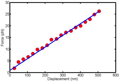
Figure 1. This figure shows force-displacement data for a single cell. The slope of this line is the bending stiffness of the cell.

Discussion

The protocol presented here is designed to quantitatively measure the bending properties of bacterial cells. The experiment setup can be applied to any rod-shaped cell that can be made to grow filamentously. We have successfully used this setup to probe the effects of cytoskeletal filaments on the bending stiffness of E. coli cells. This same technique can be used to evaluate the roles of pressure, cell wall stiffness and other intracellular components in determining overall bending stiffness of cells.

Disclosures

The authors have nothing to disclose.

Acknowledgements

We acknowledge helpful advice from Mingzhai Sun on binding cells to surfaces. We thank Ned Wingreen and Zemer Gitai for valuable discussions. This research was supported by National Institutes of Health grant P50GM07150, National Science Foundation CAREER award PHY-0844466 and the Alfred P. Sloan Foundation.

Materials

Material Name Type Company Catalogue Number Comment
cephalexin   Sigma C4895-5G  
polyethylenimine   Sigma 181978-5G  
polylysine   Sigma P8920  
0.5-μm-diameter polystyrene beads   Bangs Laboratory PS03N  
Nano-LP Series nanopositioning system   Mad City Labs NanoLP series http://www.madcitylabs.com/nanolpseries.html

References

  1. Janmey, P. A., McCulloch, C. A. Cell mechanics: integrating cell responses to mechanical stimuli. Annu Rev Biomed Eng. 9, 1-34 (2007).
  2. Morris, D. M., Jensen, G. J. Toward a biomechanical understanding of whole bacterial cells. Annu Rev Biochem. 77, 583-613 (2008).
check_url/pt/2012?article_type=t

Play Video

Cite This Article
Wang, S., Arellano-Santoyo, H., Combs, P. A., Shaevitz, J. W. Measuring the Bending Stiffness of Bacterial Cells Using an Optical Trap. J. Vis. Exp. (38), e2012, doi:10.3791/2012 (2010).

View Video